Surgical roadmap for congenital cervical spine abnormalities
Автор: Gubin Alexander V., Ulrich Eduard V., Ryabykh Sergei O., Burcev Alexander V., Ochirova Polina V., Pavlova Olga M.
Журнал: Гений ортопедии @geniy-ortopedii
Рубрика: Оригинальные статьи
Статья в выпуске: 2, 2017 года.
Бесплатный доступ
Study Design A descriptive study based on medical records of patients with congenital cervical spine abnormalities. Objective The hypothesis of the study was that patients with congenital cervical spine abnormalities could be divided according to the main pathological syndrome. Summary of Background Data Abnormalities of the cervical spine belong to embryopathies and are a very heterogeneous group. The variety of this pathology made it hard to create a general classification based on a morphological approach. Materials Medical records of 68 patients with congenital cervical spine abnormalities were analyzed and were a clinical material for working out the algorithm of their management. Computer tomography, magnetic resonance imaging (MRI) and selective angiography were used to specify the abnormality structure and preoperative planning. Use of functional positioning was an important feature in all these investigations. Various techniques of surgical treatment such as halo, anterior and posterior fusion, decompression of the cerebral, spinal cord and cervical vertebral arteries, revision of the spinal canal, neurolysis, and meningolysis were used in 28 patients aged from 2 to 47 years old. Results All patients were divided according to leading pathological syndromes. Those were instability, stenosis and brain ischemia. Each group had its own important subgroup. The main surgical steps in management of complex congenital anomalies of the cervical spine were bone fusion or (and) decompression of the brain and spinal cord. Conclusions Selection of the main pathological syndrome or combination of syndromes is a simple and effective way for making the right decision when treating patients with congenital cervical spine abnormalities. Syndromic approach can be used for prognosis as well.
Congenital spine abnormalities, cervical instability, klippel-feil syndrome, spine fusion, cervical cord decompression, cervical spine abnormalities classification
Короткий адрес: https://sciup.org/142121954
IDR: 142121954 | УДК: 616.711.1-007.1-053.1-048.445-089 | DOI: 10.18019/1028-4427-2017-23-2-147-153
Текст научной статьи Surgical roadmap for congenital cervical spine abnormalities
Abnormalities of the cervical spine belong to a very heterogeneous group of embryopathies [1]. They include all morphological types of congenital spine malformations: loss of formation and segmentation, anterior and posterior spina bifida, diastematomyelia and other tethering disorders of the spinal canal. Besides, there is an entire range of such exceptional conditions as os odontoideum, proatlas, occipitalization. Cervical abnormalities are not included into classical classifications of malformations which are valid for thoracic and lumbar spine. The abnormalities of the craniocervical junction, involving skull base and brain, should be considered as a single symptom-complex with vertebral malformations because the severity of patient’s condition is often determined by them [2]. High frequency of cervical abnormalities in the structure of genetic syndromes is a specific feature as well (Down, Larsen, Wildervank, Rokitansky-Huster-Houser, Goldenhara and other syndromes) [1, 3–5].
There is a number of vascular malformations of the cervical spine which may be of high clinical significance. Bavinsk and Weaver (1986) supposed that failure of vertebral arteries formation can play a significant role in
the etiology of malformations in Klippel-Feil, Poland, and Mobius syndromes [6].
Loss of segmentation at a single level has already been referred to a variant of Klippel-Feil syndrome [7]. The abnormalities of the craniocervical junction are more often considered separately or in the structure of various genetic syndromes. There is no unified approach to their management and no general classification of the congenital cervical spine abnormalities.
The subject of our study was to work out a surgical scheme (algorithm) of managing patients with congenital abnormalities of the cervical spine.
MATERIAL AND METHODS
Medical records of 68 patients with congenital cervical spine abnormalities were analyzed as a clinical material for working out the algorithm. Patients’ pathology was revealed on the basis of clinical data, and in some cases – incidentally. Neck deformities, facial asymmetry, head motion restrictions, multiple combined malformations of internal organs and central nervous system, skin stigmata were the most significant symptoms in the process of identification.
Lateral view X-rays confirmed the presence of abnormalities. Computer tomography, magnetic resonance imaging (MRI) and selective angiography were used to specify the abnormality structure and to make preoperative planning. Use of functional positioning was an important feature in all these investigations.
Various techniques of surgical treatment such as halo cast application, anterior and posterior fusion, decompression of the cerebral, spinal cord and cervical vertebral arteries, revision of the spinal canal, neurolysis, and meningolysis were used in 28 patients, aged from 2 to 47 years.
RESULTS
We have developed our own working scheme of congenital abnormalities of the cervical spine on the basis of our experience and the data of world literature. The main principle was the selection of a leading pathological syndrome. The potential for its development was also taken into account, thereby determining the prognosis for the patient (Fig. 1).
The definite conclusion on the neutral character of a congenital abnormality could be achieved only after a comprehensive assessment of its clinical picture and the results of additional investigations obtained dynamically.
An abnormality can be referred to a neutral one but the patient needs cosmetic surgery. We performed five cosmetic «cervicalizations» in three patients with a neutral abnormality type.
All destabilizing abnormalities were divided into primary and secondary instability groups. The instability in the primary group was caused by the structure of malformation itself and located at the level of abnormality (Fig. 2). It was presented practically from birth. The secondary instability developed in the adjacent to malformation (commonly loss of segmentation) segments due to their degenerative changes (Fig. 3). Such division was absolutely necessary as far as the first group requires stabilization as early as possible. As for the second group, its dynamic observation was used and physiotherapy was possible. Surgical interventions were performed if conservative treatment for neck pain or neurologic deficit had failed. Neurological instability prevention is the main task in treatment of all cervical destabilizing abnormalities.
All the abnormalities reducing the lumen of the spinal canal and intervertebral foramena from C0 to C7 were included into the group of stenosis. Decompression of the brain or spinal cord is the main intervention type in these patients (Fig. 4).
Stenosis abnormalities are divided into two groups: Extracanal stenosis or tethering
-
1. Progressive scoliotic and kyphotic deformities due to the loss of segmentation and formation;
-
2. Congenital narrow spinal canal;
-
3. Developmental arc abnormalities with spinal cord and (or) roots compression;
-
4. Basilar impression, platibasia, convexobasia;
-
5. Occipitalisation with narrowing of the foramen magnum.
Most of these anomalies are non-symptomatic in childhood but can become a problem in the third to fourth decades of patients’ life [8, 9].
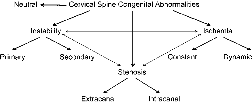
Fig. 1. Congenital cervical spine abnormalities. Scheme based on the main pathological syndrome

Fig. 2. 3-year old boy with congenital torticollis, right hand and leg mild hypoplasia and heart rhythm disorder when trying to correct head position: a – MRI. Cervical kyphosis and torticollis due to lateral posterior C4 hemivertebrae (primary instability site); b – Posterior fusion after one week of halo-cast head reposition under heart rhythm control; c – 6 years follow-up. Full sagittal plane restoration and good bone fusion. No heart rhythm problems after surgery

Fig. 3. 9-year old boy with Klippel-Feil syndrome. Tetraparesis occurred after jump from 1 m height: a – functional X-ray (medium, flexion, extension). Instability in only one not congenitally fused segment C3-C4 (secondary instability); b – MRI C3-C4 disk degeneration and cord compression; c – anterior and posterior fusion. Full neurological recovery at 14-year follow-up
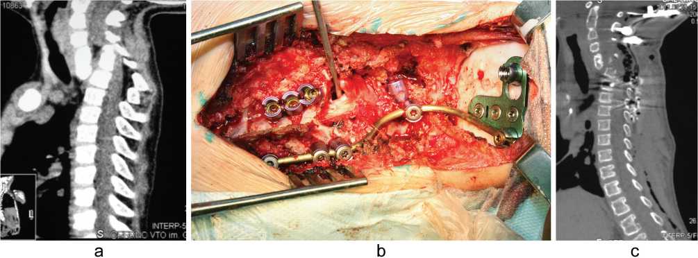
Fig. 4. 9-year old boy with tetraparesis from the age of 1.5 years. Malformation was diagnosed only at age of 9: a – CT. Cord compression by anterior inclination due to C7 body aplasia; b – full anterior and posterior decompression and fusion from posterior approach. Intraoperative view; c – postoperative CT
Intracanal stenosis or tethering
-
1. Diastematomyelia;
-
2. Dermoid cysts.
Not only clinical symptoms but negative dynamics of neurological methods of study was an indication for surgical treatment in case of stenosis congenital cervical abnormalities.
Ischemic abnormalities were divided into two groups: constant (persistent) and dynamic. Hypoplasia and aplasia of the main cervical vessels and vascular malformations were referred to constant ischemic abnormalities. All variants of cervical vessel compression by abnormal spine structures at definite head positions were referred to dynamic ischemic abnormalities. Such division was very necessary due to different tactics of managing these patients. Most of constant ischemic abnormalities were identified accidentally by ultrasound or MRI and CT angiography. Since this blood flow asymmetry was present from birth, adaptation to it was very high. These patients did not require surgical treatment. But this information had to be taken into consideration if they needed posterior screw fixation for any other reason.
We consider dynamic ischemia to be the main cause of vertebrobasilar insufficiency with clinical manifestations. We have performed surgical treatment of two boys with complete compression of one vertebral artery in rotation movement (Bow’s hunter stroke) in the complex of other congenital cervical abnormalities (Fig. 5). Occipitospondylodesis was performed in one case. Surgical decompression of vertebral artery with simultaneous fusion of malformed C2-C3 joint was made in another. The disappearance of headache and dizziness was observed in both cases.
Restoration of blood flow has been confirmed by control functional selective angiographies.
The working scheme proposed highlights the leading pathological syndrome, which requires specific diagnostic measures and, depending on their results, its correction. Nevertheless, a great number of abnormalities could migrate between the groups singled out or were combined. Thus, some patients after decompression for stenosing abnormality would require stabilization because the decompression itself leads to destabilization of the segment operated. When patients with destabilizing and (or) stenosing anomalies were prepared for surgery, the condition of the cervical vessels was assessed for ischemic abnormality. In our opinion, the combination of mechanical instability with spinal canal stenosis was an adverse one, requiring simultaneous decompression and stabilization (Fig. 6).
It was possible to highlight the leading pathological syndrome in all our patients (Table 1).
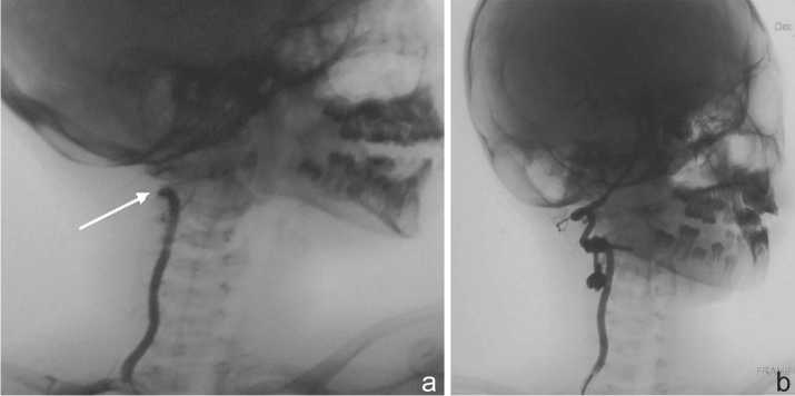
Fig. 5. 9-year old boy with chronic headache and vertigo after head movements: a – Functional selective angiography with head rotation. Blood circulation lock at C3-C2; b – Functional selective control angiography after art. vertebralis decompression and C2-C3 monolateral instrumentation. 5-year follow-up, no vertigo and reduction in headaches
Fig. 6. 8-year old boy with mild tetraplegia and chronic headache: a – combination of stenosis and instability due to Kiari syndrome and dens hypoplasia; b – C0-C1 posterior decompression and C0-C2 fusion. 5-year follow up, no complaints or neurological deficit
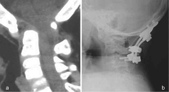
Table 1
Patients with congenital cervical abnormalities divided into groups by the main pathological syndrome. Types and number of surgeries
|
Abnormality group |
Number of patients |
Number of surgically treated patients |
Number of surgical sessions |
Surgery types |
|
Neutral |
16 |
2 |
3 |
«cervicalization» |
|
Instability |
24 |
16 |
19 |
Halo Posterior fusion Anterior fusion Anterior and posterior cord decompression + fusion |
|
Stenosis |
22 |
8 |
8 |
Posterior brain and cord decompression Anterior cord decompression 360º cord decompression from posterior approach Anterior and posterior fusion Dura reconstruction |
|
Ischemia |
6 |
2 |
2 |
C0-C4 fusion Art. vertebralis decompression |
|
All |
68 |
28 |
32 |
DISCUSSION
A unified congenital cervical spine abnormalities classification is a challenging problem due to a great variety and different combinations of malformations in the unique neck anatomy. The cervical vertebral segmentation loss or Klippel-Feil syndrome was subjected to systematization most thoroughly.
In 1919, Feil gathered 13 patients and proposed to divide them into three types [10]:
I – Massive bone fusions of the cervical and thoracic spine;
-
II – Fusion of one or two spinal-and-motion segments in combination with half-vertebrae, occipitalization and other anomalies of the cervical spine;
-
III – Segmentation disorders of the cervical spine associated with the lower thoracic and lumbar fusion.
Thomsen et al. (1997) divided their 57 patients according to Feil’s classification [10]. The following ratio has resulted in them:
I – 23 (40 %);
II – 27 (47 %);
-
III – 7 (13 %).
The authors made an important conclusion that Types I and III were the most dangerous as they correlated with a course of progressive scoliotic deformity. Type II differed by the greatest variety of bone anomalies. Type I was typical for syndromes.
Feil’s classification did not become popular. It was mainly focused on revealing the combinations of axial skeletal abnormalities but was not informative for making a useful prognosis for a patient [11].
On the basis of the criteria, worked out by McRae, Hensinger popularized the classification based on different combinations of cervical vertebral segmentation disorders [12, 13]:
Type I – occipitalization and C2-C3 fusion;
Type II – great-extent disorder and craniocervical anomaly;
Type III – two fused segments with the preserved disc between them.
In the authors’ opinion, Type III was especially dangerous in terms of instability development.
Pizzutillo et al. (1994) proposed their functional classification of Klippel-Feil syndrome according to X-ray criteria [2]. The research technique was tested on 25 patients without any pathology in the cervical spine. The authors analyzed functional spondylographies of 111 subjects with Klippel-Feil syndrome. The following types were distinguished:
I – normal range of movements in the upper and lower cervical spine;
-
II – intrasegmental hypermobility in the upper cervical spine, basilar impression or inencephaly;
-
III – intrasegmental hypermobility in the lower cervical spine, X-ray signs of degenerative process;
-
IV – combination of Types II and III.
Types II and III represented a risky group for development of neurologic complications and required observation, special regimen of working and sport activities, as well as preventive treatment.
Dino Samartzis et al. (2006) proposed their X-ray classification, based on functional images as well [11, 14]. The authors considered that it was more suitable for analyzing the Klippel-Feil syndrome in children:
Type I – single congenital bone blocks (fusion);
Type II – multiple non-adjacent congenital bone blocks;
Type III – multiple adjacent congenital bone blocks.
The authors’ clinical experience was 28 patients at a mean age of 8 years. According to their classification, the ratio of the types was 25 %, 50 %, 25 %, respectively. The clinical manifestations of myelo- and radiculopathy required surgical treatment in 3 patients of Types II and III. In their review, 36 % had complaints. Different complaints prevailed in different types. Headache, cervical pain and stiffness were characteristic symptoms of Type I. Types II and III were at risk of neurological instability development. The mean age for myelopathy development was found to be 10 years, for pain syndrome in the cervical spine – 13 years, and for radiculopathy – 18 years and is of importance as well.
Abnormalities of the craniovertebral spine were reflected in some anatomical classifications. In 2007, Gholve proposed to divide all types of occipitalization into 3 groups according to the zones of C1 fusion with the occipital bone [15]. The classification resulted from the examination of 30 patients in the mean age of 6.5 years. The fusion in Zone 1, along the anterior arch of the atlas, was observed in 6 children, in Zone 2 along the lateral masses, – in 5 children, in Zone 3, along the posterior arch, – in 4 cases. Mixed variants were observed in the remaining 15 children. It was the most important that the signs of atlanto-axial instability were observed in 57 % of cases, especially when combined with C2-C3 fusion. There was spinal cord compression in 37 %, and its highest rate (63 %) in the presence of occipitalization in Zone 2.
Dubousset described three types of atlas abnormality [16]:
Type I – isolated half atlas;
Type II – complete or partial aplasia of one atlas half with segmentation disorders in the cervical spine;
Type III – partial or complete occipitalization with partial or complete ipsi- or contralateral atlas aplasia and the anomalies of the odontoid bone and other cervical vertebrae.
The author emphasized the importance of these abnormalities in the genesis of congenital non-myogenic torticollis.
As far as clinical observations associated with blood flow impairment due to the anomalies in cervical vessels were presented in single works, the existing classifications were, in fact, descriptions of the variants of vessel structures or arrangement. So, there are 5 variants of the vertebral artery entrance into the canal of cervical vertebral transverse processes [17]. Three types of vertebral artery structure and position at C1-C2 level were described, and it is important to take it into consideration during surgical fixation of this zone [18, 19].
Despite the diversity of the above classifications the main surgical steps in complex developmental abnormalities of the cervical spine remain stabilization or decompression of the brain and spinal cord [20].
CONCLUSION
Treatment of congenital abnormalities of the cervical spine is a complex problem. The decision making involves various specialists. This is possible only in large surgical centers. Contemporary spine surgery has a wide range of methods and tools for examination and treatment of these patients. Neurological instability is a definitive indication for surgical treatment at any age. Mechanical instability requires special examination, observation and surgical treatment as far as there is a threat of its progression, risk of associated neurological instability or pain that cannot be arrested conservatively. Cervical spine deformities and malformations associated with “short neck” and dysembryogenesis stigmas are indications for using the techniques of “cosmetic spine surgery”. This conditions result in the same moral suffering as traditionally correctable deformities of the thoracic spine and chest.
The use of modern screw systems for cervical spine fixation requires the understanding of the condition and position of vertebral arteries before surgery. Thus, the detection of constant ischemic abnormalities is an important aspect of preoperative planning.
We suppose that the majority of patients with dynamic ischemic developmental abnormalities of the cervical spine should be under supervision of neurologists with the diagnosis of vertebrobasilar insufficiency. Active involvement of spine surgeons in rendering medical care for this group of patients will undoubtedly result in new statistical data and solving a number of medical problems.
Since most patients do not need surgical interventions, it is necessary to determine the prognosis and an individual scheme of observation and conservative treatment for them. Physiotherapists can also participate in the management of these patients.
Список литературы Surgical roadmap for congenital cervical spine abnormalities
- Kusumi K., Turnpenny P.D. Formation errors of the vertebral column//J. Bone Joint Surg. Am. 2007. Vol. 89, Suppl. 1. P. 64-71.
- Risk factors in Klippel-Feil syndrome/P.D. Pizzutilo, M. Woods, L. Nicholson, G.D. MacEwen//Spine. 1994. Vol. 19, No 18. P. 2110-2116.
- McLay K., Maran A.G. Deafness and the Klippel-Feil syndrome//J. Laryngol. Otol. 1969. Vol. 83, No 2. P. 175-184.
- Symptomatic atlantoaxial subluxation in persons with Down syndrome/S.M. Pueschel, J.H. Herndon, M.M. Gelch, K.E. Senft, F.H. Scola, M.J. Goldberg//J. Pediatr. Orthop. 1984. Vol. 4, No 6. P. 682-688.
- Sherk H.H., Nicholson J.T. Cervico-oculo-acusticus syndrome. Case report of death caused by injury to abnormal cervical spine//J. Bone Joint Surg. Am. 1972. Vol. 54, No 8. P. 1776-1778.
- Bavinck J.N., Weaver D.D. Subclavian artery supply disruption sequence: hypothesis of a vascular etiology for Poland, Klippel-Feil, and Möbius anomalies//Am. J. Med. Genet. 1986. Vol. 23, No 4. P. 903-918.
- The cervical spine in the Klippel-Feil syndrome. A report of 57 cases/H. Baba, Y. Maezawa, N. Furusawa, Q. Chen, S. Imura, K. Tomita//Int. Orthop. 1995. Vol. 19, No 4. P. 204-208.
- Dyste G.N., Menezes A.H., VanGilder J.C. Symptomatic Chiari malformations. An analysis of presentation, management, and long-term outcome//J. Neurosurg. 1989. Vol. 71, No 2. P. 159-168.
- The natural history of Klippel-Feil syndrome: clinical, roentgenographic, and magnetic resonance imaging findings at adulthood/J.T. Guille, A. Miller, J.R. Bowen, E. Forlin, P.A. Caro//J. Pediatr. Orthop. 1995. Vol. 15, No 5. P. 617-626.
- Scoliosis and congenital anomalies associated with Klippel-Feil syndrome types I-III/N.M. Thomsen, U. Schneider, M. Weber, R. Johannnisson, F.U. Niethard//Spine. 1997. Vol. 22, No 4. P. 396-401.
- Classification of congenitally fused cervical patterns in Klippel-Feil patients: epidemiology and role in the development of cervical Spine-related symptoms/D.D. Samartzis, J. Herman, J.P. Lubicky, F.H. Shen//Spine. 2006. Vol. 31, No 21. P. E798-E804.
- Hensinger R.N., Lang J.E., MacEwen G.D. Klippel-Feil syndrome; a constellation of associated anomalies//J. Bone Joint Surg. Am. 1974. Vol. 56, No 6. P. 1246-1253.
- Hensinger R.N. Congenital anomalies of the cervical spine//Clin. Orthop. Relat. Res. 1991. No 264. P. 16-38.
- 2008 Young Investigator Award: The role of congenitally fused cervical segments upon the space available for the cord and associated symptoms in Klippel-Feil patients/D. Samartzis, P. Kalluri, J. Herman, J.P. Lubicky, F.H. Shen//Spine. 2008. Vol. 33, No 13. P. 1442-1450 DOI: 10.1097/BRS.0b013e3181753ca6
- Occipitalization of the atlas in children. Morphologic classification, associations, and clinical relevance/P.A. Gholve, H.S. Hosalkar, E.T. Ricchetti, A.N. Pollock, J.P. Dormans, D.S. Drummond//J. Bone Joint Surg. Am. 2007. Vol. 89, No 3. P. 571-578.
- Dubousset J. Torticollis in children caused by congenital anomalies of the atlas//J. Bone Joint Surg. Am. 1986. Vol. 68, No 2. P. 178-188.
- The vertebral arteries (segments V1 and V2)/C. Argenon, J.P. Francke, S. Sylla et al.//Anat. Clin. 1980. Vol. 2. P. 29-41.
- Magnetic resonance imaging of C2 segmental type of vertebral artery/K. Sato, T. Watanabe, T. Yoshimoto, M. Kameyama//Surg. Neurol. 1994. Vol. 41, No 1. P. 45-51.
- Anomalous atlantoaxial portions of vertebral and posterior inferior cerebellar arteries/K. Tokuda, K. Miyasaka, H. Abe, S. Abe, H. Takei, S. Sugimoto, M. Tsuru//Neuroradiology. 1985. Vol. 27, No 5. P. 410-413.
- Губин А.В., Ульрих Э.В., Рябых С.О. Перспективы оказания помощи детям младшего и ювенильного возраста с хирургической патологией позвоночника//Гений ортопедии. 2011. № 2. С. 123-127.

