The effect of hypokinesia on the dynamics of the formed elements of blood during the embryonic period of prenatal development in pregnant rabbits

Author: Hamidova J., Kazimov A., Aghayeva E.
Journal: Бюллетень науки и практики @bulletennauki
Section: Кратко: новые достижения
Article in issue: 6 т.10, 2024.
Free access
The erythroformula and leucoformula indicators changed in the rabbits that experienced hypokinesia during the embryonic stage of pregnancy. Compared to the rabbits that spent the pregnancy period under normal conditions, changes in concentrations of leukoformula indicators such as leukocytes, lymphocytes, monocytes, and granulocytes were detected in rabbits exposed to hypokinesia. While significant changes in erythroformula indicators: erythrocytes, average corpuscular volume of erythrocytes and hemoglobin capacity, erythrocyte sedimentation rate did not occur. The results of our studies have shown that prenatal hypokinesia causes noticeable changes in the blood coagulation system. On the other hand, changes in the coagulation system under the influence of prenatal hypokinesia were found to occur in the first periods of postnatal ontogenesis, had a continuous nature, and were maintained until the full maturity of postnatal development. Due to the effect of the hypokinesia factor, the changes in the mentioned indicators lead to the emergence of severe pathological diseases in most cases.
Hypokinesia, prenatal, erythroformula, leukoformula
Short address: https://sciup.org/14130509
IDR: 14130509 | UDC: 638.152/154 | DOI: 10.33619/2414-2948/103/97
Влияние гипокинезии на динамику форменных элементов крови в эмбриональном периоде пренатального развития у беременных крольчих
У крольчих, подвергшихся гипокинезии в эмбриональный период беременности, изменялись показатели эритроформулы и лейкоформулы. По сравнению с крольчихами, проводившими период беременности в обычных условиях, у крольчих, подвергшихся гипокинезии, выявлены изменения концентраций таких показателей лейкоформулы, как лейкоциты, лимфоциты, моноциты и гранулоциты. При этом существенных изменений показателей эритроформулы: эритроцитов, среднего корпускулярного объема эритроцитов и емкости гемоглобина, скорости оседания эритроцитов не происходило. Результаты наших исследований показали, что пренатальная гипокинезия вызывает заметные изменения в системе свертывания крови. С другой стороны, установлено, что изменения в системе свертывания под влиянием пренатальной гипокинезии возникают в первые периоды постнатального онтогенеза, носят непрерывный характер и сохраняются до полной зрелости постнатального развития. Изменения указанных показателей вследствие воздействия фактора гипокинезии в большинстве случаев приводят к возникновению тяжелых патологических заболеваний.
Text of the scientific article The effect of hypokinesia on the dynamics of the formed elements of blood during the embryonic period of prenatal development in pregnant rabbits
Бюллетень науки и практики / Bulletin of Science and Practice
The effect of the hypokinesia factor on the organism is an actual problem of physiology. Limitation of motor activity leads to morphofunctional displacement of systems important for life and affects cell genetics [1, 4-7]. The evidence in the literature demonstrates that hypokinesia manifests itself, especially in the long-term activity of the central nervous system, as well as in the mechanisms of formation and establishment of its main regulatory processes (Mahmudova N. Sh. 2007). Hypokinesia directly or indirectly causing many pathological processes, is still being studied today as an actual problem. Although more than half a century has passed since its systematic study, hypokinesia still remains an actual problem. Because this factor accompanies us in our daily life, it would not be correct to say that "we have adapted to it" in a certain sense, because it leaves its negative mark on our body - physiological processes and rather we are under its negative influence [2]. The 16-week effect of hypokinesia was found to create an energy deficit in myocardial and liver cells, levels of ATP and glycogen decreased [8]. Based on literature evidence about prenatal hypokinesia, the effect of hypokinesia on blood parameters has not been studied satisfactorily. A more relevant issue in this field is to study the dynamics of the formation of blood indicators whose functions, according to some researchers, reflect the normal course of postnatal development. Besides, these studies can determine the formation of blood indicators thereby contributing to timely detection of possible deficiencies in the early stages of individual development.
One area of the study of hypokinesia is abnormalities in fetal development caused in the prenatal period [3]. The impetus to investigate this problem turned out to be the development of space medicine. However, today's results are not sufficient to state whether a long-term restriction of movement affects the cervical function of a healthy person [9-11]. In laboratory animals, arrhythmia was found to occur due to the hypokinesia factor [6, 7]. Both prenatal and postnatal hypokinesia cause irreversible pathological changes in internal environmental fluids, blood parameters, and behavioral reactions in a number of cases and seriously jeopardize the normal growth and development of the body. Hypokinesia is one of the most common unfavorable factors of the environment affecting the normal functioning of the body. Hypokinesia causes versatile changes in different systems of the body and has a fundamental effect on the life activity of animals as well as humans. These factors also slightly affect the course and outcome of pregnancies. In modern times, hypokinesia, which has a damaging effect on the fetus during intrauterine development, remains relevant as a factor of clinical significance, due to its widespread occurrence and embryotoxic effect. The relevance of studying the effect of this factor comes from the possibility that living organisms may be affected by hypokinesia at any stage of their life. Hypokinesia has a complex effect on the body and causes systematic changes. Immobility lasting for a long time causes muscle weakness and decreases the functions of the cardiovascular system.
Based on the research conducted on pregnant rabbits kept under hypokinesia conditions in different periods of prenatal development, we can conclude that hypokinesia negatively affects the course and outcome of pregnancy in rabbits as an unfavorable extreme factor. It can cause resorption of the fertilized egg or embryo, as well as the birth of offspring with low viability, cannibalism, and miscarriage [3]. Besides, a high percentage of complications, premature birth, and perinatal child mortality are recorded in the initial period after birth [5]. The duration of the effect of these factors and the degree of violations observed depending on the period of pregnancy have not been determined. The influence of hypokinesia in various periods of prenatal development leads to the emergence of severe pathological diseases in most cases. The effect of hypokinesia on the blood depends on its intensity and duration of action, as well as the period of pregnancy [7].
The results of our research on the influence of the factor of hypokinesia during prenatal development revealed important deviations in the physiological indicators of hypokinesia affecting mothers during the embryonic period of pregnancy. As we know, the mother-fetus system creates optimal conditions for the development of the organism. Our research aimed at studying the dynamics of changes in the formed elements of blood before and after hypokinesia in pregnant rabbits that were affected by hypokinesia during the embryonic period of prenatal development.
Materials and methods
In the study, 16 female rabbits in the embryonic period of pregnancy were used. The body size of adult rabbits was 40-50 cm, weight was 700-1100 grams. They had the ability to reproduce actively. The pregnancy period varied between 28 and 30 days. The main goal of our research was to monitor the changes that occurred in the embryonic period of pregnant rabbits as a result of prenatal hypokinesia. The studies were carried out on intact chinchilla rabbits and those exposed to hypokinesia in the embryonic period of prenatal development. The animals were kept in a dry, heated room with good natural and artificial lighting. Female and male rabbits were kept in separate cages for the intended pregnancy. All rabbits were divided into two groups: control and experimental. The control group animals were kept under the previous conditions, while the experimental animals spent the embryonic period under hypokinesia. To create conditions of hypokinesia, special cages were prepared in which rabbits could sit individually. Water bowls and food were placed inside and firmly fixed to the cage. For this purpose, (female rabbits, 8 animals in each of the control and experimental groups) the period of fertilization was determined, and then they were exposed to hypokinesia during the embryonic period of the pregnancy. The animals belonging to the control group were kept in the usual vivarium conditions. For the analysis, blood was taken from the external vein of the ear. Leukocytes, lymphocytes, monocytes, granulocytes, erythrocytes, hemoglobin, the average amount of hemoglobin in erythrocytes, the rate of sedimentation of erythrocytes, and the change dynamics of thrombocytes were determined. Biochemical study of blood parameters was carried out according to generally accepted laboratory methods. After taking blood samples from the second group of animals, they were allowed to graze freely. For this purpose, in the first series of the experiment, the blood parameters of pregnant rabbits during the embryonic period were determined.
General blood analysis was performed on a "Mindray BC-2800Vet" Hematology analyzer. During the further course of the research, changes in the blood parameters of pregnant rabbits were monitored.
Results
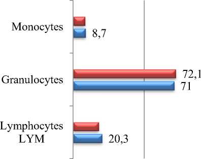
Before starting the experiments to study the effect of prenatal hypokinesia on blood parameters, we determined the blood parameters of pregnant rabbits with a normal embryonic period of prenatal development — a control variant. Then, we set up experiments to compare these indicators with the results of blood indicators of pregnant rabbits that spent the embryonic period under the condition of hypokinesia. The results were statistically processed and presented in Tables 1 and 2. Table 1 shows leukoformula indicators based on the results of the blood analysis from the intact-control group of the pregnant rabbits and those that were exposed to hypokinesia during the embryonic period. In the control group, total leucocyte count, total lymphocyte count, total granulocyte count, and total monocyte count were, respectively, (4.2±0.336)10^9/l, (0.7±0.056)10^9/l, (3.2±0.256)10^9/l, and (0.5±0.04) 10^9. The percentage of lymphocytes, granulocytes, and monocytes was 17.9±1.432%, 72.1±5.768%, and 8.3±0.664%, respectively.
The total leucoformula indicators increased significantly in pregnant rabbits that spent the embryonic period of prenatal development under conditions of hypokinesia. Thus, total leucocyte count, total lymphocyte count, total granulocyte count, and total monocyte count were, respectively, (9.8±0.784)10^9/l, (2.0±0.16)10^9/l, 97.0±0.056)10^9/l, and (0.8±0.064) 10^9/l. While the percentage of lymphocytes, granulocytes, and monocytes did not change statistically significantly and amounted to 20.3±1.624%, 8.7±0.69%, and 71.0±5.68%, respectively.
Table 1
STATISTICAL ANALYSIS OF LEUCOFORMULA INDICATORS UNDER THE EFFECT OF THE HYPOKINESIA FACTOR IN THE EMBRYONIC PERIOD OF THE PREGNANT RABBITS, (M+m) n=16
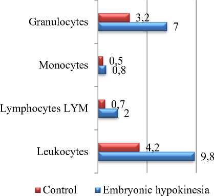
Figure 1. Statistical analysis of leucoformula
м Control И Embryonic hypokinesia
Figure 2. Statistical analysis of
|
Indicators |
Units |
Control |
Embryonic hypokinesia |
|
Leukocytes |
10^9/l |
4.2±0.336 |
9.8±0.784*** |
|
Lymphocytes LYM |
10^9/l |
0.7±0.056 |
2.0±0.16*** |
|
Monocytes |
10^9/l |
0.5±0.04 |
0.8±0.064** |
|
Granulocytes |
10^9/l |
3.2±0.256 |
7.0±0.056*** |
|
Lymphocytes LYM |
% |
17.9±1.432 |
20.3±1.624ns |
|
Granulocytes |
% |
72.1±5.768 |
71.0±5.68ns |
|
Monocytes |
% |
8.3±0.664 |
8.7±0.696ns |
Note: At P<0.01 (**), <0.05 (*), <0.001 (***), differences in mean values are significant, ns- nonsignificant.
indicators (10^9/l).
leucoformula indicators (%)
Based on the obtained results, the normal level of formed elements and other hematological indicators in the blood of pregnant rabbits (intact-control group) that underwent the embryonic period of prenatal development under normal conditions were as follows. The erythrocyte count in the blood was 5.4±0.432 (10^12/l), the erythrocyte sedimentation rate (ESR) — 4±0.32 mm/s, the concentration of hemoglobin (Hb) - 115±9.2 g/l, the mean hemoglobin (MH) — 295 ±23.6 (g/l), Mean Corpuscular Hemoglobin (MCH) — 20±1.6 pg, Mean Corpuscular Volume (MCV) -70.1±5.608 fL, and the amount of platelets was (95±7.6) 10^9/l. Red cell Distribution Width
(RDW) was 14.7±1.176%, Hematocrit (HTC) was 38.9±3.112%, and no significant changes were observed. Analysis of the parameters of blood platelets showed that the number of thrombocytes increased and reached the level of (271±21.68)10^9/l due to the hypokinesia factor. The Mean Corpuscular Volume (MCV) of platelets increased to 7.2±0.576 fL.
Table 2.
STATISTICAL ANALYSIS OF THE ERYTHROFORMULA CHANGES UNDER THE EFFECT OF HYPOKINESIA FACTOR IN THE EMBRYONIC PERIOD OF THE PREGNANT RABBIT, (M+m) n=16
|
Indicators |
Units |
Control |
Embryonic hypokinesia |
|
Erythrocytes |
10^12/l |
5.4±0.432 |
5.09±0.4072ns |
|
Hemoglobin (Hb) |
g/l |
115±9.2 |
109±8.72ns |
|
Hematocrit (HTC) |
% |
38.9±3.112 |
37.0±2.96ns |
|
Mean Corpuscular Volume (MCV) of erythrocytes |
fL |
70.1±5.608 |
72.7±5.816ns |
|
Mean Corpuscular Hemoglobin (MCH) |
pg |
20±1.6 |
21.4±1.712ns |
|
Mean Hemoglobin (MH) in erythrocytes |
g/l |
295±23.6 |
294±23.52ns |
|
Red cell Distribution Width (RDW) |
% |
14.7±1.176 |
14.2±1.136ns |
|
Platelets (PLT) |
10^9/l |
95±7.6 |
271±21.68*** |
|
Mean Corpuscular Volume (MCV) of platelets |
fL |
4.7±0.376 |
7.2±0.576** |
|
Erythrocyte Sedimentation Rate (ESR) |
mm/s |
4±0.32 |
6±0.48ns |
Note: At P<0.01 (**), <0.05 (*), <0.001 (***), differences in mean values are significant, ns- nonsignificant.
No significant changes were observed in the following parameters: Erythrocytes-5.09±0.4072 (10^12/l), ESR 6±0.48 (mm/s), Hemoglobin-109±8.72 (Hb) (g/l), Mean Hemoglobin (MH) in erythrocytes-294±23.52 (g/l), Red cell Distribution Width-14.2±1.136 (RDW) %, Mean Corpuscular Hemoglobin (MCH) of erythrocytes-21.4±1.712 pg, Hematocrit (HTC)-37.0±2.96 % and Mean Corpuscular Volume (MCV) of erythrocytes-72.7±5.816 fL.
Discussion
Our studies revealed changes in the blood coagulation system caused by the effect of the hypokinesia factor. The level of these changes depends on the embryonic development stage in which the effect of hypokinesia occurs. Thus, the statistical analysis showed that the difference in the indicators observed in the experimental groups emerged in the embryonic stage of the fetus development. Thus, the activated effect of prenatal hypokinesis on the hemostasis system was detected. Abnormalities observed in the development of the fetus in most (90%) cases are related to placental insufficiency caused by impaired blood circulation. Disruption of the blood coagulation system due to the hypokinesia factor plays a key role in delaying the whole development dynamics of the body and causes a weakening of the functions of the placental vessels that provide nutrition to the fetus. In general, the response of the blood coagulation system to certain effects is significantly determined by the functional state of the body. On the other hand, the level of these changes is directly related to the degree of influence of extreme factors (including hypokinesia), duration, as well as the period of the body's development. In this regard, the changes observed as a result of the effect of prenatal hypokinesis are more pronounced, especially in the embryonic stage. The main reason for these changes can be the indirect effect of hypokinesia on the developing organism. Thus, during the formation of embryonic layers at the initial stage of embryonic development, exposure of the mother to the influence of hypokinesis causes serious changes in the hemostasis of the intrauterine environment, which results in dynamic dysfunctions in the hematopoietic tissues of the fetus in the later stages. Besides, the changes in leukoformula indicators show that the rise of blood cells belonging to the leukocyte group has a negative effect on the body's defense reactions against pathogenic viruses, microorganisms, primitive parasites, microbial poisons, and foreign protein substances found in food. Significant changes in leukocytes due to the hypokinesia factor and this state of immobility also affect the general immunity of organisms - the ability to resist causative agents and foreign substances.
As a result of the effect of hypokinesia during the embryonic period, a decrease in some blood parameters and an increase in others were observed. Thus, under hypokinesia, vitamins, and microelements necessary for the normal development of the fetus are not formed, the nutrition of the fetus is disturbed, and fat accumulation occurs. Accumulation of excessive adipose tissue especially threatens the blood vessels, and therefore, insufficient blood reaches the tissues and the brain. Under hypokinesia, changes in blood parameters of pregnant rabbits cause various problems including faster heart failure. When in motion, the heart muscles meet the body's demand for blood (oxygen and energy substrates) by squeezing strongly. Immobility leads to muscle passivation, and the heart muscle has to make more contractions for the same job. Thus, the hypokinesia factor leads to more work and weakening of the heart muscles. Due to the hypokinesis factor, first of all, the volume of blood circulating in the body through the heart decreases. Weakening of the muscular system eventually leads to impaired blood circulation in the cardiovascular system. The weakening of the muscles in the lower limbs due to the effect of hypokinesia has a negative effect on the factor of venous blood rising. As seen in the table, there are changes in the number of leukocytes, lymphocytes, monocytes, granulocytes, and platelets in the blood of pregnant rabbits from the experimental group that spent the embryonic period under the condition of hypokinesia. The study of erythroformula and leucoformula indicators of pregnant rabbits that are exposed to hypokinesia during the embryonic period of pregnancy can be useful for taking preventive measures to eliminate the complications of hypokinesia.
References The effect of hypokinesia on the dynamics of the formed elements of blood during the embryonic period of prenatal development in pregnant rabbits
- Agaeva, E. N. (2019). Obzor istorii izucheniya gipokinezii kak aktual'noi problem. In Mezhdunarodnaya nauchnaya konferentsiya po aktual'nym problemam sovremennykh estestvennykh i ekonomicheskikh nauk, 133-135. (in Azerbaijani).
- Agaeva, E. N. (2023). Izuchenie vliyaniya faktora prenatal'noi gipokinezii yavlyaetsyaaktual'noi problemoi. In Mezhdunarodnaya nauchnaya konferentsiya po aktual'nym problemam sovremennykh estestvennykh i ekonomicheskikh nauk, 153. (in Azerbaijani).
- Agaeva, E. N. (1992). Vliyanie gipokinezii na razvitie plodov krolika v raznye sroki beremennosti: avtoref. dis. ... kand. biol. nauk. Baku. (in Russian).
- Belozerova, V. N., Matveeva, O. A., Nemirovskaya, T. L., Kuznetsov, S. L., Korol'kov, V. I., & Shenkman, B. S. (2001). Strukturnye i metabolicheskie kharakteristiki m. vastus lateralis obez'yan vo vremya 30-dnevnoi gipokinezii: effekty profilakticheskogo Gz-uskoreniya. Moscow. (in Russian).
- Bogdashkin, N. G. (1989). Gigienicheskaya aktivnost' kak prichina oslozhnenii vo vremya beremennosti, rodov i poslerodovogo perioda. Khar'kov. (in Russian).
- Makhmudova, N. Sh., Gadzhieva, G. Sh., & Abdullaeva, G. M. (2023). Vliyanie prenatal'noi gipokinezii na dinamiku EEG kory golovnogo mozga krysyat rannego vozrasta. In Neironauka dlya meditsiny i psikhologii: Materialy XIX Mezhdunarodnogo mezhdistsiplinarnogo kongressa, Moscow, 196-197. https://doi.org/10.29003/m3308.sudak.ns2023-19/196-197
- Mal'tseva, N. G., & Kuznetsova, T. G. (2008). Vliyanie gipokinezii na strukturu miokarda. Problemy zdorov'ya i ekologii, (2 (16)), 113-118. (in Russian).
- Tarakin, P. P., Gasnikova, N. M., & Shenkman, B. S. (2006). Vliyanie antiortostaticheskogo vyveshivaniya na techenie distroficheskogo protsessa v myshtsakh zadnikh konechnostei 12-mesyachnykh myshei linii MDX. Byulleten' eksperimental'noi biologii i meditsiny, 141(6), 702-705. (in Russian).
- Tkachenko, A. V. (2011). Metabolicheskie protsessy v serdtse i pecheni krys pri eksperimental'noi gipokinezii i ikh korrektsiya fitosiropom Valeoton. Vіsnik Kharkіvs'kogo natsіonal'nogo unіversitetu іmenі VN Karazіna. Serіya: bіologіya, (14), 177-184. (in Russian).
- Volodina, A. V., & Pozdnyakov, O. M. (2006). Ultrastructure of skeletal muscle capillaries under conditions of space mission. Bulletin of Experimental Biology and Medicine, 141, 755-759. (in Russian).https://doi.org/10.1007/s10517-006-0271-4
- Zaripova, R. I., Andrianov, V. V., Yafarova, G. G., Gainutdinov, Kh. L., Khabibrakhmanov, I. I., & Zefirov, T. L. (2014). Vliyanie gipokinezii razlichnoi dlitel'nosti na dinamiku produktsii oksida azota v serdtse, spinnom mozge i pecheni krys. Rossiiskii fiziologicheskii zhurnal im. IM Sechenova, 8, 926-935. (in Russian).
