The first experience of using 99mTc-1-thio-D-glucose for single-photon emission computed tomography imaging of lymphomas

Author: Chernov Vladimir I., Dudnikova Ekaterina A., Zelchan Roman V., Kravchuk Tatyana L., Danilova Albina V., Medvedeva Anna A., Sinilkin Ivan G., Bragina Olga D., Goldberg Victor E., Goldberg Alexey V., Frolova Irina G.
Journal: Сибирский онкологический журнал @siboncoj
Section: Клинические исследования
Article in issue: 4 т.17, 2018.
Free access
Introduction. The purpose of this study was to evaluate the feasibility of using 99mTc-Tg SPECT in the detection and staging of malignant lymphoma. materials and methods. Fifteen patients with newly diagnosed malignant lymphoma underwent 99mTc-Tg SPECT. Six patients had Hodgkin’s lymphoma and 9 patients had aggressive forms of non-Hodgkin’s lymphoma (NHL) : diffuse large b-cell lymphoma (7 cases), b-cell follicular lymphoma (1 case), and lymphoma from b cells in the marginal zone (1 case). Stage IIA was diagnosed in 5 patients, stage IIb in 1, stage IIIA in 1, stage IVA in 4 and stage IVb in 4 patients. results. Pathological 99mTc-Tg uptake in lymph nodes was observed in 14 (93 %) of the 15 patients. In one patient, the enlarged submandibular lymph node (16 mm in size) detected by CT was not visualized by 99mTc-Tg SPECT. This false-negative result was likely to be associated with increased accumulation of 99mTc-Tg in the oropharyngeal region. There were difficulties in the visualization of paratracheal, para-aortic and paracardial lymph nodes. These difficulties were associated with a high blood background activity, which persisted even 4 hours after intravenous injection of 99mTc-Tg. Software-based SPECT and CT image fusion allowed visualization of these lymph nodes. The pathological 99mTc-Tg accumulation in axillary, supraclavicular, infraclavicular and cervical lymph nodes was observed most often. Extranodal involvement was seen in 9 patients. 99mTc-Tg SPECT identified extranodal hypermetabolic lesions in 7 (78 %) of these patients. In one patient, hypermetabolic lesion in the lung detected by 99mTc-Tg SPECT was not detected on CT image. CT identified bone marrow involvement in the pelvic and scapula in 1 patient. The use of 99mTc-Tg SPECT allowed the visualization of hypermetabolic bone tissue lesions in this patient (Figure 4). In addition, in a patient with intact bone tissue on CT, 99mTc-Tg SPECT detected hypermetabolic lesions in the iliac bone. Conclusion. 99mTc-1-Thio-D-glucose demonstrated increased uptake in nodal and extranodal sites of lymphoma. The results indicate that SPECT with 99mTc-1-Thio-D-glucose is a feasible and useful tool in the detection and staging malignant lymphoma.
Lymphomas, hodgkin's lymphoma, single-photon emission computed tomography, 99mtc-1-thio-d-glucose
Short address: https://sciup.org/140254204
IDR: 140254204 | UDC: 616-006.441-073.756.8 | DOI: 10.21294/1814-4861-2018-17-4-81-87
Однофотонная эмиссионная компьютерная томография с 99mTc-1-тио-D-глюкозой в диагностике и стадировании злокачественных лимфом: первый опыт использования
Целью исследования явилась оценка возможности применения однофотонной эмиссионной компьютерной томографии (ОФЭКТ) с инновационным отечественным радиофармпрепаратом (РФП) 99mТс-1-тио-D-глюкоза (99mТс-ТГ) в диагностике и стадировании злокачественных лимфом. материал и методы. В исследование включены 15 пациентов (средний возраст 50,7±18,3 года) с впервые диагностированными злокачественными лимфомами. Сцинтиграфическое исследование проводили на гамма-камере E.CAM 180 фирмы “Siemens” (Германия) через 4 часа после введения РФП в дозе 500 МБк. Результаты. По данным ОФЭКТ, патологическое включение 99mТс-ТГ в лимфатические узлы наблюдалось у 14 (93 %) из 15 пациентов. У одной пациентки не удалось визуализировать определяемый с помощью компьютерной томографии (КТ) единичный, увеличенный поднижнечелюстной лимфатический узел. Этот ложноотрицательный результат исследования связан с физиологичным усилением аккумуляции 99mТс-ТГ в орофарингеальной области. Сложности, связанные с высокой фоновой активностью крови, отмечались при визуализации паратрахеальных, парааортальных и паракардиальных лимфатических узлов. Наиболее часто патологическое накопление РФП отмечалось в аксиллярных, над- и подключичных, а также шейных лимфатических узлах. С помощью ОФЭКТ с 99mТс-ТГ экстралимфатические гиперметаболические участки определялись у 7 (78 %) из 9 пациентов с ранее диагностированным экстранодальным поражением. Кроме того, у одной пациентки по данным ОФЭКТ был выявлен гиперметаболический очаг в легком, не обнаруженный по результатам КТ. В исследуемой выборке пациентов по данным КТ и ОФЭКТ с 99mТс-ТГ наличие поражения костного мозга наблюдалось в одном случае. Кроме того, сцинтиграфическое исследование позволило выявить гиперметаболические участки в лопатке у пациента с интактной по результатам КТ костной тканью. Заключение. Результаты исследования позволяют рекомендовать использование 99mТс-ТГ ОФЭКТ в качестве дополнительного диагностического метода для обследования пациентов со злокачественными лимфомами для стадирования заболевания.
Text of the scientific article The first experience of using 99mTc-1-thio-D-glucose for single-photon emission computed tomography imaging of lymphomas
Tumors of the hematopoietic and lymphoid tissues are relatively frequent in Russia, accounting for approximately 5 % of all cancers in men and 4.6% in women, and 5 % of all cancer deaths. In 2016, the incidence rate for tumors of the hematopoietic and lymphoid tissues was 19.58 per 100 000 population. The average annual growth rate is 1.78 % [1]. Among lymphoid and hematopoietic tissue tumors, Hodgkin lymphoma (HL) is observed most frequently (30 %) [2]. Among non-Hodgkin's lymphomas (NHLs), large B-cell lymphoma (33 %) and B-cell follicular lymphoma (22 %) are the most common lymphomas. Other types of lymphomas occur with a frequency of less than 10 % [3].
Accurate staging for lymphoma enables better prognostication and choice of the most appropriate treatment [4]. Thus, the estimated recurrence-free 10-year survival is highest in patients with stage I disease (90–95 %), slightly lower (80–85 %) in patients with stage II, significantly lower (70 %) in stage III and does not exceed 30–50 % in stage IV [4]. The Ann Arbor staging system for the assessment of lymphoma includes computed tomography (CT)
scans [5]. However, diagnostic capabilities of CT are limited when visualizing lymph nodes of normal size. Moreover, in patients with retroperitoneal lymph nodes measuring from 1 to 3 cm, the likelihood of detection of a specific lesion with CT is only 50 %. Even a significant increase in the lymph nodes size (more than 3 cm) is associated with their benign hyperplasia in 25 % of cases. CT has low sensitivity in the diagnosis of diffuse lesions of the liver, spleen and bone marrow [6]. 18-F-fluorodeoxyglucose (18F-FDG) – positron emission tomography (PET), and more recently PET/ computed tomography (CT), is the most sensitive and specific imaging technique currently available for patients with lymphoma.
Numerous studies indicate that PET has proved very useful for staging and therapy response of lymphoma [4–10]. The widespread use of PET in our country is limited due to the high cost and low number of PET centers, which are located mainly in the central regions of the Russian Federation. At the same time, in Russia there are more than 200 nuclear medicine departments equipped with SPECT scanners. Therefore, the use of radiopharmaceuticals labeled with gamma-emitting nuclides remains relevant. 99mTc-1-thio-D-glucose
(99mTc-TG) is the promising radiopharmaceutical for lymphoma imaging. This radiopharmaceutical is a glucose derivative complex in the form of 1-thio-D-glucose and 99mTc, in which 1-thio-D-glucose works as a radioisotope label transport (99mTc). A special feature of the pharmacokinetics of the radiopharmaceutical is the absence of its accumulation in the brain and myocardium. Tomsk NIIC and Tomsk Polytechnic University successfully completed the project «Preclinical studies of radiopharmaceutical on the basis of 99mTc labeled glucose derivative for tumor imaging» (№ 14.N08.11.0033). The laboratory regulations for 99mTc-TG preparation were developed, the analytical methods for 99mTc-TG quality control were elaborated and samples of the radiopharmaceutical were synthesized and analyzed in accordance with established methodology.
It was proved that 99mTc-TG was characterized by high accumulation in tumor cells in vivo and in vitro [11–15].
The purpose of this study was to evaluate the feasibility of using 99mTc-TG SPECT in the detection and staging of malignant lymphoma.
Materials and Methods
The study included 15 patients (mean age 50.7 + 18.3, range: 25 to 75 years) with newly diagnosed malignant lymphomas. None patients had previously received chemotherapy. The diagnosis was verified by immunohistochemical study. Six patients had HL and nine had aggressive forms of NHL: diffuse large B-cell lymphoma (7 cases), B-cell follicular lymphoma (1 case), and lymphoma from B cells in the marginal zone (1 case). Stage IIA was diagnosed in 5 patients, stage IIB in 1, stage IIIA in 1, stage IVA in 4 and stage IVB in 4 patients.
Single photon emission computed tomography was performed using a double-head gamma-camera (E.CAM 180, Siemens) equipped with parallel high energy collimators. The injection of 99mTc-TG at a dose of 500 MBq was made intravenously into the antecubital vein. Planar imaging began 4 hours after injection. Images were obtained with the patients lying in the supine position with the arms raised over the head and with the face, skull, chest, neck, abdomen, pelvis and groin included into the field of view. A total of 32 projection images were recorded into a 64×64 matrix (30 seconds per projection) without hardware magnification. The scan images were analyzed using the manufacturer software (e.soft, Siemens, Germany). Three-dimensional images of the chest, sagittal, transverse and coronal sections were obtained. Single photon emission computed tomography scans were visually assessed. Images of the contralateral areas were compared, and asymmetrically increased radiotracer uptake was considered pathological.
Results and Discussion
The pathological 99mTc-TG uptake in lymph nodes was observed in 14 (93%) of the 15 patients. In a patient with diffuse large B-cell lymphoma, a single enlarged submandibular lymph node (16 mm in size) detected by CT was not visualized by 99mTc-TG SPECT. This false-negative result is likely to be associated with the physiological enhancement of 99mTc-TG accumulation in the oropharyngeal region. There were difficulties in the visualization of paratracheal, para-aortic and paracardial lymph nodes. These difficulties were associated with a high background activity, which persisted even 4 hours after intravenous injection of 99mTc-TG. Software-based SPECT and CT image fusion allowed visualization of these lymph nodes. The pathological 99mTc-TG accumulation in axillary, supraclavicular, infraclavicular and cervical lymph nodes was observed most often Extranodal lesions were observed in 9 patients (lung in 3 patients, liver in 2, spleen in 2, stomach in 2 and parotid salivary gland in 1). 99mTc-TG SPECT identified extranodal hypermetabolic lesions in 7 (78%) of these patients. 99mTc-TG SPECT detected lesions in the lungs in 2 cases, in the liver in one, in the spleen in one, in the stomach in one and in the parotid salivary gland in one (Figure 2). In one patient, 99mTc-TG SPECT identified hypermetabolic lesion in the lung that was not detected by CT (Figure 3).
18F-FDG PET/CT was found to be superior to CT in the detection of bone marrow lesions in patients with lymphomas [16]. CT allows visualization of such pathological lesions only in the presence of bone destruction. CT detected bone marrow involvement in the pelvic and scapula in 1 patient. The use of 99mTc-TG SPECT allowed the visualization of hypermetabolic
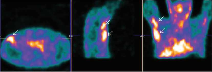
Figure 1. 99mTc-TG SPECT images of a patient with Hodgkin’s lymphoma, nodular sclerosis, stage IIA, lesions of right axillar and right pectoral lymph nodes (arrows)
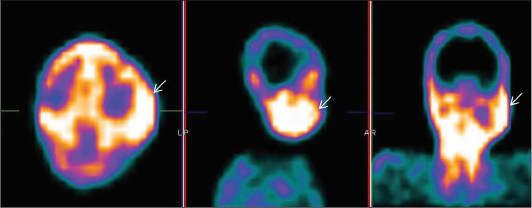
Figure 2. 99mTc-TG SPECT images of a patient with diffuse large B-cell lymphoma, germinogenic type, stage IIA (bulky) with lesions of the left parotid gland (arrows)
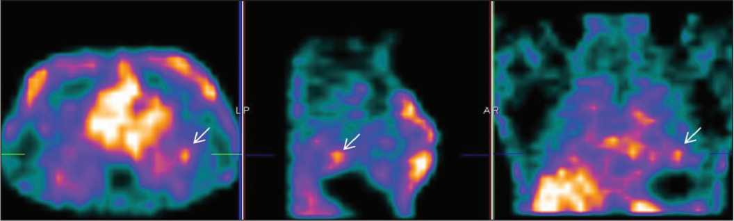
Figure 3. 99mTc-TG SPECT images of a patient with Hodgkin’s lymphoma, nodular sclerosis, IIA stage with lesions of the left lung (arrows)
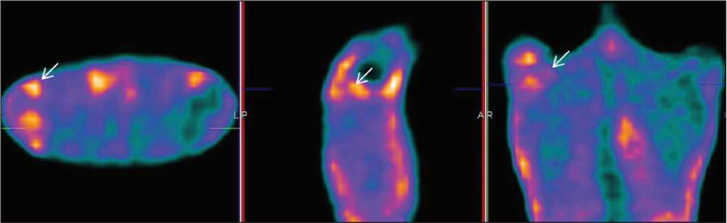
Figure 4. 99mTc-TG SPECT images of a patient with B-diffuse large-cell lymphoma, stage IVB, with lesions of cervical lymph nodes, pelvic bones and right scapula.
Hypermetabolic lesion in the right scapula is seen (arrows)
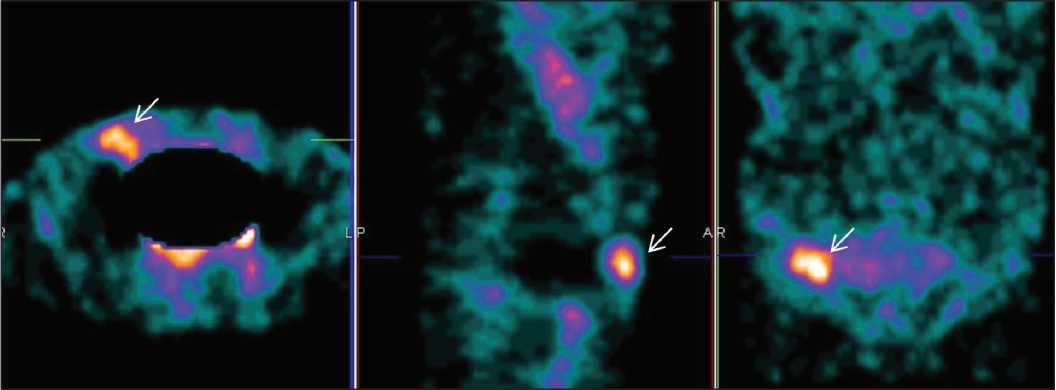
Figure 5 99mTc-TG SPECT images of a patient with diffuse large B-cell lymphoma, germinogenic type, stage IVB with lesion of the left submandibular salivary gland, right inguinal lymph node, lymph nodes of the abdominal cavity, spleen. Hypermetabolic lesion the iliac bone is visualized (arrows)
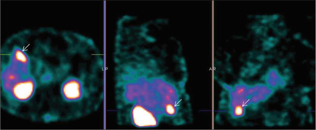
Figure 6. 99mTc-TG SPECT images of a patient with diffuse large B-cell lymphoma, germinogenic type, stage IIB with lesion of the left submandibular and paratracheal lymph nodes. Concomitant diagnosis: chronic cholecystitis. Hypermetabolic lesion in the gallbladder is visualized (arrows)
bone tissue lesions in this patient (Figure 4). In addition, in a patient with intact bone tissue on CT, 99mTc-TG SPECT detected hypermetabolic lesions in the iliac bone (Figure 5).
18F-FDG is characterized by its active accumulation in inflammation lesions, reducing the specificity of 18F-FDG PET/CT in cancer patients. In our study, two patients had cholecystitis as a concomitant disease; all of them had pathological 99mTc-TG uptake in the gallbladder (Figure 6). These observations indicate that it is necessary to take into account clinical patient data for the correct analysis of 99mTc-TG SPECT findings.
The mechanism of cellular uptake of colorectal carcinoma cell line HCT-116 and human lung adenocarcinoma cell line A549 was studied [17]. The levels of 99mTc-TG and 18F-FDG accumulation in HCT-116 cells were nearly similar, while the 18F-FDG uptake in A549 cell line was almost twice as much as 99mTc-TG. 99mTc-TG and 18F-FDG cellular accumulation decreased when the glucose concentration increased, and was enhanced in the presence of insulin. These facts suggest that both 18F-FDG and 99mTc-TG penetrate into the cell with the participation of glucose transport proteins. Physiologically, glucose is transported by the facilitated sodium-independent glucose transporters (GLUT1-GLUT6, and GLUT8) and by the sodiumdependent transporters (SGLT1 and SGLT2), with a variable expression level of these transporters in different human tissues. Most solid tumors exist in a hypoxic environment and prefer anaerobic glycolysis rather than aerobic glycolysis, converting glucose to lactate and producing less ATP with a smaller oxygen
References The first experience of using 99mTc-1-thio-D-glucose for single-photon emission computed tomography imaging of lymphomas
- Каприн А.Д., Старинский В.В., Петрова Г.В. Злокачественные заболевания в России в 2014 году (заболеваемость и смертность). М.:ФГБУ «МНИОИ им. П.А. Герцена Минздравсоцразвития России». 2018. 250.
- Kaprin A.D., Starinskii V.V., Petrova G.V. Zlokachestvennye zabolevaniya v Rossii v 2014 godu (zabolevaemost' i smertnost'). M.: FGBU «MNIOI im. P.A. Gertsena Minzdravsotsrazvitiya Rossii». 2018. 250.
- Рукавицын О.А. Гематология: национальное руководство. М.: ГЭОТАР-Медиа. 2015; 776.
- Rukavitsyn O.A. Gematologiya: natsional'noe rukovodstvo. M.: GEOTAR-Media. 2015; 776.
- Armitage J.O. A clinical evaluation of the International Lymphoma Study Group classification of non-Hodgkin’s lymphoma. Blood. 1997; 89(11): 3909 3918.
- Новиков С.Н., Гиршович М.М. Диагностика и стадирование лимфомы Ходжкина. Проблемы туберкулеза и болезней легких. 2007; 8(2): 65 72.
- Novikov S.N., Girshovich M.M. Diagnostika i stadirovanie limfomy Khodzhkina. Problemy tuberkuleza i boleznei legkikh. 2007; 8(2): 65 72.
- Kwee T.C., Kwee R.M., Nievelstein R.A. Imaging in staging of malignant lymphoma: a systematic review. Blood. 2008; 111: 504-16.
