The role of CT scan in imaging and diagnosing kidney diseases

Author: Qader K.M., Abdulkhaleq N.A., Dawood S.K., Yakoob S.
Journal: Cardiometry @cardiometry
Section: Original research
Article in issue: 32, 2024.
Free access
Kidney stones are solid deposits that usually form in the kidneys. These deposits can be very small, the size of a grain of sand, or be larger than that. They may take different shapes and cause several symptoms. The pain is usually caused by the movement of stones into the urinary tract, causing irritation and swelling of the surrounding tissue. The research was conducted in At Al-Sadr Teaching hospital The number of patients was 150 samples, 100 of which were males and 50 of which were females. The age of the patients was 40.141±1.77 for males and 2.7±44.234 for females. The clinical results for symptoms of fever were 38.% yes, while 62% there was no fever and The clinical findings. of Vomiting and Nausea 49% were yes, while 51% there was no Vomiting and Nausea as well as The clinical findings of Pain and Severe back spasms 13% were yes, while 87% there was no Pain and Severe back spasms and also The clinical findings of Severe pain in the Right flank.12% were yes, while 88% there was no Pain and 87 Severe back spasms. Clinical results for symptoms of Pain in the Ribs and Rib Cage were 38% yes, while 62% there was no Pain in the Ribs and Rib Cage while The clinical findings of Burning During Urination 12% were yes, while 88% there was no Burning During Urination, the clinical findings of Frequent Urination a Number of Times 50% were yes, while 50% there was no Frequent Urination a Number of Times symptoms and clinical findings of Urinating a Small Amount of Urine and The clinical findings of Dark or Red Urine in the same percentage, while The clinical findings of Does Urination Smell bad 12.5% were yes, while 87.5% there was no Does Urination Smell bad and The clinical findings of Feeling pain when Urinating 14% were yes, while 86% there was no feeling pain when urinating. This shows that kidney stones have a clear clinical effect.
Kidney stones, ct scan, kvp, adult patients, al-sadr teaching hospital, iraq
Short address: https://sciup.org/148329309
IDR: 148329309 | DOI: 10.18137/cardiometry.2024.32.2124
Text of the scientific article The role of CT scan in imaging and diagnosing kidney diseases
Imprint
Kzal Mohammed Qader, Nour A. Abdulkhaleq, Salwah Kareem Dawood, Sally Yakoob. The role of CT scan in imaging and diagnosing kidney diseases. Cardiometry; Issue No. 32; August 2024; p. 21-24; DOI: 10.18137/cardiometry.2024.32.2124; Available from:
-
1 INTRODUCTION
kidney disease is increasing at an alarming rate. It’s like this all over the world Approximately 10% of the population suffers from chronic kidney disease [1] . However, it ranks 16th among the most common causes of death. By 2040, this disease is likely to reach 5th place. cyst, Kidney stones mainly cause kidney dysfunction and cause pain. A renal cyst is basically a fluid-filled sac located on the kidney The surface and thin wall of the kidney are bounded by it [2]. Otherwise, kidney Stones are concentrated concentrations of crystals in the kidneys and affect 12% of people worldwide [3]. Kidney failure leads to a higher risk of dialysis or transplant-associated with it. The causes of kidney stones are complex. Calcium oxalate, that is the most typical kidney stone and forms on the surface of kidney stones at Randall’s plaques. Renal papillae. The formation mechanism of stones is a complex process is caused by a series of physical and chemical processes such as supersaturation and nuclear formation, growth, accumulation, and retention of intratubular urolithic contents cell. This ultimately leads to the formation of stone. These processes are made by differences Between Ingredients That Promote and Inhibit CrystallizationUrine. Additionally, focus should be placed on particle retention it promotes damage on the surface of the renal papilla. Oxalate exposure triggers a signaling cascade of reactions culminating in apoptosis via p38 mitogen-activated protein kinase signaling pathways in renal epithelial cells. There is currently no effective drug treatment to eliminate kidney stones or prevent their recurrence. Therefore, it is necessary to study and improve understanding the biology of kidney stone formation to treat urolithiasis with new drugs. Therefore, the purpose of this study is to consolidate the latest re
-
2.2 Statistical Analysis
For the comparison of measured parameters among subdivided groups, the widely recognized statistical system GraphPad Prism ver. 5 was utilized. The analysis of the variance table, specifically the one-way ANOVA with Tukey’s multiple comparisons test, was employed. The results were presented as Mean ± Standard Error. To assess the correlation between markers and parameters, correlation coefficients were calculated. Descriptive statistics and correlation coefficients were obtained using MegaStat (Version 10.12) for Excel 2010 (Motulsky, 2003).
search findings on the pathophysiology, etiology, and prevention of kidney stones. Kidney tumors, or renal cell carcinoma (RCC), are one of the ten most common diseases tumors worldwide. Course of disease and cardiovascular morbidity and mortalityThis can be avoided if chronic kidney disease is diagnosed early. so detect it earlyTechnology in public health is becoming increasingly important. Pathological examination has-Commonly used in imaging procedures such as X-ray and computed tomography(CT) [4]. Noncontrast spiral computerized tomography (CT) can detect nearly 95% of renal, ureteric, and bladder stones, particularly those made up of calcium [5]. Therefore introduction of CT imaging at the hospital not only helps in identifying most types of stones but also provides basic information about their composition which guides the manage-ment[6]. Preoperative CT imaging findings significantly determine surgical outcomes– more significantly in cases where incomplete resections are involved and peritumoral fat was not visualized in 87% of cases, as opposed to a 66% rate among operable cases. [7]. This emphasizes how crucial precise and thorough CT imaging is for directing renal calculi therapy choices. Furthermore, CT imaging provides urolithiasis patients with a less invasive means of treating the condition by precisely localizing and characterizing stones in order to make targeted treatments [8]. Unenhanced helical CT is a useful technique for early diagnosis and therapy of urolithiasis since it has shown good sensitivity (>95%) and specificity (>96%) in detecting the condition [9]. Notwithstanding these drawbacks, further developments in CT technology and imaging procedures offer hope for resolving these issues and improving the usefulness of CT in the identification and treatment of renal calculi.
2 MATERIALS AND METHOD 2.1 Characteristics of Patient Groups
Of the patients who receive care at Al-Sadr Hospital in the Governorate of Najaf, 150 patients were carefully chosen. There were 100 patients in the female category and 50 in the male category. Table 1 shows the distribution of sociodemographic factors among patients participating in this study. This anthropometric data (gender, age, weight) must be provided. The full set of measurements was obtained from 150 adult men and women. In addition, the age range ranged from 25 to 60 years.
|
Radiography |
Sex |
Age (years) |
Weight (kg) |
|
Kidney stones |
100 M 50 f |
(25-60) |
(59-110) |
M: male. F: female.
-
3 THE RESULTS
-
3.1 the patient’s age is divided according
-
-
3.2 Division of the number of patients according to the gender of the patient.
to the patient’s gender (years).
The picture shows the numbers of male and female patients (Figure 1).
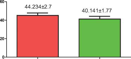
(F. 1): shows the ages of both males and females Female is high than male
The picture shows the numbers of male and female patients (Figure 2).
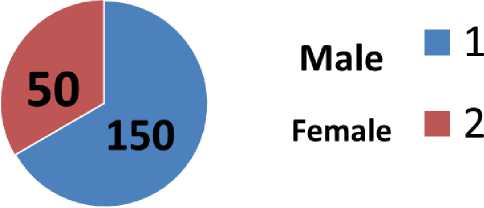
(F. 2): shows the numbers of both males and females
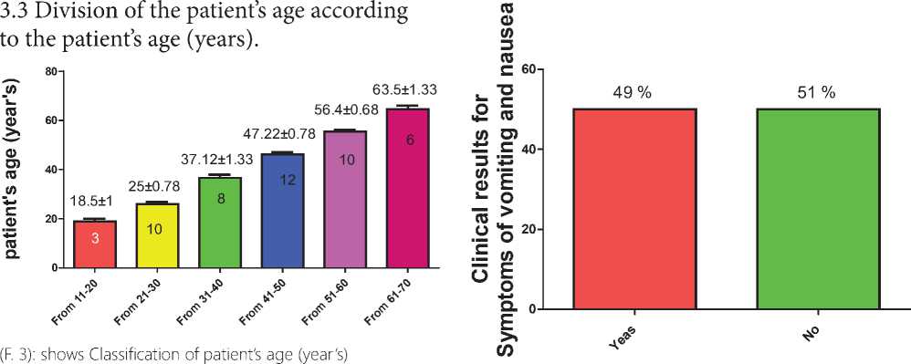
Number of patients according to age
From 11-20 = 3
From 21-30 =10
From 31-40 =8
From 41-50 = 12
From 51-60 =10
From 61-70 =6
Total number =50
-
3.4 Division of the patient according
-
3.4.1 Clinical findings of fever symptoms
-
3.4.2 Clinical findings of fever symptoms
-
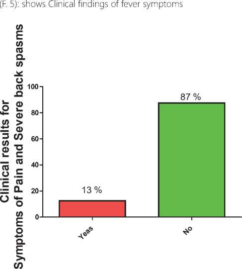
3.4.3 : Clinical results for Symptoms of Pain and Severe back spasms
to Symptoms of patient.
The clinical results for symptoms of fever were
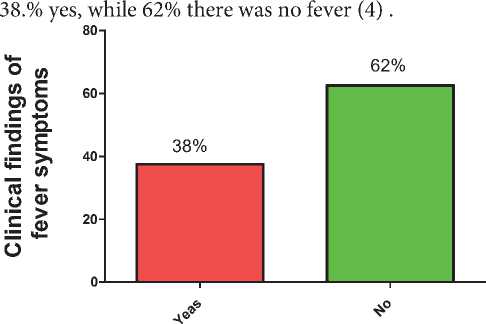
(F. 4): shows Clinical findings of fever symptoms
The clinical findings of Vomiting and Nausea 49 % were yes, while 51 % there was no Vomiting and Nausea (F. 5).
The clinical findings of Pain and Severe back spasms 13 % were yes, while 87 % there was no Pain and Severe back spasms (F. 6).
(F. 6): shows Clinical findings of Pain and Severe back spasms symptoms
-
5 THE DISCUSSION
Through the results in (F. 1) and (F. 2) the effect of gender on stone formation may be because the size of kidney stones and their distribution can differ between males and females based on several factors, including nutrition and genetics. and certain diseases. However, there are no precise and universal details about the size of kidney stones based on gender. Kidney stone sizes can range from a few millimeters to a few centimeters, and the sizes can be smaller or larger depending on previously mentioned factors [10].
From the results of (F. 3), it is clear that age has an effect on the production of stones in the body, and the cause can usually be due to kidney stones when they become large enough to cause symptoms such as severe pain in the back or side area, urinary congestion, or blood clots. In urine. Ultrasound or X-rays can be used to detect the presence of kidney stones and estimate their size and distribution[11]. From the results (F. 4) it is clear that fever has an effect, as well as from the results pain has an effect through the results observed in (F.4-5) and also Pain and Severe back spasms observed in [12].The pain is usually caused by the movement of the stones into the urinary tract, causing the surrounding tissues to become irritated and swell, leading to pain. Pain resulting from kidney stones is usually described as sharp and extending from the back to the side or to the lower abdomen, often on one side. The discomfort can come and go or linger consistently, with triggers or alleviators that are contingent upon activity in the bladder[13]. In addition to these symptoms, the pain may come alone or with other symptoms like nausea, vomiting, or changes in urination. High fever is also a sign of kidney stones particularly those that result in infection along the urinary tract. At times, this high temperature may be ushered by acute back or side pangs plus nausea and vomiting; look out for redness or swelling around your kidney area to confirm this suspicion. [14].
-
6 CONCLUSIONS
it is concluded by the current study, that:
-
1. In the age group of 41-50 years, the most elevated infection rate was observed
-
2. The manifestation of clinical symptoms demonstrated distinct impact: fever and vomiting along with other symptoms were different for each individual based on their body constitution..
-
7 RECOMMENDATIONS:
-
1. A suggestion made by the researcher is to consume a healthy diet.
-
2. Commit to drinking water regularly: it’s one of the biggest contributing factors.
-
3. Conducting a lot of research on kidney stones and observing other symptoms
References The role of CT scan in imaging and diagnosing kidney diseases
- Himelfarb J, Lakhani A, Shelton D. Appropriate use of CT for patients presenting with suspected renal colic: a quality improvement study. BMJ open quality. 2019;8(4):e000470.
- Ganesan V, et al. Accuracy of ultrasonography for renal stone detection and size determination: is it good enough for management decisions. BJU international. 2017;119(3):464–9.
- Adwan A, Binsaleh S. The accuracy of noncontrast spiral computerized tomography in detecting lucent renal stones: A case report and literature review. Urology annals. 2015;7(1):109–11.
- Ganesan V, et al. Accuracy of ultrasonography for renal stone detection and size determination: is it good enough for management decisions?. BJU international. 2017;119(3):464–9.
- Adwan A, Binsaleh S. The accuracy of noncontrast spiral computerized tomography in detecting lucent renal stones: A case report and literature review. Urology annals. 2015;7(1):109–111.
- Hillman BJ, et al. Computed tomographic analysis of renal calculi. AJR. American journal of roentgenology. 1984;142(3):549–52.
- Ottlakan A, et al. The Effect of Diagnostic Imaging on Surgical Treatment Planning in Diseases of the Thymus. Contrast media & molecular imaging. 2017; 9307292.
- Al-Shawi MM, et al. The Role of Radiological Imaging in the Diagnosis and Treatment of Urolithiasis: A Narrative Review. Cureus. 2022;14(12):e33041.
- Andrabi Y, et al. Advances in CT imaging for urolithiasis. Indian journal of urology. 2015; 31(3): 185–93
- Stamatelou K, Goldfarb DS. Epidemiology of kidney stones. Healthcare, MDPI. February 2023; 11(3): 424).
- Benly ML, Liman M. Bilateral staghorn renal calculi. Science Midwifery. 2023;10(6):4877-81.
- Malhi M, Hanumanthrao P, Kumari PR. A Update On Current Diagnostic Modalities And Treatment Of Urolithiasis Review. Journal of Survey in Fisheries Sciences. 2023; 710-7.
- Ahn JS, Harper JD. Acute Kidney Stone Management. A Clinical Guide to Urologic Emergencies. 2021; 64-82.
- Montatore M, et al. Current Status on New Technique and Protocol in Urinary Stone Disease. Current Radiology Reports. 2023; 11(12):161-76.
