
Therapeutic Potential of a Novel Nitroheterocyclic Derivative in Experimental Tuberculosis Models in C57BL/6 Mice Infected with Biologically Diverse Mycobacteria
Журнал: Juvenis scientia @jscientia
Рубрика: Оригинальные исследования
Статья в выпуске: 3 т.12, 2026 года.
Бесплатный доступ
Introduction. Drug-resistant Mycobacterium tuberculosis remains a major challenge for tuberculosis treatment. This study evaluated the therapeutic efficacy of the novel nitroheterocyclic compound LK 1674 in murine models of drug-susceptible (DS) and multidrug-resistant (MDR) tuberculosis. Materials and Methods. Male C57BL/6 mice were infected intravenously with either the DS strain H37Rv or the clinical MDR Beijing strain 5582. LK 1674 was administered orally as monotherapy (10 mg/kg) and compared with ethambutol (E), moxifloxacin (Mfx), and linezolid (Lzd). Treatment efficacy was assessed by microbiological, morphometric, and histopathological analyses. Results. In the DS model, LK 1674 significantly increased pulmonary bacterial clearance (2.1-fold; p < 0.02), reduced lung and spleen weights, decreased lung pathology scores, and attenuated inflammatory lesions. Enhanced local immune responses were associated with expansion of inducible bronchus-associated lymphoid tissue (iBALT). Its efficacy was comparable to Mfx but lower than E and Lzd. In the MDR model, LK 1674 significantly reduced pulmonary mycobacterial burden (p < 0.003) and lung pathology scores (p < 0.004) and promoted iBALT proliferation. Across most evaluated parameters, its activity was comparable to that of Mfx. Conclusion. LK 1674 exhibited antitubercular activity in both DS and MDR tuberculosis models and warrants further preclinical investigation as a potential anti-tuberculosis agent.
Короткий адрес: https://sciup.org/14139175
IDS: 14139175 | DOI: 10.32415/jscientia_2026_12_3_27-43
Терапевтический потенциал нового нитрогетероциклического производного в экспериментальных моделях туберкулёза у мышей C57BL/6, инфицированных биологически разнородными микобактериями
Введение. Лекарственно-устойчивая Mycobacterium tuberculosis остаётся серьёзной проблемой для лечения туберкулёза. В данном исследовании оценивали терапевтическую эффективность нового нитрогетероциклического соединения LK 1674 на мышиных моделях лекарственно-чувствительного (ЛЧ) и мультирезистентного (МЛУ) туберкулёза. Материалы и методы. Самцов мышей C57BL/6 инфицировали внутривенно либо ЛЧ штаммом H37Rv, либо клиническим МЛУ штаммом Beijing 5582. LK 1674 вводили перорально в виде монотерапии (10 мг/кг) и сравнивали с этамбутолом (E), моксифлоксацином (Mfx) и линезолидом (Lzd). Эффективность лечения оценивали с помощью микробиологического, морфометрического и гистопатологического анализов. Результаты. В модели ЛЧ LK 1674 значительно увеличивал элиминацию бактерий из лёгких (в 2,1 раза; p < 0,02), снижал массу лёгких и селезёнки, уменьшал показатели патологии лёгких и ослаблял воспалительные поражения. Усиление местных иммунных реакций было связано с увеличением объёма индуцибельной бронхоассоциированной лимфоидной ткани (iBALT). Эффективность LK 1674 была сопоставима с Mfx, но ниже, чем у E и Lzd. В модели МЛУ LK 1674 значительно снижал микобактериальную нагрузку в лёгких (p < 0,003) и показатели патологии лёгких (p < 0,004), а также стимулировал пролиферацию клеток iBALT. По большинству оцениваемых параметров его активность была сопоставима с таковой у Mfx. Заключение. LK 1674 продемонстрировал противотуберкулёзную активность как в моделях ЛЧ, так и в моделях МЛУ туберкулёза и заслуживает дальнейшего доклинического изучения в качестве потенциального противотуберкулёзного средства.
Текст научной статьи Therapeutic Potential of a Novel Nitroheterocyclic Derivative in Experimental Tuberculosis Models in C57BL/6 Mice Infected with Biologically Diverse Mycobacteria
Despite substantial advances in tuberculosis (TB) control, progress remains too slow to meet the milestones set by the WHO End TB Strategy for 2030 [1].
In 2024, an estimated 10.7 million people developed TB and 1.23 million died from the disease worldwide [2]. The growing impact of antimicrobial resistance has emerged as a major threat to effective TB treatment and control [3]. Over the past decade, drug-resistant forms of TB have accounted for approximately 3–4% of new cases and 18–21% of previously treated cases [2]. These include rifampicin-resistant TB (RR-TB) and multidrug-resistant TB (MDR-TB), the latter characterized by resistance to at least isoniazid and rifampicin [4]. MDR-TB is responsible for almost one-third (29%) of deaths associated with antimicrobial resistance globally [5].
Globally, treatment success among patients with MDR-TB reached 64% in 2023 [6]. In the Russian Federation, the success rate was markedly lower, at 51%. Treatment outcomes also remain unsatisfactory in patients with pre-extensively drug-resistant tuberculosis (pre-XDR-TB): according to 2022 data, successful treatment was achieved in only 46% of cases [6]. Continued transmission of MDR- and XDR-TB threatens progress towards the WHO End TB Strategy targets for 2030. The strategy calls for a 90% reduction in TB mortality and an 80% reduction in TB incidence relative to baseline levels [7]. Further expansion of drug-resistant tuberculosis may lead to the emergence of strains resistant to first-line, second-line, and reserve anti-TB agents [4, 8]. Such a scenario would have profound public health consequences and could substantially increase the global mortality burden associated with tuberculosis.
The prospect of further expansion of drug-resistant tuberculosis underscores the urgency of addressing drug resistance. This challenge has become a key driver of efforts to identify and develop novel compounds with antimycobacterial activity [9–13]. In recent years, heterocyclic scaffolds have attracted considerable attention as a source of potential anti-tuberculosis agents [14–16]. Much of the research conducted over the past decade has focused on azole derivatives, including imidazoles, triazoles and their conjugates [17–19], triazolopyrimidines [20], as well as oxadiazole-based compounds, including furazans [21]. The recently introduced nitroimidazoles delamanid and pretomanid have become key components of treatment regimens for multidrug-resistant tuberculosis [22]. Considerable research efforts have also been directed toward the identification of anti-tuberculosis agents based on other nitroheterocyclic classes, including nitrofurans [23].
Spirocyclic scaffolds, which are increasingly employed in the design of biologically active compounds, are widely regarded as privileged structures in medicinal chemistry [24]. Their favorable structural features prompted us to synthesize a series of nitroheterocy-clic derivatives based on the 7 ' H-spiro[azetidine-3,5 ' -furo[3,4-d]pyrimidine] framework [25, 26].
In subsequent in vitro studies, one of these compounds, designated LK 1674 (1-[(5-nitrofuran-2-yl) carbonyl]-2 ' -phenyl-1H,7 ' H-spiro[azetidine-3,5 ' -furo[3,4-d]pyrimidine], see Fig. 1), demonstrated potent inhibitory activity against the reference strain Mycobacterium tuberculosis H37Rv (MIC = 0.05 ц д/ mL). The compound was also active against multidrugresistant clinical isolates of the Beijing subtype, with MIC values ranging from 0.015 to 0.08 ^ g/mL. In addition, LK 1674 exhibited low acute toxicity (LD so = 2250 mg/kg, oral administration in mice) and low cytotoxicity (CC 5o = 184 n g/mL) [27].
Based on these findings, the present study aimed to evaluate the therapeutic potential of 1-[(5-nitrofuran-2-yl)carbonyl]-2 ' -phenyl-1H,7 ' H-spiro[azetidine-3,5 ' -furo[3,4-d]pyrimidine] in experimental models of tuberculosis induced by mycobacterial strains with distinct biological characteristics.
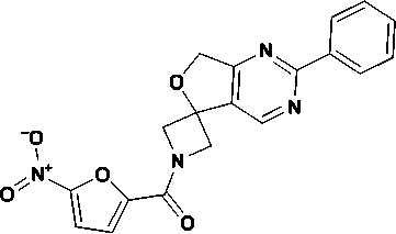
Fig. 1. Chemical structure of LK 1674 (1-[(5-nitro-furan-2-yl)carbonyl]-2 ′ -phenyl-1H,7 ′ H-spiro[aze-tidine-3,5 ′ -furo[3,4-d]pyrimidine])
MATERIALS AND METHODS
Physicochemical Assessment of Lipophilicity
Lipophilicity of compound LK 1674 was determined by reversed-phase thin-layer chromatography (RP-TLC) on glass plates coated with RP-18F254s silica gel containing a fluorescent indicator (Merck Millipore). The mobile phase consisted of isopropanol (analytical grade, Merck) and water at volumetric fractions of 50%, 60%, 70%, and 80% organic solvent. LK 1674 was dissolved in dichloromethane (1 mg/mL) and applied to the plates using a capillary, with four applications per solution. Chromatography was performed in a chamber pre-saturated with the mobile phase for 1 h at room temperature, with a migration distance of 10 cm. After development, spots were visualized under UV light at 254 and 365 nm. For each mobile phase composition, measurements were performed in triplicate to reduce experimental error. The obtained Rf values were used to calculate the RM parameter (reversed-phase TLC lipophilicity parameter) according to the Bate–Smith and Westall equation [28]. RM values were plotted against the volumetric fraction of the organic modifier, followed by extrapolation to zero organic solvent content.
Strains of Mycobacterium tuberculosis used for in vivo experiments
Two Mycobacterium tuberculosis strains representing different phylogenetic lineages and genotypes were used for the development of experimental tuberculosis models. The reference strain Mycobacterium tuberculosis H37Rv (TBC #1/47) was originally obtained from the Institute of Hygiene and Epidemiology, Prague (1976). For experimental use, it was sourced from the Federal State Budgetary Institution “Scientific Centre for Expert Evaluation of Medicinal Products” (Ministry of Health of the Russian Federation) on 07 August 2013. This strain belongs to the Euro-American Lineage 4, sublineage 4.9, and is classified as drug-susceptible, with no mutations associated with drug resistance detected. The clinical strain No. 5582 belongs to the Beijing Central Asian/ Russian sublineage (Clade A) and originates from the collection of the St. Petersburg State Research Institute of Phthisiopulmonology of the Ministry of
Health of the Russian Federation (SPbSRIP). This strain exhibits multidrug resistance (MDR) due to four high-confidence mutations associated with resistance to first-line antituberculosis drugs, including rifampicin, isoniazid, streptomycin, and pyrazinamide (rrs C517T, rpoB S450L, katG S315T, pncA T135P).
Experimental procedure for preparation of a viable Mycobacterium tuberculosis culture for animal
The protocol used to obtain a pure, viable M. tuberculosis culture with sufficient biomass for subsequent animal infection consisted of four stages: thawing, culture growth, collection of bacterial biomass, and homogenization and resuspension. Three weeks prior to the experiment, cryovials containing a mycobacterial suspension were thawed at room temperature and inoculated onto L o wenstein-Jensen solid medium (Becton Dickinson, USA). On the day of animal infection, the bacterial mass harvested from the solid medium was transferred into a dry sterile glass tube containing 8–10 glass beads (3 mm in diameter) and homogenized using a vortex shaker for 15–20 s. Three drops of physiological saline were added to the bacterial material, followed by thorough grinding to obtain a homogeneous suspension. Subsequently, 5–6 mL of physiological solution was added, and the mixture was again homogenized by vortexing. The resulting suspension was transferred into a centrifuge tube and centrifuged at 1,000 rpm for 10 min to sediment bacterial clumps. The upper portion of the supernatant was carefully collected, avoiding disturbance of the sedimented clumps in the lower layers, and transferred into a polystyrene tube for turbidity measurement. The suspension was adjusted to 1.5 McFarland units (corresponding to approximately 5 x 10 s cells/ mL) using a DEN SILAMETER densitometer. The resulting suspension was then diluted with physiological saline to the concentration required for infection, corresponding to 10 6 mycobacterial cells/0.2 mL. The absence of bacterial clumps, which may effectively increase the infective dose to some extent, was confirmed microscopically (an example is shown in Fig. 2).
Experimental animals
A controlled experimental study was conducted using a murine model of tuberculosis infection. All

Fig. 2. Microscopic characterization of the M. tuberculosis H37Rv suspension (5 * 10е CFU) used for inoculation of experimental animals. Ziehl–Neelsen staining. Light microscopy under oil immersion (100 × objective, 10 × eyepiece).
procedures involving experimental animals were reviewed and approved by the Local Ethics Committee of the SPbSRIP (Protocol No. 80 dated 23 June 2021, Extract No. 80.1). Approval was granted in accordance with GOST R 33044–2014 and Resolution No. 4 of the Chief State Sanitary Physician of the Russian Federation dated 28 January 2021, approving SanPiN 3.3686–21 “Sanitary and Epidemiological Requirements for the Prevention of Infectious Diseases”.
Study design
Animals that successfully completed the 14-day quarantine period were randomized into two experimental series. The first series (n = 50) involved infection with the drug-susceptible international reference strain M. tuberculosis H37Rv. The second series (n = 50) involved infection with the clinical multidrugresistant strain No. 5582 of the Beijing sublineage. We followed the general methodological approach that was previously described [29, 30, 31]. A mycobacterial suspension for infecting mice was prepared ex tempore from three-week-old second-generation strains. A total of 100 mice of the C57/black/6 line were infected by inoculation into the lateral tail vein of a suspension of three-week-old M. tuberculosis strains (1x10⁶ CFU/mouse in 0.2 mL of saline solution). According to the classical experimental model for the evaluation of new anti-tuberculosis drugs developed by Pershin et al.[31], a group of intact animals was not included in the study. However, data on the lung and spleen mass coefficients were obtained and were 0.95 (95% CI: 0.74–1.00) for the lungs and 0.51 (CI: 0.45– 0.57) for the spleen.
On day 3 post-infection, animals in both series were allocated into five groups of 10 animals each. From this time point, mice in group 5 of both series received the investigational compound LK 1674 (1-[(5-nitrofuran-2-yl)carbonyl]-2 ′ -phenyl-1H,7 ′ H-spiro[azetidine-3,5 ′ -furo[3,4-d]pyrimidine]) at a dose of 10 mg/kg. As reference drugs in both infection models, ethambutol (E; Pharmsynthesis OJSC) at 20 mg/kg (group 2), moxifloxacin (Mfx; PharmConcept LLC) at 10 mg/kg (group 3), and linezolid (Lzd; ROZOLEX PHARM LLC) at 10 mg/kg (group 4) were used. The investigational compound and comparator drugs were administered as monotherapy. We used doses recommended for the treatment of tuberculosis in humans according to Russian national guidelines, which have also been tested in our studies using experimental tuberculosis models caused by pathogens with different biological properties [32–38].
Drug solutions were prepared ex tempore using Tween-80 as a solubilizer and administered per os via atraumatic gavage (0.2 mL), once daily for 30 administrations. Each experimental series included an infected untreated control group (group 1; M. tuberculosis culture control), which received an equimolar volume of physiological saline containing the same concentration of Tween-80 as used in the treatment groups, administered per os.
Following completion of the treatment course, mice from both experimental series were euthanized on day 47 post-infection by cervical dislocation. The following procedures were subsequently performed: macroscopic assessment of lung involvement, as previously described [39, 40]; determination of lung and spleen weights, followed by calculation of organ weight coefficients (organ weight/body weight × 100); and collection of lung tissue samples for microbiological and histological analyses.
Microbiological analysis
The microbiological analysis included the following procedures: weighing of lung tissue samples (100 mg); preparation of lung homogenates under aseptic conditions; quantitative plating onto L o wenstein-Jensen egg medium (Becton Dickinson, USA) using the serial dilution method; and assessment of M. tuberculosis growth after 4 weeks of incubation at 37 °C by determining the number of colony-forming units (CFU) per unit mass of lung tissue. The microbial burden was expressed as a percentage of the maximum CFU count in the infected untreated control group.
Histological analysis
For histological analysis, lung tissue samples were fixed in 10% neutral buffered formalin, embedded in celloidin–paraffin–oil, sectioned, and stained with hematoxylin and eosin. Microscopic examination was performed using an Olympus BX45 microscope (Olympus Corp., Tokyo, Japan) equipped with a digital camera and Olympus DP-Soft software. The extent of the exudative component of specific inflammation, the presence of tissue destruction, and indicators of local immune activity in the lungs were evaluated. The results were expressed as semiquantitative scores for the principal histopathological features observed in the lung tissue. Visual patterns of specific inflammation were assessed using Fisher’s exact test based on the presence or absence of the feature in small samples. Differences were considered statistically significant when p ≤ 0.05.
Assessment of therapeutic efficacy
The treatment efficacy index was calculated using the following mathematical formulation. For each disease severity parameter, efficacy was determined as:
= -2---1 x 100%, XCj where Xcj is the mean value of parameter in the infected untreated control group, the Xij represents the mean value for treatment group i whith i ranging from 2 to 5, and j= 1,2,3 corresponds to the lung weight coefficient, lung lesion index, and microbial burden in the lung tissue, respectively. The overall treatment efficacy index for each group was then calculated as the arithmetic mean of the three parameter-specific efficacy values:
TEIi-^Eij, 3 j=i
Data processing
Statistical analysis was performed using Statistica 7 software (StatSoft Inc., USA). Data normality was assessed using the Shapiro–Wilk test. Variables that did not follow a normal distribution ( p ≤ 0.05) are presented as the median and interquartile range, Me (Q1–Q3). Comparisons between two independent groups were performed using the Mann–Whitney U test, whereas comparisons among three or more groups were conducted using the Kruskal–Wallis test. Multiple comparisons were adjusted using the Bonferroni correction. Differences were considered statistically significant at p ≤ 0.05.
RESULTS
Lipophilicity Assessment
The experimental lipophilicity data obtained for LK 1674 were analyzed by linear regression of the retention parameter (R) against the isopropanol concentration in the mobile phase. The resulting regression slope was –1.7941, which is in close agreement with the value reported for the reference compound
(a)
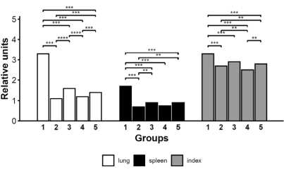
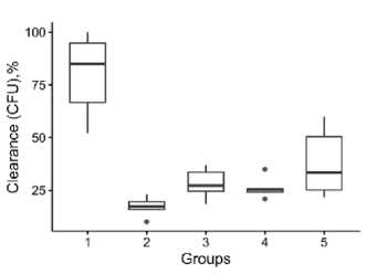
Fig. 3. Disease severity and pulmonary M. tuberculosis burden in C57BL/6 mice infected with the international reference drug-susceptible M. tuberculosis H37Rv strain, evaluated 47 days after initiation of anti-tuberculosis therapy. (a) Comparison of disease severity parameters among the experimental groups. Statistical significance: p > 0.05; ** p < 0.05; *** p < 0.001; **** p < 0.0001. (b) Pulmonary M. tuberculosis burden, expressed as a percentage of the CFU count in the infected untreated control group. Group 1, infected untreated control; Groups 2–5, animals treated with ethambutol (E, 20 mg/kg), moxifloxacin (Mfx, 10 mg/kg), linezolid (Lzd, 10 mg/kg), or LK 1674 (10 mg/kg), respectively.
(b)
omeprazole (–1.763). As expected for reversed-phase chromatographic systems, increasing the proportion of the organic modifier reduced analyte retention. The similarity between the regression slopes of LK 1674 and omeprazole indicates comparable chromatographic lipophilicity.
Evaluation of Therapeutic Efficacy in the H37Rv Infection Model
In the murine tuberculosis model established with the international reference drug-susceptible Mycobacterium tuberculosis strain H37Rv, infected animals were allocated to several experimental groups, including an untreated control group (Group 1). At the end of the observation period, mice in the untreated group developed generalized tuberculous process, with the lungs representing the primary site of disease involvement. Gross pathological examination revealed numerous coalescing miliary tuberculous lesions, accompanied by high lung lesion index. Biometric analysis demonstrated increased lung and spleen weight coefficients. Microbiological analysis showed a high pulmonary mycobacterial burden, reaching 5.91 ± 0.05 log CFU (Figs. 3a, b). Compared with the untreated control group, LK 1674 monotherapy significantly reduced multiple indicators of disease severity, including the lung weight coefficient (by 45.8%, p < 0.004), lung lesion score (by 15.5%, p < 0.03), and spleen weight coefficient (by 48.0%, p < 0.004). In addition, the pulmonary mycobacterial burden was reduced 2.1-fold (53%; p < 0.02). Relative to the reference drugs, LK 1674 showed efficacy comparable to that of moxifloxacin across most measures of disease severity, although both ethambutol and linezolid remained substantially more effective (Fig. 3a). Notably, analysis of colonyforming unit (CFU) counts, an integrated measure of therapeutic efficacy, demonstrated a reduction in pulmonary mycobacterial burden with LK 1674 that was comparable to that achieved with moxifloxacin. No significant difference in CFU counts was observed between LK 1674 and linezolid, whereas LK 1674 was less effective than ethambutol in promoting pulmonary clearance of M. tuberculosis (Fig. 3b).
Histological examination of the lungs in the control group mice revealed lesions characterized by extensive confluent foci of inflammation, disruption of distinct spatial cellular organization, decreased aeration of the lung tissue affecting more than 30% of the histological section area, and the presence of neutrophilic infiltration in 100% of cases (Fig. 4a). Histological analysis confirmed the efficacy of the LK 1674 compound in attenuating the severity of specific inflammatory alterations compared
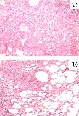
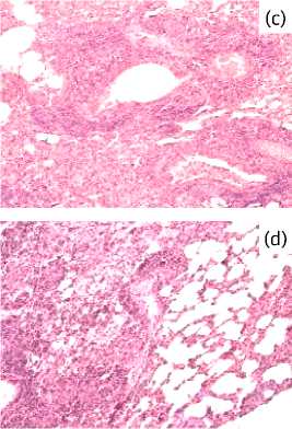
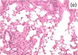
Fig. 4. Representative micrographs of lung sections from mice infected with the drug-susceptible reference strain M. tuberculosis H37Rv, 47 days after treatment initiation. Foci of specific infiltration are shown for the study groups: 1, control, untreated (a); 2–5, experimental groups treated with E (b), Mfx (c), Lzd (d), and LK 1674 (e). Hematoxylin and eosin staining; magnification × 300.
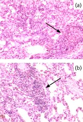
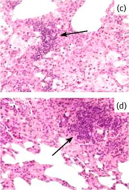
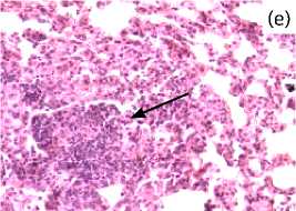
Fig. 5. Representative micrographs of lung sections from C57BL/6 mice infected with the drug-susceptible reference strain M. tuberculosis H37Rv, 47 days after treatment initiation. iBALT aggregates are shown in the study groups: 1, control, untreated (a); 2–5, experimental groups treated with E (b), Mfx (c), Lzd (d), and LK 1674 (e). Hematoxylin and eosin staining; magnification × 600.
to the infected control group. Treatment with the compound LK 1674 resulted in a reduction of the exudative component: inflammatory infiltrates were non-confluent in the majority of cases (4 out of 6 vs.
0 out of 6 in the infected control group; p = 0.005) and were restricted to an area of 20–40 alveoli (Fig. 4e); furthermore, a marked suppression of neutrophilic infiltration, a key marker of tissue destruction, was observed. The state of local immunity was assessed by evaluating the extent of perivascular and peribronchial infiltration, as well as the cellular composition of inducible bronchus-associated lymphoid tissue (iBALT), which forms during chronic inflammation, including tuberculosis [40, 41].
Monotherapy with compound LK 1674 led to qualitative changes in iBALT structure, characterized by a reduction in the number of conglomerates with large accumulations of epithelioid cells and the appearance of structures containing only scattered cells (in 6 of 6 cases versus 0 of 6 in the untreated mouse group; p < 0.0005, Fig 5). Furthermore, a decreased frequency of large perivascular and peribronchial lymphohistiocytic infiltrates was documented (in 6 of 6 cases versus 2 of 6, p < 0.006). Comparative histopathological evaluation of lung tissue revealed that treatment with compound LK 1674 induced milder inflammatory alterations than those observed in the moxifloxacin (Mfx) group. The test compound exhibited efficacy comparable to that of linezolid (Lzd) and ethambutol (E). Statistical comparison of histological parameters between treatment groups revealed no significant differences between LK 1674 and the comparator drugs (p > 0.05 for all pairwise comparisons), except for a greater extent of confluent infiltrates in the Mfx group compared with LK 1674, E, and Lzd (p < 0.05). In the LK 1674, E, and Lzd monotherapy groups, most animals showed only limited infiltration, with lesions encompassing 20–40 alveoli (Fig. 4b, d). By contrast, large confluent infiltrative foci with accompanying loss of pulmonary aeration were seen in 5 of 6 Mfx-treated specimens (Fig. 4c).
Features common to all experimental groups included: the absence of neutrophilic granulocytes within the infiltrates, remodeling of iBALT conglomerates toward a reduction in epithelioid cell numbers (Fig. 5b–e), and the presence of marked perivascular and peribronchial lymphohistiocytic infiltration (Fig. 5b–e). Counting of iBALT conglomerates with a predominantly epithelioid composition revealed intergroup differences: the highest detection rate was in the Mfx group (3/6 cases), the lowest in the E group (1/6), and intermediate rates in the LK 1674 and Lzd groups (2/6 cases each).
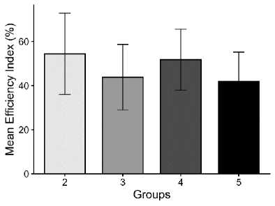
Fig 6. Therapeutic Efficacy Index (TEI, %) for each group in C57BL/6 mice infected with the international reference drug-susceptible M. tuberculosis H37Rv strain, evaluated 47 days after initiation of antituberculosis therapy. Group 1, infected untreated control; Groups 2–5, animals treated with ethambutol (E, 20 mg/kg), moxifloxacin (Mfx, 10 mg/kg), linezolid (Lzd, 10 mg/kg), or LK 1674 (10 mg/kg), respectively.
Since increases in all indices of internal organ involvement in mice were unidirectional and reflected an increase in disease severity, a composite endpoint — the Therapeutic Efficacy Index (TEI) — was used to compare the effects of the investigated compounds across experimental groups (Fig. 6). The TEI was calculated based on lung weight coefficient, lung lesion index, and pulmonary mycobacterial burden (see Materials and Methods for the exact definition). In ascending order of TEI, the investigated compounds were ranked as follows: LK 1674 (+41.8%), moxifloxa-cin (+43.8%), linezolid (+51.8%), and ethambutol (+54.4%). Thus, with respect to the severity of tuberculosis caused by the international reference drug-susceptible Mycobacterium tuberculosis H37Rv strain, the therapeutic efficacy of LK 1674 was comparable to that of moxifloxacin monotherapy across most parameters but lower than that of ethambutol and linezolid.
Evaluation of Therapeutic Efficacy in Clinical Strain Infection Model
LK 1674 monotherapy significantly reduced disease severity in mice infected with multidrug-resistant clinical Mycobacterium tuberculosis strain 5582 of the Beijing genotype. As in the drug-susceptible tu-
(a)
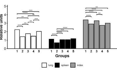
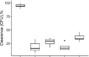
Fig. 7. Disease severity and pulmonary M. tuberculosis burden in C57BL/6 mice infected with a clinical multidrug- resistant M. tuberculosis strain 5582 of the Beijing genotype, evaluated 47 days after initiation of anti-tuberculosis therapy. (a) Comparison of disease severity parameters among the experimental groups. Statistical significance: p > 0.05; ** p < 0.05; *** p < 0.001; **** p < 0.0001. (b) Pulmonary M. tuberculosis burden, expressed as a percentage of the CFU count in the infected untreated control group.
1 2 3 4 5
Groups
(b)
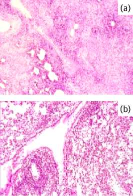
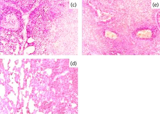
Fig. 8. Representative histological sections of the lungs of C57BL/6 mice infected with the multidrug- resistant Beijing genotype clinical strain 5582, 47 days after treatment initiation. Foci of specific infiltration in the study groups: 1 (control), untreated; 2–5 (experimental), treated with the test compounds: E (b), Mfx (c), Lzd (d), LK 1674 (e). Hematoxylin and eosin staining. × 300 magnification.
berculosis model, this was reflected by a decrease in the lung lesion index (p < 0.004) and improved pulmonary mycobacterial burden (p < 0.003) compared with the infected untreated control (Fig. 7a, b). Histological analysis of lung tissues showed marked proliferation of the iBALT system in LK 1674-treated mice, accompanied by the formation of perivascular and peribronchial lymphohistiocytic inflammatory infiltrates relative to the infected control group (Fig. 8a-e).
In the murine drug-resistant tuberculosis model, comparison of the investigated treatments demonstrated that LK 1674 was significantly less effective than ethambutol (E) and linezolid (Lzd) with respect to lung weight coefficient (p < 0.003) and pulmonary mycobacterial clearance (p < 0.01; Fig. 7a, b).
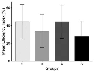
Fig 9. Therapeutic Efficacy Index (TEI, %) for each group infected with a clinical multidrug- resistant M. tuberculosis strain 5582 of the Beijing genotype, evaluated 47 days after initiation of anti-tuberculosis therapy. Group 1, infected untreated control; Groups 2–5, animals treated with ethambutol (E, 20 mg/kg), moxifloxacin (Mfx, 10 mg/kg), linezolid (Lzd, 10 mg/ kg), or LK 1674 (10 mg/kg), respectively.
However, no significant differences were observed between LK 1674 and moxifloxacin (Mfx) for most evaluated parameters. Histopathological changes in the lungs were broadly comparable across all treated groups (E, Mfx, Lzd and LK 1674) (Fig. 8b–e). Across all treatment groups, inflammatory lesions remained highly active and were characterized by extensive cellular infiltration together with nuclear debris resulting from macrophage and neutrophil degeneration.
Assessment of the mean Therapeutic Efficacy Index (TEI) at 47 days after treatment initiation (Fig. 9) showed the highest values in mice treated with ethambutol (+44.1%) and linezolid (+44.1%), followed in descending order by moxifloxacin (+33.8%) and LK 1674 (+27.6%). Thus, LK 1674 promoted a more favorable course of tuberculosis caused by the clinical multidrug-resistant Mycobacterium tuberculosis strain 5582 of the Beijing genotype compared with the infected untreated control. The therapeutic activity of LK 1674 was comparable to that of moxifloxacin monotherapy across most evaluated parameters but was significantly lower than that of ethambutol and linezolid.
DISCUSSION
Drug resistance in Mycobacterium tuberculosis is one of the major factors limiting the efficacy of chemotherapy. The emergence of resistance to currently used anti-tuberculosis drugs is inevitable, driving the search for and development of new compounds with antimycobacterial activity [10]. In this study, the therapeutic potential of the newly synthesized nitroheterocyclic compound LK 1674 was evaluated in murine experimental tuberculosis models induced either by the international reference drug-susceptible (DS) M. tuberculosis strain H37Rv or by a clinical multidrug-resistant (MDR) M. tuberculosis strain 5582 of the Beijing genotype. Comparator drugs were selected according to the resistance phenotype and genotype of the M. tuberculosis strains and included next-generation anti-tuberculosis agents. The effects of the investigational drugs on the course of experimental tuberculosis infection were assessed using a comprehensive clinical and laboratory approach, with the rate of mycobacterial elimination from the lungs serving as an integral criterion of therapeutic efficacy.
In a murine model of drug-susceptible tuberculosis, LK 1674 promoted a more favorable course of infection compared with the infected untreated control, as evidenced by a significant reduction in mycobacterial burden, decreased extent of specific pulmonary lesions, absence of markers of tissue destruction, and increased activity of local lung immunity, reflected by the presence of iBALT aggregates containing single epithelioid cells as well as large lymphohistiocytic infiltrates. According to the literature, the formation of iBALT structures during tuberculosis infection is associated with an effective immune response and a favorable disease outcome [42, 43]. Comparison of LK 1674 with established anti-tuberculosis drugs (E, Lzd, and Mfx) showed that its effects on inflammatory parameters and local lung immune activity were significantly weaker than those of ethambutol, comparable to those of linezolid, and more pronounced than those of moxifloxacin.
The findings obtained in the drug-susceptible model were further examined in a model of drug-resistant tuberculosis. In this model, LK 1674 monotherapy, compared with infected untreated controls, likewise resulted in a significant reduction in disease severity, as assessed by pulmonary mycobacterial clearance and lung lesion score. These effects were comparable to those of moxifloxacin (Mfx) but significantly less pronounced than those observed with ethambutol (E) and linezolid (Lzd). Histopathological examination of lung tissues from mice treated with LK 1674 and the reference anti-tuberculosis drugs (E, Mfx, and Lzd) revealed a broadly similar pattern across all groups. However, no significant differences were observed between LK 1674 and moxifloxacin (Mfx) for most evaluated parameters. In the groups of mice treated with E, Mfx, Lzd, and LK 1674, the cellular characteristics of iBALT did not differ and were more polymorphic and less specific, owing to the greater activity of the inflammatory process that developed, compared with animals infected with the drug-susceptible M. tuberculosis strain. A more pronounced and more mature response was also observed with the use of the drugs under study compared with the infected control group.
Taken together, these findings demonstrate the therapeutic activity of LK 1674 in both drug-susceptible and drug-resistant experimental tuberculosis. The effects of LK 1674 were comparable to those of the reference drugs for several of the evaluated parameters, although their magnitude varied depending on the tuberculosis model and the parameter assessed.
An important limitation of the present study is the absence of pharmacokinetic data for LK 1674. Based on the chromatographic lipophilicity determined in the present study and its similarity to that of omeprazole, it may be hypothesized that LK 1674 has pharmacokinetic characteristics comparable to those of omeprazole, including a relatively short elimination half-life (approximately 40 min) and predominantly renal excretion of its metabolites (approximately 80%). However, these assumptions cannot be confirmed in the absence of direct pharmacokinetic data. Dedicated pharmacokinetic studies are therefore required to characterize the pharmacokinetic profile of LK 1674 and to further interpret the therapeutic effects observed in the present study.
Another methodological limitation concerns the verification of the infectious dose. Although the study was designed according to the classical Pershin model, in which optical density measurement is used for inoculum standardization, the administered dose was not retrospectively verified by quantitative titration. Nevertheless, the absence of bacterial clumps was confirmed microscopically (Fig. 2), and the preparation protocol was strictly standardized. Thus, while these measures support the reproducibility of the infection procedure, the lack of quantitative CFU confirmation of the actual administered dose should be considered when interpreting the findings.
CONCLUSION
The results obtained from the evaluation of the efficacy of 1-[(5-nitrofuran-2-yl)carbonyl]-2’-phe-nyl-1H,7’H-spiro[azetidine-3,5’-furo[3,4-d]pyrimidine], designated LK 1674, in murine models of drug-susceptible and drug-resistant tuberculosis indicate its potential as a standalone therapeutic agent. Overall, these findings suggest that LK 1674 may represent a promising alternative in cases where moxifloxacin is insufficiently effective, as well as a potential component of combination anti-tuberculosis therapy, although further in-depth preclinical studies are required.
Funding: This study is supported by the State Assignment of the Ministry of Health of the Russian Federation, “Development and Preclinical Evaluation of Antimycobacterial Agents Using Machine Learning Approaches for the Treatment of Tuberculosis Caused by Multidrug- and Extensively Drug-Resistant Pathogens” (2024–2026), No. 226012924796–0.
Conflict of interest: The authors declare no conflict of interest.
Compliance with Ethical Principles: The study was conducted in accordance with Directive 2010/63/ EU and the ARRIVE 2.0 guidelines and was approved by the Independent Ethics Committee of the Saint Petersburg Research Institute of Phthisiopulmonology, Ministry of Health of the Russian Federation.