A comparative analysis of paints on the Karakol burial slabs

Author: Pakhunov A.S., Devlet E.G., Molodin V.I., Lazin B.V., Karateev I.A., Dorovatovsky P.V., Kaloyan A.A., Podurets K.M., Senin R.A., Blagov A.E., Yatsishina E.B.
Journal: Archaeology, Ethnology & Anthropology of Eurasia @journal-aeae-en
Section: Art. The stone age and the metal ages
Article in issue: 3 т.45, 2017.
Free access
Short address: https://sciup.org/145145327
IDR: 145145327 | DOI: 10.17746/1563-0110.2017.45.3.056-068
Text of the article A comparative analysis of paints on the Karakol burial slabs
About 40 years have passed since the time of the excavations at the burial sites of the Karakol culture near the Ozernoye village, the Ongudaysky District, in the Altai Republic, one of which accommodated a slab with an anthropomorphic image (Pogozheva, Kadikov, 1979). More than 30 years ago, at the center of the Karakol village in the Ongudaysky District of the Altai Republic, burials in stone cists, constructed
using slabs and stelas with petroglyphs, were discovered. During secondary use, graphical images were often placed horizontally, superimposed with impressive anthropo-zoomorphic figures depicted with paint (Kubarev V.D., Soenov, Ebel, 1992; Kubarev V.D., 1988, 2013; and others). The Karakol images are among the most vivid manifestations of artistic and ritual human activities of the Bronze Age in this region (Molodin, 2006); taken together, these constitute a unique example of painting in prehistoric art of Eurasia. Therefore, the eagerness of researchers to clarify the technical and technological peculiarities of the images, using new methods and techniques, is comprehensible.
The pigments used in the burial rite have always been of interest to archaeologists and anthropologists in terms of both technologies and burial rite semantics. In recent years, analytical studies of the use of red and other pigments in funerary practices and decoration of rock surfaces at the archaeological sites of Russia have changed meaningfully, owing to the involvement of numerous analytical methods (Esin et al., 2014; Pakhunov et al., 2014; Mednikova, 2015; Trifonov et al., 2015).
Substantial supplementation of our understanding of the artistic activities of the Karakol culture bearers, including the use of red pigment in the decoration of Karakol and Ozernoye slabs, has become possible with the application of digital image enhancement, reflection transformation imaging, etc. (Devlet E.G., Pakhunov, Devlet M.A., 2016). The objective of this study was to conduct a comparative analysis of paints on slabs from the same burial and from different burials and burial mounds, in order to determine the principles of selecting paint materials, differing in their tone and composition, by scanning electron microscopy with X-ray spectrometry and synchrotron powder X-ray diffraction methods. A comprehensive analysis has allowed us to draw a reliable distinction between the ferruginous crusts and the surface areas colored with pigment; to refine the details of images and the composition of pigments used to perform figurative and non-figurative elements; and to compare the composition of the paints used at the various stages of decoration of slabs from different burials and burial mounds. In total, the color decoration has been analyzed of five slabs from three Karakol burials and of one slab from Ozernoye, the slabs being exhibited in the Museum of History and Culture of the Peoples of Siberia and the Far East of the Institute of Archaeology and Ethnography of the SB RAS*.
According to the historical and cultural concept common for Western and Southern Siberia, the sites of the Karakol culture belong to the group of Early and Middle Bronze Age cultures contemporaneous with Okunev and Krotovo. Consequently, taking into account the calibrated radiocarbon dates, these sites can be assigned to the second half of the 3rd to the beginning of the 2nd millennium BC (Molodin, Epimakhov, Marchenko, 2014: 145, fig. 2).
Materials and methods
The composition of pigments is determined by various analytical methods, which are selected depending both on the research goal and the specimen’s size. Usually, scanning electron microscopy with X-ray spectrometry is applied for elemental analysis of paint samples. The possibilities of this method are used to obtain images of samples at high magnifications, in order to provide a comparative analysis of pigment particles based on their morphology, characteristic sizes, and distribution in a sample (Clottes, Walter, 1990; Vignaud et al., 2006; Balbín Behrmann, de, González, 2009; Iriarte et al., 2009; Podurets et al., 2016). A complementary method for elemental analysis is X-ray diffraction (Beck et al., 2014). Elemental analysis provides information only on the quantity and distribution of elements, while X-ray diffraction allows mineral phases identification. The methods can be used simultaneously (Scott, Scheerer, Reeves, 2002; Dayet, d’Errico, Garcia-Moreno, 2014). Analysis of X-ray diffraction patterns makes it possible to determine some characteristics of pigment, such as the presence or absence of calcination, particle size distribution, and degree of their crystallinity (Pomies, Morin, Vignaud, 1998; Pomies, Menu, Vignaud, 1999; Gialanella et al., 2011; Salomon et al., 2015; Podurets et al., 2016). Powder X-ray diffraction of paint samples is often performed using synchrotron radiation sources (Wess et al., 2001; Huntley et al., 2014; Zubavichus, Slovokhotov, 2001) or specially designed laboratory devices (Wainwright et al., 2002; Salomon et al., 2012; Kovalchuk et al., 2016). Both methods allow operation with minimal quantities of pigment, which is the crucial factor when studying rock art. Analysis of the Karakol pigments was conducted by the methods of scanning electron microscopy coupled with X-ray spectrometry, and powder X-ray diffraction at synchrotron radiation facility, which allow us to analyze grains of pigment.
The studies have been carried out under the agreement on cooperation between the National Research Center “Kurchatov Institute” (NRC KI) and the Institute of Archaeology of the RAS for joint research of the application of analytical methods to humanities in the NRC KI. The studies were conducted using a specialized synchrotron radiation source, the only one in the former Soviet Union, and a unique facility of the Nanozond resource center of the Kurchatov Complex of NBICS (Nano-, Bio-, Information, Cognitive, Socio-Humanistic) technologies: a scanning electron-ionic microscope Versa 3D DualBeam (FEI, USA) equipped with a nitrogen-free silicon sdd-detector Octane Plus (EDAX, USA) with the resolution of 128 eV. The microscope camera allows operation both in high-vacuum mode and in natural-environment mode (pressure up to 2700 Pa). Back-scattered electrons are detected using segmented solid-state detectors (CBS for high vacuum, Annular GAD for environment operations); and Helios Nanolab 600i (FEI, USA)—a scanning electron-ionic microscope equipped with an energy-dispersive X-ray microanalysis system (EDAX, USA), with the Omniprobe micromanipulator, and with gas injection systems (GIS) allowing to spray Pt, W, C films on the sample surface. The device resolution is 0.8 nm with an accelerating voltage of 30 kV. Samples were studied using electron microscopes after evaporating a gold layer less than 1 nm thick.
Studies of samples by powder X-ray diffraction were conducted at the Belok station at NRC KI synchrotron radiation facility, using the Rayonix SX165 two-dimensional detector, with wavelength λ = 0.9752Å. A small sample of pigment was secured under the microscope in a polymerous loop using Apiezon grease. Data accumulation was conducted for 10 minutes, while the sample was rotating through 360°. The use of synchrotron radiation facility allowed to analyze minute samples (Kheiker et al., 2007). In order to preserve the initial particle-size distribution of the pigment, the sample was not ground for analysis. Formally, this method is nondestructive; however, it is almost impossible to use samples for additional analyses because of their contamination with the grease by means of which the samples are secured in a vertically oriented holdingtool. Analysis can be carried out using samples of approximately 40 μm; however, the paint layers on the slab from burial 4, mound 1 in Ozernoye, and on slab 1 from burial 5 in Karakol were too thin, so it was impossible to prepare samples for study.
The Ozernoye and Karakol slabs were documented using Nikon D800 and OM-D E-M10 Mark II cameras, Nikkor 60/2,8 Micro, Nikkor 105/2,8 Micro lenses, and a remote flash.
The colors of paints were determined visually using the NCS S Index 1950 palette. Conversion to the Munsell color system was performed by means of the translation keys (NCS-Munsell…, 2008); the differences of the pairs did not exceed 2 ∆E. Daylight fluorescent lamps and a LED-panel with the color-rendering index of more than 88 units were alternately used as illuminating devices. The colors of paints on slab 1 from burial 3, mound 2 at the Karakol site were also determined under the bright diffused daylight*.
Samples
At both sites, the images are depicted on schist slabs. The inward decorated surface of the burial has a weathering layer, non-uniform in color and composition, and differing from the host rock.
To obtain a reliable result, at least two samples of each paint were taken. Also, the following criterion was fulfilled: the sampling-places should be small, and located within inconspicuous areas. In total, 22 samples were analyzed: 14 samples of red paints from six slabs, 1 sample of black paint, and 7 samples from the surface of slabs without paint (Fig. 1, 1–4 , 6 , 7 , a ). Samples were taken mainly by scratching with a small steel ophthalmic scalpel. The samples of pigments were placed into
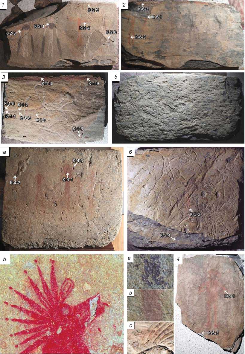
Fig. 1. Slabs from the Karakol ( 1–3 , 5–8 ) and Ozernoye ( 4 ) sites, with designation of sampling locations.
1 – slab 1 from burial 2, mound 2, a raking light image; 2 – slab 2 from burial 2, mound 2, a raking light image; 3 – slab 1 from burial 3, mound 2, a raking light image; 4 – a slab from burial 4, mound 1, a diffused-light photograph; 5 – slab 2 from burial 2, mound 2, the side without drawings; 6 – slab 3 from burial 2, mound 2, a raking light image; 7 – slab 1 from burial 5: a – a raking light image; b – image of the central figure’s head after color enhancement; 8 – macrophotographs of areas of the paint layer on various Karakol slabs: a – slab 1 from burial 3, mound 2; b – slab 1 from burial 3, mound 2; c – slab 3 from burial 2, mound 2.
Characteristics of slabs
|
Slab No. * |
Size, cm* |
Color of the outline / color of drawings on the slab* |
Color of paint |
Color according to NCS S |
Color according to Munsell |
|
1 from burial 3, |
67 × 46 × 3 |
Dry dark red ocher |
Karakol Red |
NCS S 5030-R – |
5 R 3/6–5 R 3/4 |
|
mound 2 1 from burial 2, |
97 × 57 × 3 |
– Dark red ocher |
Reddish brown arc-shaped line Light red |
S 6020-R NCS S 8010-Y90R – S 8010-R10B NCS S 2070-Y80R |
7.5 R 2/2–5 R 2.5/2 8.75 R 4/12 |
|
mound 2 |
Red paint |
" |
NCS S 2070-Y80R |
8.75 R 4/12 |
|
|
2 from burial 2, |
109 × 58 × 3 |
Dark red ocher |
" |
NCS S 2070-Y80R |
8.75 R 4/12 |
|
mound 2 |
Bright red ocher |
" |
NCS S 2070-Y80R |
8.75 R 4/12 |
|
|
3 from burial 2, |
71 × 61 × 5 |
Dark red ocher |
" |
NCS S 2070-Y80R |
8.75 R 4/12 |
|
mound 2 |
" |
" |
NCS S 2070-Y80R |
8.75 R 4/12 |
|
|
1 from burial 5 |
120 × 89 × 5 |
Absent |
– |
– |
– |
|
Slab from burial 4, |
54 × 39 × 3 |
Dark crimson ocher Absent |
Red Ozernoye – |
NCS S 5030-R – |
2.5 R 3/6 – |
|
mound 1 |
Dry ocher |
Red |
NCS S 3560-Y90R |
10 R 3/10 |
*Data by V.D. Kubarev (2013).
the sterile micro-tubes, rock fragments without paints were placed into polyethylene zip bags. In view of the fact that the sample preparation imply their contamination, the samples were split into several sub-samples, so as to use a unique sample for each analysis.
Results
The colors of paints determined according to the NCS Atlas belong to four groups, with different ratios of the yellow and red hues (the content of yellow component is from 20 % to zero (see Table )). The color of paints on all slabs from burial 2, mound 2 is stable and typical for red ocher. The red line on slab 1 from burial 2, mound 2, and the figures on slab 1 from burial 5 are made with a paint of another tone, which color does not contain the yellow component; it was probably pure hematite. The arc-shaped line on slab 1 from burial 3, mound 2 is reddish brown. This probably indicates the complex composition of the material—a mixture of various substances and to its non-uniform distribution (Fig. 2, 1 , a ).
Karakol site
Slab 1 from buria l 2, mound 2. The decorated surface of the slab is flat; the losses in the lower right part were formed before drawing the images (see Fig. 1, 1 ). The spalls in the upper left part are modern*.
Three figures are depicted on the slab, using pecking and engraving techniques. The slab was placed in the burial horizontally, following which, four characters were depicted on it with black and red paints, and by engraving. The black pigment layer is uniform and dense. On the rightmost figure, streaks underneath the rock layer overhanging by 1–2 mm have been found. The upper edge of the slab is marked with a red line. The combination of RTI and color enhancement has allowed us to reveal a partial sketch of the figure.
The largest aggregates of hematite particles reach 5 μm in samples 2-4, 2-8. The iron content
*“The left upper part was spalled during the opening of the mound before starting the excavation” (Kubarev V.D., 2013: 15).
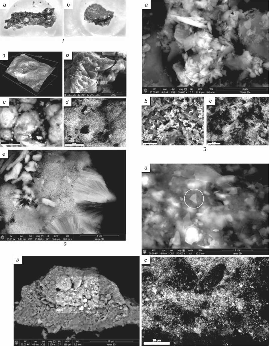
Fig. 2. Microphotographs of samples, back-scattered electron photographs, and iron-distribution maps of three samples.
1 – microphotographs of paint samples: a – K-1-7, b – K -1-9; 2 – sample K-1-7: a – 3D model of surface; b – a back-scattered electron image of spherulites; c – back-scattered electron image of the analyzed area; d – iron-distribution map in the 2c image area; e – needle-shaped particles of goethite; 3 – sample K-1-9: a – a back-scattered electron image; b – a back-scattered electron image of the analyzed area; c – iron-distribution map in the 3b image area; 4 – sample K-4-3: a – a back-scattered electron image; b – a back-scattered electron photo of the analyzed area; c – iron-distribution map in the 4b image area.
in the red paint samples is about 20% wt. The paints from the figures are similar to each other both in the elemental composition and in the size of pigment particles. The sample from the red line shows a somewhat smaller iron content: visually, the line color looks less saturated, and the paint layer is thinner.
Slab 2 from burial 2, mound 2. The slab’s surface is flat, with some losses (exfoliation) of the weathered layer, which had been formed before the images were made: the head of the second figure on the left overlaps the boundary of the decay (see Fig. 1, 2 ). In the right part of the slab, the engravings are partially overlapped by a black-paint drawing; the layer is uniform and dense. In the left part of the slab, only red images appear. The upper boundary of the slab is marked with a red line. The back shows different-depth engravings representing bull-headed figures (see Fig. 1, 5 ).
The main fraction of red pigment is up to 2.5– 3.5 μm. Large aggregates of more than 10 μm are actually absent. The paint sample from the red line has no particles of more than 4 μm.
Slab 3 from burial 2, mound 2. The slab’s surface is flat, the loss in the lower right part had been formed before the slab was placed in the burial. Numerous pecked and engraved images pertain to the initial period of art activities (see Fig. 1, 6 ). The central figure is made by engraving (the head with horns) and painting (the body). The upper boundary of the slab is marked with a red line. The figure is hardly discernible due to the small thickness of paint layer, the line is very thin too.
The drawings are made with liquid paint covering the slab surface with embossed engravings (see Fig. 1, 8 , c ). The iron content in the analyzed samples from the figure and the line is within 10– 20% wt, and depends in a greater degree on the sample’s characteristics (its size and orientation) than on the paint’s consistency. The calcium content in the weathered layer is considerably increased as compared to the host rock, which determines the specific hardness of the stone surface, and allows the possibility of deep engraving.
Slab 1 from burial 3, mound 2. This slab has a strong relief. This is not related to the arrangement of the figures. Two completed images and one sketch (a partial figure of a bull-headed figure, a spectacleshaped sign, and two short parallel lines) are made by the pecking technique. Inside the burial, the slab was turned around in such a way that the figures were placed horizontally; the upper edge of the slab is marked with a red line.
The slab’s surface color is non-uniform, and only the upper right part, limited by an arc-shaped line, is uniformly yellow. The arc-shaped reddish brown line is very dense, and fully covers the stone surface; three samples were taken from it. X-ray microanalysis has determined a large amount of iron (30–40% wt), which is indicative of an iron-containing mineral forming a compact layer on the surface; using elemental mapping, a uniform distribution of iron throughout the entire analyzed area has been revealed, which is not typical for paints*, and suggests natural pigmentation of this line (see Fig. 2, 2 , d ).
The surface morphology of selected samples was assessed by constructing a 3D model in secondary electrons. It has been shown that the colored material is composed of spherical particles, with some losses at the center (see Fig. 2, 2 , a ). These particles have the divergent structure typical for goethite (see Fig. 2, 2 , b ). The ends of needle-like goethite crystals in the back-scattered electron mode look like bright spots; however, at high magnification, it can be seen that they are not separate particles (see Fig. 2, 2 , e ). The orientation of spherical particles is consistent throughout the entire layer, which tightly adjoins the rock’s surface (see Fig. 2, 2 , c ).
Regular-shaped hematite crystals are encountered in the paint sample from the bounding line. The typical size of pigment particles is 0.5–0.9 μm, though large elongate particles 1.2–3.3 μm in size and small crystals less than 0.3 μm are also encountered. The paint contains up to 0.6% wt of manganese, which is atypical of the paint itself; manganese is also present in the weathered layer, but absent in the host rock. Weathered layers on the slab’s surface differ in their content of iron and manganese; the yellowish layer contains iron, while manganese is almost absent.
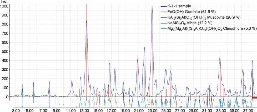
The powder X-ray diffraction was employed to study two samples: K-1-1 and K-1-3 (Fig. 3). Samples К-1-1, -2, and -8 were taken from the arc-shaped line; these are similar to each other, and therefore the most representative diffraction pattern of sample К-1-1 is published.
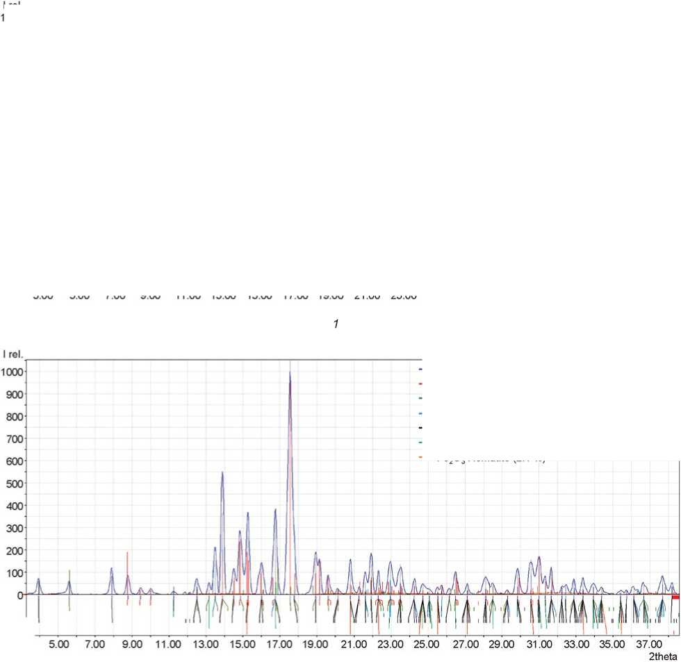
Fig. 3. X-ray diffraction patterns of samples K-1-1 ( 1 ) and K-1-3 ( 2 ).
2theta
— K-1-3 sample
— NaAISi3Os Albite (69.2 %)
— KAI2(Si3AI)O10(OH,F)2 Muscovite (17.7 %)
— FeO(OH) Goethite (6.0 %)
— Mg3(Mg2AI)((Si3AI)O10)(OH)2O3 Clinochlore (2.5 %)
— SiO2 Quartz (2.5 %)
— Fe2O3 Hematite (2.1 %)
In the samples from the dark arc-shaped line, the main components are goethite, albite, muscovite, and clinochlore. In goethite, iron ions fill a half of the octahedral cavities formed by oxygen ions in hexagonal packing. Parameters of an elementary cell (in angstroms) of goethite are a = 4.57; b = 9.93; c = 3.036 in the dark paint, and a = 4.56; b = 9.96; c = 3.04 in the red paint. The natural diversity of goethite reveals itself in a varying degree of hydration and substitution. Weakly hydrated particles have a grain-oriented fibrous-needle-like structure, and are strongly anisotropic. But strongly hydrated particles are usually isotropic, while the size of crystals is smaller. The shift in elementary-cell parameters, which can be observed in the X-ray photographs, is related to partial replacement of iron in the goethite lattice by other elements. A high value of parameter b is indicative of high isomorphic substitution for iron in the lattice; this results in the formation of an aggregate of irregular conglutinated particles instead of needle-like individuals, typical for goethite. Then the color gets brown tones (Belenky, Riskin, 1974; Vodyanitsky, 2003).
The absence of quartz in the samples argues for the following presumption against the dark-line nature: the goethite layer was formed as a result of natural processes during the formation of the rock, i.e. it was not a weathering product; the layer was easily separated from the slab during sampling.
Hematite, which ensures an intense red color, has been identified in the sample from the red line, in addition to quartz, albite, and mica group minerals (Mas et al., 2013).
Slab 1 from burial 5. The slab is flat, with local defects on the surface. The weathered layer of the slab surface, including the pecking area, is uniform in color (see Fig. 1, 7 , a ). The stone texture can be seen under the paint layer, which is not very dense. Owing to the high resolution of the obtained photographs, new additional details of the image have been revealed, e.g. teeth in the mouth of the central figure. The artist probably worked using a thin brush (see Fig. 1, 7 , b ).
The pigment particles of small size (0.1–0.5 μm) are distributed uniformly in the paint. The hematite crystals have a sharp shape, so we have discovered plate-like particles of various sizes. Obviously, the artist used paint with a high content of hematite to be grinded during the paint preparation, which resulted in formation of small crystal fragments (see Fig. 2, 4 , a ). Fig. 2, 4 , a , b shows a schist particle with a paint layer in the upper part of it*.
Ozernoye site
A slab from burial 4, mound 1. Unlike the Karakol slabs, the surface of this slab is non-uniform. An anthropomorphic figure is made by a thin layer of red paint (see Fig. 1, 4 ). Apart from this figure, no other traces of drawing have been revealed on the slab. The fragmented yellow outlining is modern, and no pecking or engraving traces on the surface are found by RTI.
The aggregates of hematite particles in the bulk of the paint reach a size of less than 1 μm, while separate particles are indiscernible. The difference between the iron content in the weathered layer and in the paint is less than 50 %: 5 and 9% wt., respectively. Large calcite inclusions have been found in the sample.
Discussion and conclusions
Red and black are the basic colors in prehistoric art. In nature, the most widespread and available red pigment is red ocher. The color of iron-containing paints is determined by their composition. Iron compounds determine yellow (goethite, lepidocrocite, jarosite), red (hematite), or black (magnetite) color. The presence of these chromatic components, as well as quartz, calcite, gypsum, and other minerals, in various ratios, and also variations of the shape and size of goethite and hematite crystals (the color of large crystals and aggregates of goethite and hematite is black) ensure diversity of the tones of natural materials (Torrent, Schwertmann, 1987; Schwertmann, 1993; Elias et al., 2006; Froment, Tournie, Colomban, 2008; Mastrotheodoros, Beltsios, Zacharias, 2010).
Comparison of the descriptions of the paint colors contouring lines and figures on the slabs from burial 2, mound 2 at Karakol, presented by V.D. Kubarev (who mentions dark red, dark crimson, red, and bright red paints) (see Table ) allows us to presume that figures were done using various red tones, while the line was always made by dark red paint (see, e.g., (Kubarev V.D., 2013: 15–16)). However, determining the color with the use of the NCS color chart has allowed us to establish that the lines were made with the same paint as the figures. Notably, bodies of the deceased were drawn using paints of various tones; and also for painting and drawing, different paints were selected (Ibid.: 17).
Images on the Karakol slabs are made by various red pigments. Analysis of samples showed the similarity between the paints used for painting of all three slabs from burial 2, mound 2, not only in color, but in their elemental composition as well. The light red paint is red ocher with a hematite content of 10–20% wt. The particles of pigment are small, in the form of aggregates up to 5 μm in size; however, larger particles of more than 10 μm are also encountered. Taking into account that no differences have been found in the composition of the paints, the differences in the size of hematite aggregates can be related to the degree of pigment grinding: pigment that was not fully ground might have remained at the bottom of the vessel in which the paint was prepared.
The dark red paint sampled on slab 1 from burial 5 contains small hematite particles. They are uniformly distributed in the bulk of the samples taken from the central figure, whereas the sample from the small figure in the right part of the composition shows aggregation of particles forming local areas with a high content of hematite particles. This can be related to the technical process of paint preparation and the procedure for drawing the images: at first, two large central figures were made, after which, two small ones were depicted in the left part of the slab using the remaining paint. All figures were probably depicted with the same paint, since no differences in color and elemental composition are observed.
The color of the paint that was used to make the red outline on slab 1 from burial 3, mound 2, is the same as that of the paint on slab 1 from burial 5; however, the shape of hematite particles is different: it contains more crystals of elongate shape and naturally faced crystals.
The red line extending along the upper boundary of the slabs from the majority of Karakol burials was applied after the placing the slabs in the grave. This conclusion was prompted by the displaced slabs, for example, those constituting the northern wall in burial 3 of mound 2 (Kubarev V.D., 2009: 155, fig. 75): the line on them was made in the same paint that was used for the figurative images.
In the Paleolithic, black pigment was provided most frequently by carbon-containing materials such as charcoal or soot (Prinsloo et al., 2008; Iriarte et al., 2013), or by manganese minerals such as pyrolusite, romaneshite, etc. (Chalmin, Menu, Vignaud, 2003; Lahlil et al., 2012; Pitarch et al., 2014). Analysis of black paint from slab 1 from burial 2, mound 2, has demonstrated that this was soot. The paint forms a very thin and dense layer, which is typical for fine pigments: separate particles cannot be seen at 2000x magnification, and the images made by the scanning electron microscope also show that the pigment layer uniformly covers the painted surface.
Gypsum (Mawk, Nobbs, Rowe, 1996; Wainwright et al., 2002), calcium carbonate (Scott, Hyder,
1993), white clay (Ward et al., 2001), or their mixtures (Koski, McKee, Thomas, 1973; Hall, Meiklejohn, Arocena, 2007) were probably used as white pigments by the artists who made open-air rock art images. No white paint was used in the Karakol paintings; however, in order to render white color for the depicted figures, the stone was scraped off on slab 1 from burial 2, mound 2 (Kubarev V.D., 2013: 15). As a result, small rock crystals were formed on the surface. They scattered light in all directions, which determined the white color of the surface after scraping-off.
Thus, the results of analysis point to the natural origin of coloring of the arc-shaped line on slab 1 from burial 3, mound 2, to the use of one red paint for decorating slabs from burial 2, mound 2, to the application of soot as a black pigment, and to the absence of white pigments. The differences in color and composition of red pigments suggest that in order to prepare paints, the available material was selected rather than a specific one. This conclusion is confirmed by the features of pigments at the Karakol and Ozernoye sites.
Subsequently, on the basis of the imaging and analytical studies performed, a reconstruction of the operating procedure can be carried out, implying the repeated, intentional symbolical use of some stelae and slabs with images for decoration of the Karakol funerary stone cists. Studies of the pigments that were used by the bearers of the Chemurchek culture, contemporaneous with the Karakol culture, appear to be promising (Kovalev, Erdenbaatar, 2014: 277–279; and others). We are talking about the first and the second Turochak rock art sites near the Biya River, which are very likely to belong to the Karakol culture (Molodin, 2016), and about the Okunev culture imagery (see, e.g., (Pyatkin, Martynov, 1985)). Comparison of the said pigments with those from geographically and chronologically close images from the Tom River (Tomskaya Pisanitsa rock art site) (Kovtun, Rusakova, 2014; Rusakova, 2015) and the Altai Mountains (Kurmantau) (Kubarev G.V., 2003) may provide important scientific results. Another promising direction of studies may be provided by analysis of the stone cist slabs from the studied Okunev culture burial grounds by means of optical microscopy, which could reveal remains of decayed colorants.
Acknowledgements
This study was supported by the Russian Foundation for Basic Research (Projects 16-01-00418 and 17-29-04172). We express our gratitude to A.A. Velikzhanin (National Research Center “Kurchatov Institute”) for his help in processing of X-ray diffraction data.
