A digital X-ray analysis of Middle Bronze Age skeletal samples from the Baraba forest-steppe

Author: Marchenko Z.V., Kishkurno M.S., Grishin A.E., Reinhold S., Zhuravkov F.V.
Journal: Archaeology, Ethnology & Anthropology of Eurasia @journal-aeae-en
Section: Anthropology and paleogenetics
Article in issue: 3 т.49, 2021.
Free access
We present the results of a comparative analysis of skeletal and dental pathologies in Middle Bronze Age individuals buried at Late Krotovo and Andronovo (Fedorovka) cemeteries in southwestern Siberia. This was the period when the Andronovo steppe tradition in Northern and Central Asia expanded in various directions, including the foreststeppe. Growth arrest lines on tibiae (Harris lines) and dental pathologies (enamel hypoplasia and caries) were recorded. To evaluate developmental anomalies in the bone tissue, digital X-ray analysis was used. The principal sample includes representatives of various sex and age groups buried at the largest cemetery in the region, Tartas-1 (Baraba forest-steppe). Harris lines and enamel hypoplasia result from a broad range of factors such as infections, occasional malnutrition, traumas, vitamin defi ciencies, etc. Caries is caused by a high amount of carbohydrates in the diet, accompanied by low standards of oral hygiene. These pathologies occur at different ages: Harris lines and enamel hypoplasia evidence adverse factors during infancy and adolescence, whereas caries is typical of adulthood. Late Krotovo and Andronovo groups differ in terms of occurrence and combination of pathologies. Enamel hypoplasia is less frequent in the Andronovo sample, indicating a lesser stress level in children. Harris lines are less frequent in the Late Krotovo group, suggestive of lower stress level during adolescence. These differences may be tentatively attributed to various models of subsistence and cultural adaptation.
Digital X-ray analysis, Harris lines, enamel hypoplasia, Tartas-1 cemetery, Andronovo (Fedorovka) culture, Late Krotovo (Cherno-Ozerye) culture
Short address: https://sciup.org/145146293
IDR: 145146293 | DOI: 10.17746/1563-0110.2021.49.3.136-146
Text of the article A digital X-ray analysis of Middle Bronze Age skeletal samples from the Baraba forest-steppe
Human skeletal samples are a unique biological archive of individual features of life-long trends in health and development in ancient populations. Changes in the bone surface may provide information on occupational or craft activities, as well as on trauma and diseases striking bone tissue. The internal structure of bones is also a valuable source of information about morbid conditions and the status of physiological development.
This structure is studied using various diagnostic techniques: X-ray, magnetic resonance imaging (MRI), and 3D computed tomography (CT). All these methods are non-invasive and thus are broadly applied in the study of normal and pathological conditions in mummified human remains from Egypt, China, Europe, and South America (Murphy et al., 2003; Jackowski, Bolliger, Thali, 2008; Mai et al., 2016; Licata, Pinto, 2020).
The fixation of transversal (or Harris) lines of growth arrest in the cortical layer of the long bones, mainly at the distal tibia, is a popular method of studying the health and physiological development of ancient populations. The lines can be detected only via radiation diagnostics. They have been observed on X-ray images starting from the late 19th century, predominantly in patients who suffered rickets (Hughes, Heylings, Power, 1996). A special study of the lines was carried out by H.A. Harris during the First World War. The researcher called them “tombstones” that point to the illness suffered by the individual in the past (Ibid.). The factors leading to the appearance of Harris lines—systemic disorders, nutritional and vitamin (A, C, D) deficiency, physiological and psychological stress—have been subsequently studied using various experimental and clinical methods (Park, 1964; Huss-Ashmore, 1981; Hughes, Heylings, Power, 1996). It was shown that the time of the influence of a negative factor, i.e. the period during which the individual was stressed, rather than simply its presence, was the more influential on the formation of the lines. However, a number of studies have shown that Harris lines can be present even in the absence of those adverse conditions. In such cases, these can be considered sensitive signals during normal growth (Alfonso, Thompson, Standen, 2005; Papageorgopoulou et al., 2011). The experiments on the influence of nutritional deficiency on the formation of transversal lines in rabbits have demonstrated that the frequency of their appearance was higher in the group of periodically starving animals as compared to the permanently malnourished group (Alfonso-Durruty, 2011).
From a histological point of view, Harris lines are formed during the periods when cessation of growth in the epiphysis coincides with the continuing growth of the diaphysis (dissociation of the rates of chondroplasia and osteogenesis). With time, after the traumatic or stress episode, the pace of growth of both elements eventually recovers (Follis, Park, 1952). As a result, the medullar trabeculae form condensations of increased mineral density. The formation of the lines is associated with three periods of the most intense growth of the body, i.e. the first year of life, 5–7 years, 11–12 years (girls) and 15–16 years (boys). This is a physiological reaction of bone tissue on a spectrum of negative factors (Gindhart, 1973). According to some clinical studies, Harris lines can disappear in adults and elderly people as a result of remodeling of the cortical layer (Garn, Schwager, 1967).
The link between the appearance of Harris lines and adverse developmental conditions was established clinically, thus providing a theoretical base for X-ray studies of this marker in archaeological collections. The lines have been assumed to be “indicators or ‘memory’ of previous growth disruption and stress in an archeological population” (Goodman, Clark, 1981: 35) and were employed for assessing the health conditions of skeletal individuals during their childhood and adolescence (McHenry, 1968; Goodman, Clark, 1981; Hughes, Heylings, Power, 1996; Buzhilova et al., 2013; Mednikova, Engovatova, Tarasova, 2015). Thus, Harris lines have been used as markers of dietary and/or physiological stress. The number and frequency of the lines in the tibia can be utilized to determine the time of their formation during growth and to model the periods of physiological stress in individuals (Hummert, Van Gerven, 1985; Byers, 1991; Ameen et al., 2005).
In a study comparing the ancient and modern populations of the same area of Switzerland (Ameen et al., 2005), Harris lines were present in individuals older than 50 years in both samples. On the basis of this observation, the authors of the study hypothesized that the lines could form later in life (not during childhood or adolescence) and be related, not to growth arrests, but to chronic diseases (degenerative changes in the cortical layer, including osteoarthritis, osteoporosis, etc.) or trauma (lower limb fractures, etc.) (Ibid.). That study only reported the fact of detection of Harris lines in adults, but not the mechanism of their formation in such cases. Later, a comparison of X-ray images of the modern people of the Republic of Korea and the Joseon dynasty skeletal collection (16th–18th centuries AD) revealed a higher prevalence of the marker in the medieval sample (Beom et al., 2014). The frequency of Harris lines was higher in females, which was related to their poorer nutrition due to lower social status (Ibid.).
Notably, the assessment of health status in ancient populations should not be based solely on tracing Harris lines (Hughes, Heylings, Power, 1996), but other stress markers as well. These include dental diseases, e.g. enamel hypoplasia (Clarke, 1982; Alfonso, Thompson, Standen, 2005), which forms as a reaction to morbid conditions or malnutrition (El-Najjar et al., 1976; El-Najjar, De Santi, Ozebek, 1978; Goodman, Armelagos, Rose, 1980; Duray, 1996).
The skeletal stress-indicators are employed in archaeological studies of the adaptive strategies in populations with various modes of subsistence. For example, two populations from Central Europe, representing the Neolithic and Bronze Ages, were analyzed in order to detect the biological changes in the human body during the transition to agriculture (Krenz-Niedbala, 2014). The population of the Linear Pottery culture (Neolithic) was purely agricultural, while the population of the Corded Ware culture (Bronze Age) was practicing a mixed subsistence economy based on agriculture, pastoralism, hunting, and gathering. The analysis of Harris lines, enamel hypoplasia, and cribia orbitalia has shown a higher prevalence of the pathological markers in the agriculturalists. This can be explained by the influence of adverse social conditions (high population density and a relatively sedentary lifestyle led to the rapid spread of infections) and poor nutrition (invariability of diet, dependence on a single food source).
Thus, while there are different views on the factors in the formation of Harris lines, the polyetiology of this lesion is broadly accepted. Many researchers suggest that physiological stress suffered during childhood, a maladaptive process, is the main cause. But Harris lines have also been interpreted as a marker of dietary disturbances and subadult injuries. The presence of the lines in elderly individuals (>50 years of age) was hypothesized to be explained by recent traumatic lesions (fractures) and other pathologies of the musculoskeletal system (Ameen et al., 2005). But in our opinion, such an interpretation is poorly based at present, and comprehensible additional studies of the postcranial skeleton are required. Thus, the main aim of the present study was to describe the paleopathological markers in the skeletons of the samples representing the Late Krotovo (Cherno-Ozerye) and Andronovo (Fedorovka) archaeological cultures of the Baraba forest-steppe from the Tartas-1 cemetery in the Vengerovo District of the Novosibirsk Region. The study protocol included various pathological manifestations of physiological stress and dietary disturbances associated with changes in a subsistence economy, both at the individual and population levels.
Tartas-1 site
The site has been studied since 2003 by the Institute of Archaeology and Ethnography SB RAS, in cooperation with the German Archaeological Institute, under the leadership of V.I. Molodin from the Russian part (Fig. 1). At present, it is the largest multi-temporal burial ground in Western Siberia (about 800 burials); the majority of graves belong to the Middle Bronze Age (Molodin et al., 2020). The studied sample of human remains includes two cultural groups that formed at Tartas-1 their separate necropolises: the Late Krotovo and Andronovo (Fedorovka) groups.
The Late Krotovo (Cherno-Ozerye) culture was identified by V.I. Molodin and described by him as the latest form of existence of the autochthonous Krotovo culture, developed under the influence of
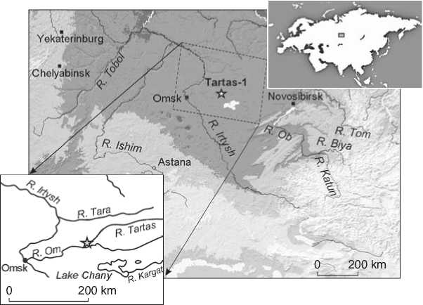
Fig. 1. Location of the Tartas-1 site.
the Andronovo steppe realm, which was reflected in the appearance of products of the Timber Grave-Andronovo affinity (bronze weapons and jewelry) (2014). In the anthropological features of the population and their ritual practice, this influence is not so vivid (Ibid.; Chikisheva, 2012: 109–110). In the spatial distribution of graves at the Tartas-1 cemetery, two clusters of the Late Krotovo burials are distinguished: northern and southern (Marchenko et al., 2021). Hypothetically, this reflects different micropopulations within the same cultural group. The Late Krotovo burial practice is characterized by shallow graves, most often individual ones. The buried were placed in an extended supine position, with their heads to the northeast. Ceramic vessels were placed in graves quite rarely. Men were usually accompanied by weapons (bronze daggers, dart-heads, bone arrowheads); women with bronze ornaments and awls. Horse phalanges and metacarpals of small ruminants have been occasionally found in burials.
The Late Krotovo people practiced a complex economy. The populations of the Baraba settlements of Vengerovo-2 and Preobrazhenka-3 were engaged in breeding small ruminants; cattle and horses to a lesser extent (Molodin, Mylnikova, Nesterova, 2017). Isotope analysis of anthropological materials showed a significant proportion of fish in the structure of human protein nutrition (Marchenko et al., 2021). The Late Krotovo burials at Tartas-1 date back to the 19th– 17th centuries BC (Molodin et al., 2012).
The Andronovo (Fedorovka) group is the largest at the cemetery (ca 50 %) (Molodin et al., 2020: 486); it is characterized by the widespread use of ceramic vessels of the Andronovo or “syncretic” types in the ritual practice (Molodin, Mylnikova, Ivanova, 2014); the dead were placed in a flexed position on their sides (Molodin, 2011). Completely cremated remains are less common; some graves revealed cremated and non-cremated remains. The spatial distribution of the Andronovo (Fedorovka) burials is different in different parts of the cemetery: in dense rows, or sparsely, or with traces of the kurgan organization of space (Molodin et al., 2020). The following features atypical of the Andronovo tradition have been recorded: a significant percentage of communal graves, placing bronze daggers and horn dishes in graves, and the use of fish as funeral food (Ibid.). All these features together reflect the “barbarization” of the Andronovo culture in the Baraba forest-steppe and the heterogeneity of this population group at the cemetery (Ibid.). Cultures of the Andronovo affinity are traditionally considered pastoralist (Kuzmina,
1986: 32). However, in the burials of the group at Tartas-1, the use of fish in burial practice was noted in a significant number of cases, which indirectly indicates their consumption by the Baraba pastoralists (Molodin et al., 2015). The Andronovo (Fedorovka) burials of the cemetery date back to the 18th–15th centuries BC (Molodin et al., 2012).
Craniological materials from other Baraba burial grounds contemporaneous with Tartas-1 indicate that the population that left this necropolis experienced a difficult situation reflecting the “ethno-racial interaction of migrants and groups of the autochthonous population” (Chikisheva, 2012: 116, 117). The female subgroup of the Baraba forest-steppe is the most polymorphic as compared to all other Andronovo groups* (Ibid.: 116).
Material and methods
The Late Krotovo sample includes 17 individuals (9 males, 6 females, 2 sub-adults) of which 11 are the burials of the southern cluster. The Andronovo (Fedorovka) sample includes 27 individuals (16 males, 9 females, 2 sub-adults). The sexes of the deceased were determined on the basis of pelvic and cranial morphology (Alekseev, 1966: 27); the age-at-death was determined by the degree of cranial suture fusion and tooth wear (in adults), and the dental eruption status (in sub-adults) (Meindl, Lovejoy, 1985; Scott, 1979). The main age cohorts followed the standard gradations (Alekseev, Debets, 1964: 39): Infantilis I (before the eruption of the first permanent molars, ca 6–7 years); Infantilis II (before the eruption of the second permanent molars, ca 11–12 years); Juvenis (before the fusion of the spheno-occipital synchondrosis, ca 20 years); Adultus (younger than 35 years); Maturus (younger than 50–55 years); Senilis (older 55 years). The skeletal specimens were examined for the presence of Harris lines in the tibia, and some dental pathologies (caries, enamel hypoplasia).
Harris lines are transversally oriented strips observed in the growth zones of the long bones metaphyses and diaphyses. This lesion is polyetiological and can be a result of a stress episode in childhood, as well as of disturbances of endocrine and metabolic processes (Alfonso, Thompson, Standen, 2005; Shalina, Yarmolinskaya, Abashova, 2018). The
X-ray images of the tibia were obtained using the PRDU-02 device (CJSC “Eltech”, St. Petersburg) at the Institute of Archaeology and Ethnography of SB RAS under the following protocol: voltage 45 kV, amperage 0.07 μA, exposition time 10 s. Visualization of the images was carried out in QuantorMed, ver. 2.0, using the FireCR scanner. Both tibiae of the individuals were examined at the distal and proximal ends, without magnification. The observed Harris lines were not counted, only their presence or absence and severity (weak, medium, strong) were assessed. The results of the assessment were additionally checked by a practicing radiologist.
Caries is a lesion of hard dental tissue (enamel, dentine, cement). The main cause of caries is the infectious cariogenic microflora (Borovsky et al., 2001: 190; Newbrun, 1982). The conditions stimulating the development of caries lesions are various. The main of these is the frequent consumption of food rich in carbohydrates, in particular fast (e.g. sucrose) (Newbrun, 1982; Larsen, Shavit, Griffin, 1991), and a low level of oral hygiene. An increasing layer of dental calculus stimulates the reproduction of bacteria and the decrease in the strength of the tooth enamel. Other factors favoring the development of caries are hypomineralization of enamel, decrease in the antimicrobial functions of saliva, general immunodeficiency of the body (Newbrun, 1982), diseases of the gastrointestinal tract, and, in general, serious metabolic disorders (Borovsky et al., 2001: 210–211; Kanchan et al., 2015).
Enamel hypoplasia is a deficit of the enamel layer due to a decreased activity of ameloblasts during the secretory phase of enamel formation (Skinner, Goodman, 1992). The pathology develops under the influence of various diseases during the formation of permanent teeth enamel (El-Najjar, De Santi, Ozebek, 1978; Borovsky et al., 2001: 134; Groshikov, 1985: 38). The main reasons for these morbid conditions are nutritional imbalance, deficiency of vitamins A, C, D, infections and hypocalcemia causing severe diseases (El-Najjar, De Santi, Ozebek, 1978; Borovsky et al., 2001: 82).
All the pathological dental and skeletal conditions analyzed in the present study do not appear simultaneously under the influence of a single factor, since the time of the formation of each of the markers is different. Enamel hypoplasia of permanent teeth develops at the age of 7–8 years, during the period of amelogenesis of the permanent incisors, canines, premolars, first and second molars. The most active growth of the long bones and, accordingly, the highest probability of the appearance of Harris lines fall on the first year of life and 9–12 years of age (Alfonso, Thompson, Standen, 2005). Unlike these, carious lesions can form at any age. Therefore, the pathological indicators employed in the present study can be viewed as a proxy for the individual biological adaptation to the changes in occupational activity or environment throughout life. The prevalence of these indicators in different cultural groups can help, in turn, to determine features varying at the population level.
Results and discussion
Harris lines in the tibia of the Late Krotovo sample were detected in 5 cases (29 %), in the metaphyseal area of the proximal and distal ends of the bones, which points to their formation mainly during late childhood. The number of the lines varies from one to three, severity is weak.
The dental pathologies of the Late Krotovo sample from Tartas-1 were thoroughly analyzed previously (Kishkurno, 2019). The sample employed in the present study displays a very high prevalence of caries (75 %). The lesions were mainly located on the occlusal surface of the upper and lower molars, less frequently on the buccal surface, and only in single cases on the distal and lingual surfaces (Table 1). Carious cavities were absent in five individuals: two from the southern cluster (55–60-year-old female and a 9 ± 2-year-old sub-adult), and three from the northern cluster (30–40-year-old males, Adultus–Maturus). The prevalence of enamel hypoplasia in the Late Krotovo sample is very high (94 %). Linear type dominated in the anterior teeth, but single lesions were detected in the molars. Point type is much less common and only found on the canines.
In five individuals, enamel hypoplasia and Harris lines were observed simultaneously (Table 2): three of these cases were males (from 20 to 45 years of age), one female (20–25 years), and one adolescent (12 ± 2.5 years). In all five individuals, carious lesions, mainly on the occlusal and buccal surfaces, were present as well. Much more often, hypoplasia and Harris lines were not observed in the same individuals (65 %), but in only one case were both markers absent (male, Adultus–Maturus). Harris lines were detected only in the skeletons from the southern planigraphic cluster (see Table 1), while enamel hypoplasia was equally frequent in both parts of the necropolis.
In the Andronovo (Fedorovka) sample, Harris lines were detected in 14 cases (52 %), up to 3–4 in one individual. The lines were weakly developed
Table 1. Individual distribution of the frequency of the pathological markers in the Late Krotovo sample
|
Burial/ skeleton No. |
Sex |
Age, years |
Caries |
Enamel hypoplasia |
Harris lines |
||||
|
Surface |
|||||||||
|
occlusal |
buccal |
distal |
lingual |
linear |
point |
||||
|
Southern cluster |
|||||||||
|
8 |
Fem. |
30–35 |
+UМ1 |
+LМ1, LМ2 |
0 |
0 |
+UI1, UI2, UC, LI1, LI2, LC |
+LC |
0 |
|
11 |
Male |
20–25 |
+LM1 |
+LM1, LM2 |
0 |
0 |
+UC, LI2 |
0 |
+ |
|
19 |
Fem. |
20–25 |
+M1, M2 |
0 |
0 |
0 |
+I1, I2, C |
0 |
0 |
|
20 |
" |
20–25 |
+UM2 |
0 |
0 |
0 |
+UM1 |
0 |
+ |
|
25 |
Male |
25–30 |
+LM1, LM3 |
+LM1, LM2, LM3 |
+LM1 |
+UM3 |
+UI1, UC, LC |
0 |
+ |
|
29 |
– |
9 (± 2) |
0 |
0 |
0 |
0 |
+UI1, UC |
0 |
0 |
|
36/1 |
Male |
25–30 |
+UM2, UM3, LM3 |
+LM2 |
0 |
0 |
+UC, UP1, LI2, LC |
0 |
0 |
|
36/2 |
Fem. |
55–60 |
0 |
0 |
0 |
0 |
+UI1, UC, UM1, LI1, LI2, LC, LM1 |
0 |
0 |
|
39 |
Male |
40–45 |
+LM3 |
0 |
0 |
0 |
LC |
0 |
+ |
|
78 |
– |
12 (± 2.5) |
+UP2, M2 |
0 |
0 |
0 |
0 |
+UC, LC |
+ |
|
94 |
Fem. |
20–25 |
+UM1 |
0 |
0 |
0 |
+UM2 |
+LC |
0 |
|
Northern cluster |
|||||||||
|
251 |
Male |
Maturus |
+LM3 |
+LM2 |
0 |
0 |
+LC |
0 |
0 |
|
315 |
" |
20–25 |
+UM1 |
0 |
0 |
0 |
0 |
+LC |
0 |
|
318/2 |
" |
35–40 |
0 |
0 |
0 |
0 |
+UI2, UC |
0 |
0 |
|
325/1 |
" |
30–35 |
0 |
0 |
0 |
0 |
+LI1, LC |
0 |
0 |
|
325/2 |
" |
Adultus– Maturus |
+LM1 |
0 |
0 |
0 |
0 |
0 |
0 |
|
374/2 |
Fem. |
Adultus– Maturus |
+LM3 |
0 |
0 |
0 |
+LC |
0 |
0 |
Table 2. Summary data on the three pathological markers
|
Cultural group |
Caries |
Enamel hypoplasia |
Harris lines (HL) |
HL and hypoplasia |
Hypoplasia without HL |
HL without hypoplasia |
Individuals without HL and hypoplasia |
|
Late Krotovo ( n = 17) |
13 (76 %) |
16 (94 %) |
5 (29 %) |
5 (29 %) |
11 (65 %) |
0 |
1 (6 %) |
|
Andronovo (Fedorovka) ( n = 27) |
19 (70 %) |
16 (59 %) |
14 (52 %) |
8 (30 %) |
8 (30 %) |
6 (22 %) |
5 (19 %) |
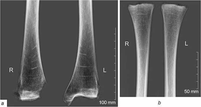
Fig. 2. X-ray images of the tibiae with Harris lines (orthogonal projection). a – distal parts (burial 324); b – proximal parts (burial 287).
and typically observed in the metaphyseal area of the distal end of the bone (Fig. 2, a ). Single cases of lines of medium or strong severity were also found in both metaphysis and diaphysis of both proximal and distal ends of the bone (Fig. 2, b ).
The frequency of caries in the Andronovo (Fedorovka) sample is high (71 %). The cavities are typically located on the occlusal tooth surface, rarely on the buccal side, and only in single cases on the lingual and disto-vestibular surfaces (Table 3). Enamel hypoplasia was detected in 16 individuals (59 %). Unlike the Late Krotovo sample, this marker occurs on the incisors and canines only. The lineal type is prevalent, while the point type was found in just three individuals.
Eight cases (30 %) of the combination of hypoplasia and Harris lines (see Table 2) were observed: in three males (from 35 to 55 years of age), four females (from 20–25 years of age to Senilis), and one sub-adult (10 ± 2.5 years of age). The prevalence of individuals displaying hypoplasia without the lines was slightly higher (30 %) than the prevalence of those with an opposite combination (22 %). In five skeletons, both markers were absent (19 %): three males from 20 to 35 years of age, female (Juvenis), and a sub-adult (10 ± 2 years of age).
Stable isotope studies have shown that the protein part of the diet of the Late Krotovo population, as well as that of the preceding Odino culture groups, was still acquired by the consumption of fish and the meat of forest-steppe mammals. No substantial increase in the proportion of vegetable proteins was detected (Marchenko et al., 2016, 2021). The present study has demonstrated some differences in the distribution of pathological markers between the Late Krotovo and Andronovo (Fedorovka) samples (see Table 2). In the latter, the prevalence of caries is slightly lower (70 vs. 76 %), and of hypoplasia substantially lower (59 vs. 94 %). This can be attributed to the influence of various factors, but we preliminarily suggest that this observation points towards better dental health in the Andronovo population. In our previous work (Kishkurno, 2019) we hypothesized that the environmental conditions of the Late Krotovo population were extreme owing to some economical or ecological changes, which led to an increase in the proportion of plants in their diet. The high prevalence of enamel hypoplasia in this sample suggests physiological stress or severe illness suffered during childhood. Some researchers point to the link between early weaning and this marker in traditional societies (Masterson, Sabbah, 2015). The same could apply to the development of caries as well.
Notably, the frequency of Harris lines in the tibia manifests an opposite trend, being much higher in the Andronovo (Fedorovka) sample: 52 vs. 29 %. In the Late Krotovo individuals, this marker is always accompanied by enamel hypoplasia (Table 2), while in the Andronovo sample it is found both with (30 %) and without (22 %) hypoplasia. No clear association of the pathological markers with sex or age was detected in either population.
Conclusions
The results of the present study led us to the following, mainly preliminary, conclusions. A high prevalence
Table 3. Individual distribution of the frequency of the pathological markers in the Andronovo (Fedorovka) sample
But for driving more solid conclusions regarding the prevalence and etiology of pathological manifestations in ancient skeletal samples, two methodological advances seem necessary. First, a larger number of markers should be studied employing state-of-the-art medical and anthropological techniques. Second, larger samples are required for a more thorough and detailed description of the intragroup paleopathological status.
The further development of the present project will be based on the complex use of radiological and osteoscopic methods for the study of the postcranial skeleton. This will help to clarify the conclusions drawn and expand the source-base for comparative interpopulation analysis.
Acknowledgements
This study was supported by the Russian Foundation for Basic Research and the German Research Foundation, Projects No. 18-509-12067a and DFG RE2688/3-1/2. We are grateful to V.I. Molodin for the access to the skeletal collections from Tartas-1, and to T.A. Chikisheva for her consultations while carrying out the study and writing the article.
References A digital X-ray analysis of Middle Bronze Age skeletal samples from the Baraba forest-steppe
- Alekseev V.P. 1966 Osteometriya: Metodika antropologicheskikh issledovaniy. Moscow: Nauka.
- Alekseev V.P., Debets G.F. 1964 Kraniometriya: Metodika antropologicheskikh issledovaniy. Moscow: Nauka.
- Alfonso M.P., Thompson J.L., Standen V.G. 2005 Reevaluating Harris lines - a comparison between Harris lines and enamel hypoplasia. Collegium Antropologicum, vol. 29 (2): 393-408.
- Alfonso-Durruty M.P. 2011 Experimental assessment of nutrition and bone growth’s velocity effects on Harris lines formation. American Journal of Physical Anthropology, vol. 145: 169-180. URL: https://doi.org/10.1002/ajpa.21480
- Ameen S., Staub L., Ulrich S., Vock P., Ballmer F., Anderson S.E. 2005 Harris lines across centuries: A comparison of two populations, medieval and contemporary in Central Europe. Skeletal radiology, vol. 34: 279-284. URL: https://doi.org/10.1007/s00256-004-0841-3
- Beom J., Woo E.J., Lee I.S., Kim M.J., Kim Y.-S., Oh C.S., Lee S.-S., Lim S.B., Shin D.H. 2014 Harris lines observed in human skeletons of Joseon Dynasty, Korea. Anatomy and Cell Biology, vol. 47: 66-72. URL: https://doi.org/10.5115/acb.2014.47.1.66
- Borovsky E.V., Ivanov V.S., Maksimovsky Y.M., Maksimovskaya L.N. 2001 Terapevticheskaya stomatologiya: Ucheb. posobiye dlya sudentov stomatolog. fakul. med. vuzov. Moscow: Meditsina.
- Buzhilova A.P., Potrakhov N.N., Potrakhov E.N., Gryaznov A.Y. 2013 Analiz markerov stressa metodami mikrofokusnoy rentgenografii (po antropologicheskim materialam epokh kamennogo veka). Biotekhnosfera, No. 2 (26): 46-51.
- Byers S. 1991 Calculation of age at formation of radiopaque transverse lines. American Journal of Physical Anthropology, vol. 85: 339-343. URL: https://doi.org/10.1002/ajpa.1330850314
- Chikisheva T.A. 2012 Dinamika antropologicheskoy differentsiatsii naseleniya yuga Zapadnoy Sibiri v epokhi neolita - rannego zheleza. Novosibirsk: Izd. IAET SO RAN.
- Clarke S.K. 1982 The association of early childhood enamel hypoplasia and radiopaque transverse lines in a cultural diverse prehistoric skeletal sample. Human Biology, vol. 54 (1): 77-84.
- Duray S.M. 1996 Dental indicators of stress and reduced age at death in prehistoric Native Americans. American Journal of Physical Anthropology, vol. 99: 275-286. URL: https://doi.org/10.1002/(SICI)1096-8644(199602)99:2-275::AID-AJPA5>3.0.CO;2-Y
- El-Najjar M., Ryan D., Turner C.H., Lozoff B. 1976 The etiology of porotic hyperostosis among the prehistoric and historic Anasazi Indians of Southwestern United States. American Journal of Physical Anthropology, vol. 44: 447-488. https://doi.org/10.1002/ajpa.1330440311.
- El-Najjar M.Y., De Santi M.V., Ozebek L. 1978 Prevalence and possible etiology of dental enamel hypoplasia. American Journal of Physical Anthropology, vol. 48: 185-192. URL: https://doi.org/10.1002/ajpa.1330480210
- Follis R.H., Park E.A. 1952 Some observations on bone growth with particular respect to zones and transverse lines of increased density in the metaphysis. American Journal of Roentgenology, vol. 68: 709-724.
- Garn S.M., Schwager P.M. 1967 Age dynamics of persistent transverse lines in the tibia. American Journal of Physical Anthropology, vol. 27: 375-378. URL: https://doi.org/10.1002/ajpa.1330270310
- Gindhart P.S. 1973 Growth standards for the tibia and radius in children aged one month through eighteen years. American Journal of Physical Anthropology, vol. 39: 41-48.
- Goodman A.H., Armelagos G.J., Rose J.C. 1980 Enamel hypoplasias as indicators of stress in three prehistoric populations from Illinois. Human Biology, vol. 52 (3): 515-528.
- Goodman A.H., Clark G.A. 1981 Harris lines as indicators of stress in prehistoric Illinois populations. In Research Report 20: Biocultural Adaptation Comprehensive Approaches to Skeletal Analysis. Boston: University of Massachusetts, pp. 35-43.
- Groshikov M.I. 1985 Nekariozniye porazheniya tkaney zuba. Moscow: Meditsina.
- Hughes C., Heylings D.J.A., Power C. 1996 Transverse (Harris) lines in Irish archaeological remains. American Journal of Physical Anthropology, vol. 101: 115-131. URL: https://doi.org/10.1002/(SICI)1096-8644(199609)101:1-115::AID-AJPA8>3.0.CO;2-U
- Hummert J.R., Van Gerven D.P. 1985 Observations of the formation and persistence of radiopaque transverse lines. American Journal of Physical Anthropology, vol. 66: 297-306. URL: https://doi.org/10.1002/ajpa.1330660307
- Huss-Ashmore R. 1981 Bone growth and remodeling as a measure of nutritional stress. In Research Report 20: Biocultural Adaptation Comprehensive Approaches to Skeletal Analysis. Boston: University of Massachusetts, pp. 84-91.
- Jackowski C., Bolliger S., Thali M.J. 2008 Common and unexpected fi ndings in mummies from ancient Egypt and South America as revealed by CT. Radiographics, vol. 28: 1477-1492. URL: https://doi.org/10.1148/rg.285075112
- Kanchan T., Machado M., Rao A., Kewal K., Garg A.K. 2015 Enamel hypoplasia and its role in identification of individuals: A review of literature. Indian Journal of Dentistry, vol. 6 (2): 99-102. URL: https://doi.org/10.4103/0975-962X.155887
- Kishkurno M.S. 2019 Zubochelyustniye patologii nositeley pozdnekrotovskoy (chernoozerskoy) kultury Barabinskoy lesostepi (po materialam mogilnika Tartas-1). Camera Praehistorica, No. 2 (3): 147-155.
- Krenz-Niedbala M.A. 2014 A biocultural perspective on the transition to agriculture in Central Europe. Anthropologie, vol. 52 (2): 115-132.
- Kuzmina E.E. 1986 Drevneishiye skotovody ot Urala do Tyan-Shanya. Frunze: Ilim.
- Larsen C.S., Shavit R., Griffi n M.C. 1991 Dental caries evidence for dietary change: An archaeological context. In Advances in Dental Anthropology, M.A. Kelley, C.S. Larsen (eds.). New York: Wiley-Liss, pp. 179-202.
- Licata M., Pinto A. 2020 Radiology in archaeology: Fundamentals and perspective - examination of the living. In Radiology in Forensic Medicine: From Identifi cation to Post-mortem Imaging. Cham: Springer, pp. 43-54. URL: https://doi.org/10.1007/978-3-319-96737-0_6
- Mai H., Yang Y., Abuduresule I., Li W., Hu X., Wang C. 2016 Characterization of cosmetic sticks at Xiaohe cemetery in Early Bronze Age Xinjiang, China. Scientifi c Reports, vol. 6: 18-39. URL: https://doi.org/10.1038/srep18939
- Marchenko Z.V., Panov V.S., Grishin A.E., Zubova A.V. 2016 Rekonstruktsiya i dinamika struktury pitaniya odinovskogo naseleniya Barabinskoy lesostepi na protyazhenii III tys. do n.e.: Arkheologicheskiye i izotopniye danniye. Vestnik arkheologii, antropologii i etnografi i, No. 3 (34): 164-178.
- Marchenko Z.V., Reinhold S., Grishin A.E., Pozdnyakov D.V., Babina K.A., Batanina O.V. 2021 Perviye rezultatu izotopnogo analiza antropologicheskogo materiala pozdnekrotovskoy (chernoozerskoy) kultury pamyatnika Tartas-1: Rekonstruktsii diyety i mobilnosti naseleniya. In Arkheologiya Severnoy i Tsentralnoy Azii: Noviye otkrytiya i rezultaty mezhdistsiplinarnykh issledovaniy. Barnaul: Izd. Alt. Gos. Univ., pp. 72-79.
- Masterson E.E., Sabbah W. 2015 Maternal allostatic load, caretaking behaviors, and child dental caries experience: A cross-sectional evaluation of linked mother-child data from the third national health and nutrition examination survey. American Journal of Public Health, vol. 105 (11): 2306-2311. URL: https://doi.org/s://doi:10.2105/AJPH.2015.30272
- McHenry H. 1968 Transverse lines in long bones of prehistoric Californian Indians. American Journal of Physical Anthropology, vol. 29: l-18.
- Mednikova M.B., Engovatova A.V., Tarasova A.A. 2015 Diakhronniye izmeneniya kachestva zhizni naseleniya Yaroslavlya v XIII-XVII vv. po dannym radiologii. Rossiyskaya arkheologiya, No. 3: 94-106.
- Meindl R.S., Lovejoy C.O. 1985 Ectocranial suture closure: A revised method for the determination of skeletal age based on the lateral-anterior sutures. American Journal of Physical Anthropology, vol. 68: 57-66.
- Molodin V.I. 2011 Migratsii nositeley andronovskoy kulturno-istoricheskoy obshchnosti v Barabinskuyu lesostep. In Drevneye iskusstvo v zerkale arkheologii: K 70-letiyu D.G. Savinova. Kemerovo: Kuzbassvuzizdat, pp. 58-69. (Trudy Sib. assotsiatsii issledovateley pervobytnogo iskusstva; iss. VII).
- Molodin V.I. 2014 The Late Krotovo (Cherno-Ozerye) Culture in the Irtysh Forest-Steppe, Western Siberia. Archaeology, Ethnology and Anthropology of Eurasia, No. 1 (57): 49-54.
- Molodin V.I., Durakov I.A., Hansen S., Nenakhov D.A., Nenakhova Y.N., Reinhold S., Kobeleva L.S., Mylnikova L.N., Selin D.V., Nesterova M.S., Efremova N.S., Shvetsova E.S., Bobin D.N., Borzykh K.A. 2020 Osobennosti pogrebalnoy praktiki nositeley andronovskoy (fedorovskoy) kultury severo-zapadnoy chasti mogilnika Tartas-1. In Problemy arkheologii, antropologiim etnografi i Sibiri i sopredelnykh territoriy, vol. XXVI. Novosibirsk: Izd. IAET SO RAN, pp. 484-492.
- Molodin V.I., Durakov I.A., Kobeleva L.S., Koneva L.A. 2015 Fish in the Burial Rite of Andronovo (Fedorovka) People, Based on Tartas-1 Cemetery. Archaeology, Ethnology and Anthropology of Eurasia, vol. 43 (3): 77-90.
- Molodin V.I., Marchenko Z.V., Kuzmin Y.V., Grishin A.E., Van Strydonck M., Orlova L.A. 2012 Radiocarbon chronology of burial grounds of the Andronovo period (Middle Bronze Age) in Baraba forest steppe, Western Siberia. Radiocarbon, vol. 54 (3/4): 737-747.
- Molodin V.I., Mylnikova L.N., Ivanova D.P. 2014 A Morphological Analysis of Vessels from Middle Bronze Age (Early 2nd Millennium BC) Burials at Vengerovo, in the Irtysh Forest-Steppe. Archaeology, Ethnology and Anthropology of Eurasia, No. 2 (58): 44-66.
- Molodin V.I., Mylnikova L.N., Nesterova M.S. 2017 Khozyaistvenno-proizvodstvenniye kompleksy na poseleniyakh krotovskoy kultury. In Trudy V (XXI) Vserossiyskogo arkheologicheskogo syezda v Barnaule- Belokurikhe, vol. I. Barnaul: Izd. Alt. Gos. Univ., pp. 307-311.
- Murphy W.A., zur Nedden D., Gostner P., Knapp R., Recheis W., Siedler H. 2003 The iceman: discovery and imaging. Radiology, vol. 226 (3): 614-629. URL: https://doi.org/10.1148/radiol.2263020338
- Newbrun E. 1982 Sugar and dental caries: A review of human studies. Science, vol. 217: 418-423. URL: https://doi.org/10.1126/science.7046052
- Papageorgopoulou C., Suter S.K., Rühli F.J., Siegmund F. 2011 Harris lines revisited prevalence, comorbidities, and possible etiologies. American Journal of Human Biology, vol. 23: 381-391.
- Park E.A. 1964 The imprinting of nutritional disturbances on growing bone. Pediatrics, vol. 33: 815-862.
- Scott E.C. 1979 Dental wear scoring technique. American Journal of Physical Anthropology, vol. 51: 214-217.
- Shalina M.A., Yarmolinskaya M.I., Abashova E.I. 2018 Vliyaniye gormonalnoy terapii na kostnuyu tkan: Mify i realnost. Zhurnal akusherstva i zhenskikh bolezney, No. 3 (67): 83-94.
- Skinner M.F., Goodman A.H. 1992 Anthropological uses of developmental defects of enamel. In Skeletal Biology of Past Peoples: Research Methods. New York: Willey-Liss, pp. 153-174.
