Cattle Sarcocystis spp. infection prevention and control
Автор: Rutaganira Joseph, Glamazdin Igor
Журнал: Вестник Воронежского государственного университета инженерных технологий @vestnik-vsuet
Рубрика: Пищевая биотехнология
Статья в выпуске: 1 (91), 2022 года.
Бесплатный доступ
Cattle Sarcocystis spp. are protozoa. They often parasitise tissues of Cattle. Few of these species are zoonoses. Therefore, they are foodborne parasites associated with consumption of raw or insufficiently thermally treated sarcocystic beef meat. Swallowing oocysts from environmental objects primarily contaminated water, garden crops, grazing on contamited pasture, etc. can cause Cattle sarcocystosis. Sarcocystis spp specific to Cattle include S.hominis, S.heydorni, S.cruzi, S.hirsuta, S.rommeli &S.bovifelis. Among them, S.hominis and S.heydorni are zoonotic and pathogenic agents. Human Intestinal/ Muscular Sarcocystosis is a disease that caused by eating raw or poorly cooked Cattle meat infected by Sarcocystis zoonoses (S.hominis&S.heydorni). Intestinal Sarcocystosis was reported almost from all corners of the world. This has been well documented but no powerful Preventive and control methods available to public yet. With the world growing population, researchers should provide or suggest practical solution to supply safe food to the consumers. During our research work we tried to compare the effectiveness of all available documented Cattle sarcocystis spp. Testing methods to recommend the best one to the public for screening health from infected Cattle before slaughter in the slaughter house. Though culture and society play a fundamental role in foodborne control, we also came up with additional control safety measures recommendations all along the beef meat supply chain.
Beef meat parasites, cattle sarcocystosis, foodborne parasites, human intestinal sarcocystosis, pcr test
Короткий адрес: https://sciup.org/140293788
IDR: 140293788
Текст научной статьи Cattle Sarcocystis spp. infection prevention and control
DOI:
The globalization of the food supply, combined with a greater risk of eating poorly cooked or raw beef meat, can expose human to the infection which may lead to the disease with chronic or acute forms of Intestinal Sarcocystosis (Macpherson and Bidaisee, 2015a).
The genus “ Sarcocystis ” is an obligate parasite in the living body of an intermediate and final host to ensure its life cycle. The genus Sarcocystis contains more than 200 species but six species among others are specific to Cattle where Cattle serve as intermediate hosts of those species. S.hominis, S.heydorni, S.cruzi, S.hirsuta, S.rommeli & S.bovifelis are so far known as sarcocystis species specific to Cattle(Fayer et al., 2015) (Lindsay and Dubey, 2020). Clinically S.cruzi is the most pathogenic species for Cattle. Clinical signs of infected Cattle are fever, anemia, emaciation, and hair loss (especially on the rump and tail of the cattle) (Lindsay and Dubey, 2020), and sometimes Cattle may die. Pregnant Cattle may abort, and growth is slowed down or arrested due to sarcocystosis. Sarcocystis species are more specific to the intermediate host than to the definitive host. S.hominis and S.heydorni are the only zoonotic agents and human serves as a definitive host to them. During S.heydorni & S.hominis lifecycle, the human beings excrete oocysts and the Cattle contain sarcocyst (Lindsay and Dubey, 2020). Oocysts are passed fully sporulated by the definitive host (predator). The sexual stages develop only in the human (definitive host) whereas asexual stages develop only in the Cattle (intermediate host). Sarcocystis species are more specific to the intermediate host than to the definitive host.
-
1 Specificity of Cattle Sarcocystis spp . From the table below, Sarcocytis spp. Share Cattle as common intermediate host but definitive hosts are different.
Sarcocystis spp. name
Intermediate host
Definitive host
S.hominis
Cattle
Human
S.heydorni
Cattle
Human
S.cruzi
Cattle
Canids (dog)
S.rommeli
Cattle
Felids (Cat,)
S.hirsuta
Cattle
Felids (Cat,)
S.bovifelis
Cattle
Felids (Cat,)
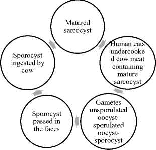
The human beings (definitive host) become infected by intestinal sarcocystosis after ingesting poorly cooked Cattle meat tissues containing S. hominis in the form of mature sarcocysts (Khieu et al., 2017). symptoms include nausea, muscle pain, stomachache, abdominal discomfort, diarrhea and itching skin. Bradyzoites liberated from the sarcocyst by ingestion in the stomach and intestinal epithelial tissue penetrate the mucosa of the small intestine and transform into male (micro) and female (macro) stages and after fertilization of a macrogamete by a microgamete a wall develops around the zygote and an oocyst is formed (Lindsay and Dubey, 2020). Oocysts of sarcocystis hominis sporulate & release the sporocysts into the intestinal lumen from which they are passed in the feces. Sporocysts are shed two weeks after ingesting the infected beef meat (Lindsay and Dubey, 2020). Cattle fed with water or grass seeded with sporocysts become infected and develop sarcocystosis disease.
Rutaganira J., Glamazdin I. Cattle Sarcocystis spp.infection Prevention Rutaganira J., Glamazdin I. Cattle Sarcocystis spp.infection Prevention and Control // Вестник ВГУИТ. 2022. Т 84. № 1. С. 40–42. and Control. Vestnik VGUIT [Proceedings of VSUET]. 2022. vol. 84

Figure 1. S.hominis Lifecycle
The diagnosis of intestinal Sarcocystis infection in human can be made by fecal examination. Since it is not possible to distinguish one species of sarcocystis from another by structure of sporocysts in the faces, PCR on sporocysts can be used to determine the species present in human.
In Cattle before slaughter diagnosis of muscular sarcocystosis can only be made by histological examination of muscle collected by biopsy or at necropsy. The finding of immature sarcocysts with metrocytes suggests recently acquired infection but if only mature sarcocysts are present then the infection is chronic. An inflammatory response associated with sarcocysts may help to distinguish an active disease process from incidental finding of sarcocysts. Serologic Test and PCR are the best for distinguishing Sarco-cystis spp in Cattle (Lindsay and Dubey, 2020).
Materials and methods
Results and discussions
Sarcocystis infection is common in Cattle worldwide. Several factors contribute to the high prevalence in muscular infections in Cattle. Six species are so far specific to Cattle as intermediate host but each of them is having its own definitive host (Rubiola et al., 2020). Each infected definitive host can shed millions of infectious sporocysts over several months contaminating the environment (Lindsay and Dubey, 2020). Sarcocystis sporocysts and oocysts remain viable for many days in the environment, they are resistant to freezing, and they can withstand winter on pasture. Sarcocystis oocysts, unlike those of many other species of coccidia, are passed in feces in the infective form freeing them from dependence on warm moist weather conditions for maturation to infectivity. There is no vaccine so far to protect Cattle against sarcocystosis (Lindsay and Dubey, 2020). Shedding of Sarcocystis oocysts and sporocysts in feces of the definitive hosts is the key factor in the spread of Sarcocystis infection; to interrupt this cycle, Religions integrate preventive measures in society. For instance, Cow is a sacred animal in Hinduism. Therefore, it is forbidden for Hindus to slaughter and consume beef meat (Matsuo et al., 2014). Feed, water and bedding for Cattle should be hygienic. Cattle should not graze on pasture. Carnivorous animals should not be allowed on the cattle pasture. people caring for animals should be examined for harboring parasites, toilets should be equipped with modern cleanliness tools on farms. Alternatively, cattle should not graze on pasture. Raw or semi-raw meat should be consumed only in expensive restaurants that buy beef on certified farms and can pay for additional studies for the presence of parasites. Beef meat can be frozen to inhibit sarco-cysts activities and prevent them to grow and develop disease. Exposure to heat at 60o C for 20 minutes kills sarcocysts (Lindsay and Dubey, 2020). Dead livestock should be buried or incinerated (Lindsay and Dubey, 2020). Above all educational programs for parasitic zoonoses have to contend with the cultural factors that favor the propagation of the infections. For example, the cultural traditions and values attached to food choices, preparation, and consumption challenge a system of beliefs and practices against health management. The educational efforts must therefore offer sustainable, and affordable solutions to inform relevant behavioral changes to traditional practices (Macpherson and Bidaisee, 2015b). Though globalisation may expose a wide range of people to the foodborne parasites, it enables research teams from a range of disciplines to combine and disseminate their knowledge and skills to serve as the greatest resource for combating these pathogens (Robertson et al., 2014).
Conclusion
Cattle meat related Foodborne parasites may have a serious economic consequences and loss of human lives. No Vaccine to the Cattle sarcocystosis known to public so far and no serious control and preventive measures have been yet taken globally. Therefore, Serological Testing method and PCR are the recommended reliable Intestinal and Muscular Sarcocystosis testing methods. More serious studies on Technological Testing methods are needed.
Список литературы Cattle Sarcocystis spp. infection prevention and control
- Fayer R., Esposito D.H., Dubey J.P. Human Infections with Sarcocystis Species. Clin. Microbiol. Rev. 2015. vol. 28. pp. 295-311. doi: 10.1128/CMR.00113-14
- Khieu V., Marti H., Chhay S., Char M.C. et al. First report of human intestinal sarcocystosis in Cambodia. Parasitology International. 2017. vol. 66. pp. 560-562. doi: 10.1016/j.parint.2017.04.010
- Lindsay D.S., Dubey J.P. Neosporosis, Toxoplasmosis, and Sarcocystosis in Ruminants. Veterinary Clinics of North America: Food Animal Practice. 2020. vol. 36. pp. 205-222. doi: 10.1016/j.cvfa.2019.11.004
- Macpherson C.N.L., Bidaisee S. Role of society and culture in the epidemiology and control of foodborne parasites, in: Foodborne Parasites in the Food Supply Web. Elsevier. 2015a. pp. 49-73. doi: 10.1016/B978-1-78242-332-4.00004-7
- Macpherson C.N.L., Bidaisee S. Role of society and culture in the epidemiology and control of foodborne parasites, in: Foodborne Parasites in the Food Supply Web. Elsevier. 2015b. pp. 49-73. doi: 10.1016/B978-1-78242-332-4.00004-7
- Matsuo K., Kamai R., Uetsu H., Goto H. et al. Seroprevalence of Toxoplasma gondii infection in cattle, horses, pigs and chickens in Japan. Parasitology International. 2014. vol. 63. pp. 638-639. doi: 10.1016/j.parint.2014.04.003
- Robertson L.J., Sprong H., Ortega Y.R., van der Giessen J.W.B. et al. Impacts of globalisation on foodborne parasites. Trends in Parasitology. 2014. vol. 30. pp. 37-52. doi: 10.1016/j.pt.2013.09.005
- Rubiola S., Civera T., Ferroglio E., Zanet S. et al. Molecular differentiation of cattle Sarcocystis spp. by multiplex PCR targeting 18S and COI genes following identification of Sarcocystis hominis in human stool samples. Food and Waterborne Parasitology. 2020. vol. 18. pp. e00074. doi: 10.1016/j.fawpar.2020.e00074
- Januskevicius V., Januskeviciene G., Prakas P., Butkauskas D. et al. Prevalence and intensity of Sarcocystis spp. infection in animals slaughtered for food in Lithuania. Veterinarni medicina. 2019. vol. 64. no. 4. pp. 149-157. doi: 10.17221/151/2017-VETMED
- Mirzaei M., Rezaei H. A survey on Sarcocystis spp. infection in cattle of Tabriz city, Iran. Journal of Parasitic Diseases. 2016. vol. 40. no. 3. pp. 648-651. doi: 10.1007/s12639-014-0551-2
- El-Kady A.M., Hussein N.M., Hassan A.A. First molecular characterization of Sarcocystis spp. in cattle in Qena Gov-ernorate, Upper Egypt. Journal of parasitic diseases. 2018. vol. 42. no. 1. pp. 114-121. doi: 10.1007/s12639-017-0974-7
- Mavi S.A., Teimouri A., Mohebali M., Yazdi M.K.S. et al. Sarcocystis infection in beef and industrial raw beef burgers from butcheries and retail stores: A molecular microscopic study. Heliyon. 2020. vol. 6. no. 6. pp. e04171. doi: 10.1016/j.heliyon.2020.e04171
- Lau Y.L., Chang P.Y., Tan C.T., Fong M.Y. et al. Sarcocystis nesbitti infection in human skeletal muscle: possible transmission from snakes. The American journal of tropical medicine and hygiene. 2014. vol. 90. no. 2. pp. 361. doi: 10.4269/ajtmh. 12-0678
- Amairia S., Amdouni Y., Rjeibi M.R., Rouatbi M. et al. First molecular detection and characterization of Sarcocystis species in slaughtered cattle in North-West Tunisia. Meat science. 2016. vol. 122. pp. 55-59. doi: 10.1016/j.meatsci.2016.07.021
- Portella L.P., Fernandes F.D.A., Rodrigues F.D.S., Minuzzi C.E. et al. Macroscopic, histological, and molecular aspects of Sarcocystis spp. infection in tissues of cattle and sheep. Revista Brasileira de Parasitologia Veterinaria. 2021. vol. 30. doi: 10.1590/S1984-29612021050
- Nourollahi-Fard S.R., Kheirandish R., Sattari S. Prevalence and histopathological finding of thin-walled and thick-walled Sarcocysts in slaughtered cattle of Karaj abattoir, Iran. Journal of Parasitic Diseases. 2015. vol. 39. no. 2. pp. 272-275. doi: 10.1007/s12639-013-0341-2
- Amairia S., Rouatbi M., Rjeibi M.R., Gomes J. et al. Molecular detection of Toxoplasma gondii and Sarcocystis spp. co-infection in Tunisian Merguez, a traditional processed sausage beef meat. Food Control. 2021. vol. 121. pp. 107618. doi: 10.1016/j.foodcont.2020.107618
- Faghiri E., Davari A., Nabavi R. Histopathological Survey on Sarcocystis Species Infection in Slaughtered Cattle of Zabol-Iran/Zabol-Iran'da Kesilen Sigirlarda Sarcocystis Turlerinin Yol Actigi Enfeksiyonlar Uzerine His-topatolojik Inceleme. Turkish Journal of Parasitology. 2019. vol. 43. no. 4. pp. 182-187.
- Mousa M.M., El Sokkary M.Y., Hamouda Abd El Naby W.S., Hegazy M.A. The Prevalence of Sarcocystis Affecting Slaughtered Cattle and Buffalo at SirsElian Abattoir in Egypt. Alexandria Journal for Veterinary Sciences. 2021. vol. 69. no. 2.
- Chauhan R.P., Kumari A., Nehra A.K., Ram H. et al. Genetic characterization and phylogenetic analysis of Sarcocystis suihominis infecting domestic pigs (Sus scrofa) in India. Parasitology Research. 2020. vol. 119. no. 10. pp. 3347-3357. doi: 10.1007/s00436-020-06857-3


