Comparative clinical analysis of the cardiometric method the thermodilution and the Fick method

Author: Rudenko M.Y.
Journal: Cardiometry @cardiometry
Section: Editorial
Article in issue: 28, 2023.
Free access
A brief comparative clinical analysis of the cardiometric method, thermodilution and the Fick method is presented below herein. It is shown that the cardiometric method makes it possible to obtain the values of a complete set of hemodynamic parameters in each cardiac cycle with recording as many cardiac cycles as required, monitor the state of the patient’s cardiovascular system in real time and obtain information about the dynamics of changes in its performance under the influence of external and internal impacts. It allows not only obtaining numerical values of hemodynamic parameters, but also qualitatively assessing the main functions of the cardiovascular system performance
ID: 148326838 Short address: https://sciup.org/148326838
Text of the article Comparative clinical analysis of the cardiometric method the thermodilution and the Fick method
Imprint
Mikhail Y. Rudenko. Comparative clinical analysis of the cardiometric method, the thermodilution and the Fick method. Car-diometry; Issue No. 28; August 2023; p. 9-12; DOI: 10.18137/ cardiometry.2023.28.912; Available from:
The Fick method
The Fick method is based on measuring the volumetric difference between the oxygen content in arterial and venous blood, followed by the calculation of the minute volume of blood circulation (l / min) from these data [1-3].
According to the classical Fick method, oxygen inhaled by the lungs is used as a substance introduced into the vascular bed. To measure the arteriovenous oxygen difference (AVDO2, ml/100 ml), blood sam- ples obtained from the pulmonary artery during catheterization of the right heart, and from the aorta or the left ventricle during catheterization of the left heart are applied for this purpose.
The absorption of oxygen by blood in the lungs per unit of time (VO2, ml/min) can be reliably determined only with the help of a spirometer. In fact, this indicator is most often calculated according to empirical formulas depending on the body surface area (KO, m2) and the age of the patient. In this case, the surface area of the body is determined by a special tabulated nomogram, produced according to the Dubois formula, depending on the height and weight of the patient’s body. This adds an error in determining the value of minute volume VO2.
Determination of the minute volume of blood by the Fick method includes the following steps:
-
• Blood sampling from the pulmonary artery to determine the oxygen saturation of mixed venous blood.
-
• Blood sampling from the left ventricle or aorta to determine oxygen saturation of arterial blood.
-
• Determination of the oxygen content in both blood samples, taking into account the hematocrit (Hb, g/dl).
Since 1 gr. hemoglobin binds 1.34 ml oxygen, then the hemoglobin content Hb is multiplied by 1.34 (Hüf-ner number).
-
• Calculation of arteriovenous oxygen difference (AVDO2).
-
• Determination of the amount of oxygen (VO2) absorbed by blood in the lungs, according to empirical formulas, taking into account the body surface area and the age of the patient:
VO2 = KO (161 – 0.54 age) (male)
VO2 = KO (147.5 – 0.47 age) (female)
Calculation of the minute blood volume MV shall be according to the formula:
MV= (VO2 / 10)AVDO2 (l/min).
Despite the fact that the Fick method has been used for a long time and is considered one of the most accurate methods for determining the minute volume of blood, there are many factors that can lead to measurement errors, in particular:
-
1. The use of empirical formulas with many conditional coefficients, namely CO, data from nomograms
-
2. The method measures an integral value of MV and cannot measure the values in more than one cardiac cycle.
-
3. This method is not suitable for use in patients with severe forms of lung disease, leading to impaired oxygen diffusion in the lungs.
-
4. Using the invasive measuring Fick principle, it is impossible to determine the minute volume in the presence of intracardiac blood discharges, since in this case part of blood bypasses the pulmonary circulation, which will also lead to an error.
-
5. The Fick method is most accurate at a low minute volume and large arteriovenous oxygen difference, and at high values of the minute volume, the error of the method may be greater.
and some other factors, is always associated the risk of obtaining erroneous results.
The thermodilution method
The thermodilution method is also based on the Fick principle. But however the role of the indicator in this case is performed not by oxygen, but by a solution cooled below body temperature, for example, an isotonic sodium chloride solution or a 5% glucose solution [1-4].
Determining the minute volume of blood by thermodilution includes the following steps:
-
• A standard amount of cooled fluid is injected into the right atrium and, using a thermistor located in the pulmonary artery, a blood temperature decrease curve (similar to the dye dilution curve) is recorded. For this purpose, multi-lumen balloon catheters are used. Through one lumen of the catheter with a hole at the level of the right atrium, a thermoindicator is inserted, through the other – the wire of the thermistor located in the pulmonary artery.
-
• Changes in the temperature of blood flowing through the pulmonary artery are recorded as a curve, the area under which is inversely proportional to the pulmonary blood flow. In this case, the calculation of the minute volume can be carried out only if there is an impeccable curve. If it is unstable, then it is excluded from the calculation.
-
• To measure the area under the temperature curve, it is automatically integrated.
-
• The value of the minute volume of blood, MV, is calculated using the simplified Stewart-Hamilton formula as given below:
where:
-
VI is the volume of the introduced indicator;
tH is blood temperature;
tI is indicator temperature;
K is an empirical coefficient that takes into account the specific density and heat capacity of blood and indicator;
S is the area under the dilution curve.
Both in case with the Fick method (for oxygen) and the thermodilution method, there are a number of factors leading to an error in measuring the minute volume of blood, which can be summarized as follows:
-
1. Uneven (jumping) introduction of the indicator, as well as too fast or too slow (> 4 seconds) introduction of the indicator, which can lead to uneven mixing with blood in the right atrium and right ventricle of the heart.
-
2. Extrasystole in the process of measurement.
-
3. In contrast to the Fick method, the thermodilution method gives the greatest error at a low minute volume of blood (< 3.5 l/min).
The cardiometric method
The functional relationship between the stroke volume SV, the minute blood volume MV and the duration of the phases of the cardiac cycle has the following form:
SV = f(a, g, S0, QRS, QS, QT) (ml) MV = 0.06 SV / TT (l)
Sources of possible errors in measuring the minute volume of blood by the cardiometric method may be as follows:
-
1. The lumen area of the ascending aorta S0 is determined empirically as the average value for adults. In this case, it participates in the calculations as a constant factor. This may introduce an error in the measurement of the absolute value of the minute and stroke volumes of blood, but will not affect the dynamics of changes in these parameters over time under the influence of internal or external impacts.
-
2. The method has a high accuracy of measuring the phases of the cardiac cycle, but at the same time, it shows high sensitivity. It captures the slightest changes in the patient’s cardiovascular system performance, including changes in the emotional state that affect hemodynamics. Anxiety, sudden movements, intermittent breathing can lead to distortion of the recorded signals. In this regard, before starting ECG and REO
recording, the patient should be brought into the rest state in order to obtain the true hemodynamic parameters characteristic of the patient’s background state.
Main conclusions
For the comparative analysis of the Fick, the thermodilution and the cardiometric methods, Bland-Altman statistical methods and Spearman’s rank correlation have been used (see Fig. 1-3 herein).
In rank correlation, the following ranges of correlation coefficients (zones of significance) are accepted:
-
• less than 0.3 – weak correlation;
-
• from 0.4 to 0.7 – moderate correlation;
-
• above 0.7 – high correlation.
A comparative analysis of the three methods has given the following results.
It can be concluded that the values of the minute blood volume measured by the cardiometric method fall into the zone of moderate correlation with the Fick and the thermodilution methods [5,6].
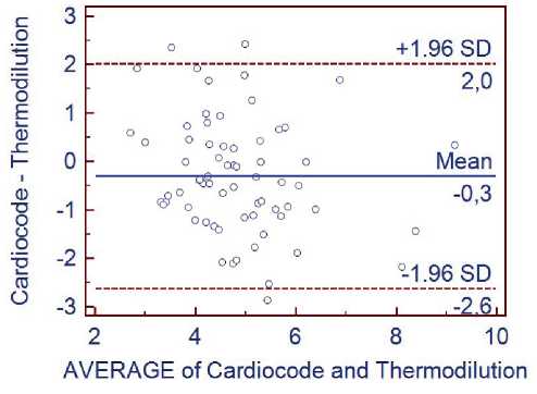
Figure 1. Cardiometric method vs. thermodilution method.
Spearman’s correlation coefficient 0.388...0.511
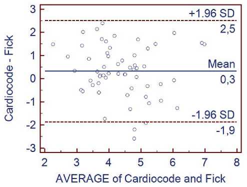
Fiure 2. The cardiometric method vs. the Fick method. Spearman’s correlation coefficient 0.333 ... 0.514
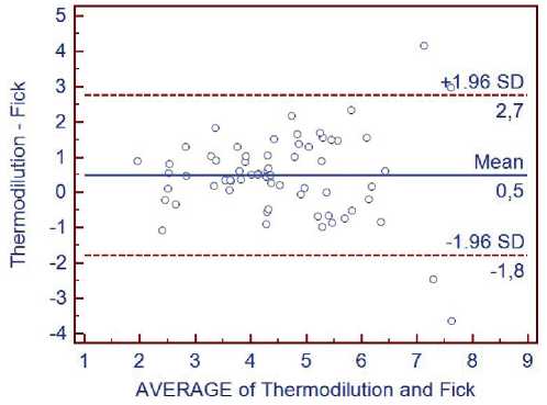
Figure 3. Thermodilution method vs. Fick method. Spearman’s correlation coefficient 0.814
When evaluating the difference in MV values, it is necessary to take into account that the above invasive methods do not directly measure blood flow, it requires also to be calculated, similar to the case with the calculation of the non-invasive cardiometric method. Various empirical coefficients are used for the conventional invasive methods of measuring. Moreover, there are even more of them applied as against the cardiometric technology, and they all are variables. This introduces an additional error in the MV measurements by the conventional invasive methods. In addition, to determine the minute volume of blood by the thermodilution and Fick methods, special conditions and special preparation of the patient are required that takes several hours. The invasive procedure itself, which is, in fact, a surgical operation, can cause serious complications.
The non-invasive cardiometric method allows, without special preparation of the patient, even under the field conditions, to obtain data on the minute volume of blood and other basic parameters of hemodynamics in just a few seconds. In this case, any complications are avoided.
Also, an important advantage of the cardiometric method is that it is very easy to implement, it is more accessible, highly cost-effective and more informative.
It is necessary to pay attention to one more fact. The methods by Fick and the thermodilution are capable of giving only the one value of the minute volume of blood circulation for one cardiac cycle in the examination of the patient, while the cardiometric method makes it possible to obtain the values of a complete set of hemodynamic parameters in each cardiac cycle (stroke, minute and phase blood volumes) with recording as many cardiac cycles as required.
This makes it possible to monitor the state of the patient’s cardiovascular system in real time and obtain information about the dynamics of changes in its functioning under the influence of external and internal impacts.
The non-invasive cardiometric method allows not only obtaining numerical values of hemodynamic parameters, but also qualitatively assessing the main functions of the cardiovascular system performance.
References Comparative clinical analysis of the cardiometric method the thermodilution and the Fick method
- Rudenko M.Yu., et al. Cardiometry. Fundamentals of theory and practice. Taganrog; Moscow: Izd-vo IKM, 2020. 215 p.[in Russian].
- Rudenko M.Y., et al. ADRENALINE HEART. Cardiometry. 2022;22:10.
- Rudenko MY, Zernov VA, Voronova OK. Cardiometry as a new fundamental scientific field in cardiology. Cardiometry. 2014;4:100-1.
- Rudenko MY, Zernov VA, Voronova OK. Cardiometry: new options in cardiology. Cardiometry. 2014; 4:139-40.
- Rudenko MY, Voronova OK, Zernov VA. Biophysical phenomena in blood flow system in the process of indirect arterial pressure measurement. In: The Cardiovascular System Physiology, Diagnostics and Clinical Implications. Rijeka, Croatia, 2012. p. 179-194.
- Rudenko MY. Strategic dissociation between systems of the higher vocational training of preparation of engineers of medical instrument making and doctors. Izvestiya SFedU. Engineering Sciences. 2009; 9(98): 219-22. [in Russian].
