Cranial traumas in a sample from the Pucar'a de Tilcara fortress (Jujuy province, Argentina)
")
Author: Zubova A.V., Dmitrenko L.M., Ananyeva N.I., Stulov I.K., Andreev E.V.
Journal: Archaeology, Ethnology & Anthropology of Eurasia @journal-aeae-en
Section: Anthropology and paleogenetics
Article in issue: 3 т.49, 2021.
Free access
We analyze injuries in the cranial sample from the Pucará de Tilcara fortress, dating to the time of the Inca conquest. Analysis of violence markers, carried out by visual examination and computed tomography, and the comparison of results with those relating to samples from the Regional Development Period of the Quebrada de Humahuaca valley, suggest that although the violence level remained high, its nature could have changed after the arrival of the Inca. The female sample reveals just two perimortal injuries, no trophy skulls were found, and the frequency of nasal bone fractures is higher than in earlier samples. This may indicate lower level of between-group fi ghting for control over resources, and higher risk of interpersonal violence. The observed pattern suggests that having arrived in the Quebrada de Humahuaca region, the Inca eased political tension by establishing control over trade routes and the distribution of arable land areas, which had previously been the main cause of local armed clashes. The infl uence of artifi cial cranial modifi cations on the pathological and traumatic status of individuals was also analyzed. Two types of modifi cation were recorded in the sample—fronto-occipital tabular oblique and fronto-occipital tabular straight. None of them caused pathological changes or a decrease in the thickness of cranial bones.
Pucará de Tilcara, Regional Development Period, Inca period, interpersonal violence, bioarchaeology, computed tomography
Short address: https://sciup.org/145146294
IDR: 145146294 | DOI: 10.17746/1563-0110.2021.49.3.147-156
Text of the article Cranial traumas in a sample from the Pucar'a de Tilcara fortress (Jujuy province, Argentina)
This article continues the series of publications of the results of the bioarchaeological study of the cranial sample from the Pucará de Tilcara fortress from the collection of the MAE RAS. The fortress is located in Northwestern Argentina, the Quebrada de Humahuaca valley (modern Jujuy Province). The results of the analytical attribution of the archaeological and anthropological collections from this site were published previously (Dmitrenko, Zubova, 2020), as well as a description of a case of surgical extraction of a lower third molar of one of the individuals (Zubova, Pikhur, Obodovsky et al.,
2020), and the data on the prevalence of chronic maxillary sinusitis in the inhabitants of the fortress (Zubova, Ananyeva, Moiseyev et al., 2020). The main aim of the present study was to analyze the prevalence and distribution of cranial trauma in the sample and their association with the socio-political situation in the region.
The Pucará de Tilcara fortress is one of the main archaeological complexes of the Quebrada de Humahuaca valley. The stratigraphic context of the site is rather complex, owing to the long history of its habitation and development. First settlements at the location of the fortress were established by the Omaguaca Indians no earlier than the 8th century AD. By the end of the 15th century, the territory of Northwestern Argentina had been colonized by the Inca, and in 1536 occupied by the Spanish conquistadors led by Diego de Almagro (Zaburlín, 2009; Greco, Otero, 2015). The functioning of the Pucará de Tilcara fortress belongs to the Regional Development Period (1000– 1430 AD) and Inca colonization (Seldes, Botta, 2014; Sprovieri, 2013: 26).
This period was one of the tensest in the history of the Quebrada de Humahuaca region. It was a time of rapid social and political changes due to the complication of political structures, intensification of agriculture and trade, and change in the population dispersal model. Besides traditional conglomerates of small villages localized near sites of concentration of natural resources, fortified settlements were emerging and becoming the centers of political control and trade. Population density and the competition for resources were growing accordingly. Thus, the number of military and interpersonal conflicts was also increasing, as suggested by both written sources and analysis of traumatic lesions in the skulls from the Yakoraite, Los Amarillos, and La Huerta sites in the northern part of the valley (Seldes, Botta, 2014: 88). These settlements were densely populated during the Regional Development Period; but with the advent of the Inca, Los Amarillos was abandoned, the population density at Yakoraite decreased, and only La Huerta retained its status as one of the regional centers of the empire.
The Pucará de Tilcara fortress did not lose its significance after the conquest by the Inca led by Túpac Yupanqui. Until the Spanish invasion in 1536, the fortress was an administrative center of the Inca Empire in the region. The Inca occupied the upper (“prestigious”) part of the hill where luxury workshops and some administrative buildings became concentrated. A square of typical Inca architecture was located nearby, at the northernmost boundary of Pucará de Tilcara (Zaburlín, 2009: 94–95).
Material and methods
The main sample employed in the present study is the collection of skulls sent to the Kunstkamera in 1910 by the Argentinean archaeologist J.B. Ambrosetti, who had carried out the excavation of the Pucará de Tilcara fortress in 1908–1910. The exact location of the specimens and their stratigraphic context are unknown. But owing to the study of field documentation (Zaburlín, Otero, 2014: 207) and register notes (General Catalogue of the Ethnographic Museum of Buenos Aires, notes 4100–7600) it was firmly established that all the materials—both anthropological and archaeological—obtained during the 1909–1910 expeditions were from the northwestern part of the site. This part was inhabited after the Inca conquest, which is suggested by its layout and the presence of ceramics of the Inca type (Otero, 2013: 107), stone knives “tumi”, various copper medical instruments, etc. in the archaeological record (Marino, Gonzales-Portillo, 2000: 947).
The sample includes 20 artificially deformed skulls: 18 adult (7 female, 11 male), one 6–8 years old subadult, and one 14–15 years old adolescent (Zubova, Ananyeva, Moiseyev et al., 2020: 146). The skulls were visually assessed for fixing ante-and perimortem lesions of the bones of the face and cranial vault. The descriptions of the lesions included the anatomical locations of the traumas with respect to the closest cranial sutures; size; and presence or absence of penetration into the cranial cavity, or signs of healing.
Computed tomography scans of all the individuals were obtained at the St. Petersburg Bekhterev Psychoneurological Research Institute, using a medical 64-channel scanner Philips Brilliance CT (PB64), under the following protocol: X-ray tube voltage 120 kV, amperage 100 μA, no filter, slice thickness 0.9 mm. Postprocessing of the images was carried out using a workstation Extended Brilliance Workspace for multiplanar (MPR) and volume (VR)
reconstructions. The localization of the detected traumas with respect to the brain structures and the pattern of possible damage to the brain tissues were described using the CT images. These data were employed to reconstruct possible clinical outcomes of the trauma for the individual.
In order to detect the influence of artificial deformation on the vulnerability of different skull regions to trauma, the thickness of the temporal, parietal, frontal, and occipital bones was measured. As the data on the variation of the skull bones’ thickness for the native populations of Argentina were absent in the literature, we used various anthropologically contrasted samples from Eurasia as a reference, including: modern Nepalese (Thulung et al., 2019) and Malaysians (Mahinda, Murty, 2009), Russians of the Tula Region (Plitnichenko, Telkov, 2011), and a population from the Lower Volga region, which also practiced artificial skull deformation (Pererva, 2015). The following total ranges of variation were accepted: for the frontal bone – 3–12 mm, temporal – 2.0–6.7 mm, parietal – 4–12 mm, occipital – 4–13 mm.
Results
The influence of artificial deformation on the cranial vault’s bone thickness. Two types of artificial modification are observed in the skulls from Pucará de Tilcara: fronto-occipital tabular straight and oblique, of which the latter is prevalent. In individuals with oblique deformation, the occipital part of the skull skews distally with respect to the vertical axis of the body, almost parallel to the axis of inclination of the frontal part (Fig. 1). This variant is observed in 65 % of all the cases, while straight deformation was found in eight male skulls.
A facial approximation of one of the individuals displaying oblique tabular deformation was carried out by D.V. Pozdnyakov from the Institute of Archaeology and Ethnography of the SB RAS (Zubova, Pikhur, Obodovsky et al., 2020: Fig. 1). This approximation was elaborated by I.G. Shirobokov (MAE RAS), using Artbreeder and Adobe Photoshop, to a photorealistic color portrait*. The Artbreeder neural network is a popular tool for generating portraits of various styles, and is not originally intended for making facial approximations. Even at the stage of uploading an original image, the facial traits can be slightly but unpredictably biased. During working with a portrait, the number of such errors grows substantially, which often leads to significant distortions of individual traits. Therefore, the image produced in Artbreeder was afterward modified in Adobe Photoshop in order to achieve a maximum similarity between the original and new portraits. Such similarity was assessed by the superimposition of reference landmarks.
No significant association between cranial deformation and any pathological conditions observed in the CT images was detected. Two individuals displayed digital impressions on the endocranial surface, while three others exhibited a deepening of the vascular grooves, mainly those of the diploic veins. But such changes are commonly found in many samples without an artificial cranial deformation; thus, these modifications cannot be a source of negative influence on the brain in the sample from Pucará de Tilcara.
The thickness of the calvarial bones in most skulls did not deviate from the norm: the deformation had only affected their shape. Skull No. 5148-7 was an exception, displaying a local thinning of the parietal bone by up to 1.5 mm, but this was not associated with a cranial modification. The thickness in the individuals with cranial traumas was normal.
Description of the traumatic lesions. As the sample is not large, we provide the descriptions of all the individual cases.
5148-2. Sex undetermined, juvenilis. A nasal bone fracture at the deepest point of the nasal bridge is observed. The blow was struck from the left side. Bone tissue at the location of the blow is sclerosed, while a right-side deviation of the internasal suture can be seen at the deepest point of the nasal bridge.
5148-3. Male, maturus. Signs of a completely healed fracture of the right nasal bone, without inflammation, are visible. A crushing perimortem fracture of the frontal bone was detected above the left browridge (Fig. 2). The traumatic defect is of a pentagonal shape and widens from the superciliary arch to the middle of the orbital margin. The compact layer is crushed across the whole area of the defect and broken down into
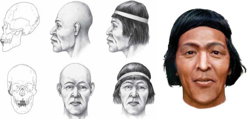
Fig. 1. Drawing of a skull with a tabular oblique deformation (No. 5148-9), and a facial approximation of the same individual.
15 fragments of an irregular shape, attached to the lower layer of the bone. The maximum length of the defect is 33 mm, width 12 mm. Five fissures radiate from the lesion. The largest pass through the browridge: one via the right superciliary arch
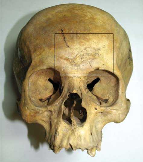
Fig. 2. General view of the lesion of the frontal bone of skull No. 5148-3.
toward the margin of the right orbit, and another crosses the left superciliary arch, terminating at the superorbital foramen.
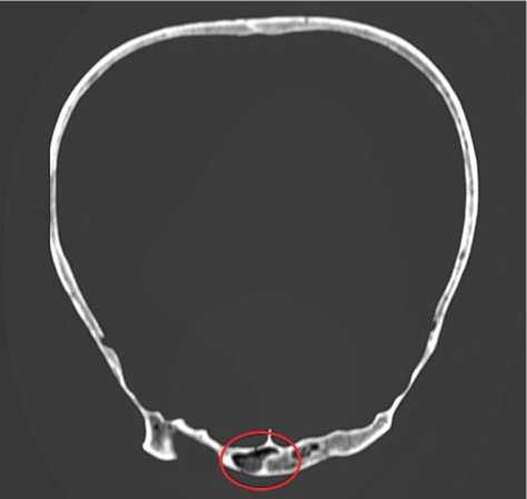
The blow was caused by a blunt object and likely through a headdress, since the fracture did not penetrate deep inside the bone, the radiating fissures are short, and there are no manifestations of direct contact with the tool at the surface of the bone. An accompanying lesion of the right frontal sinus—a linear defect of the right half of its posterior wall—was detected in the CT scan (Fig. 3). Signs of healing are absent, thus the individual died shortly after being injured.
Theoretically, this trauma could have had different consequences for the individual. If the brain was crushed or meningeal hematomas emerged, the injury might have been accompanied by loss of consciousness, and have ultimately led to death because of an axial interception of the brain complicated by the hematoma and impairment of its functions. If the brain was not seriously damaged, the trauma might have provoked pseudocerebellar static and coordination disorders—dizziness and difficulty concentrating. These would have made the individual less defensive, and then he could have received another trauma that led to death. This probable additional trauma could not be detected, owing to the absence of postcranial remains from Pucará de Tilcara in the collection of the MAE RAS.
Fig. 3. Computed tomography scan of skull No. 5148-3 (the area where the line of the fracture reaches the posterior wall of the right half of the frontal sinus is marked).
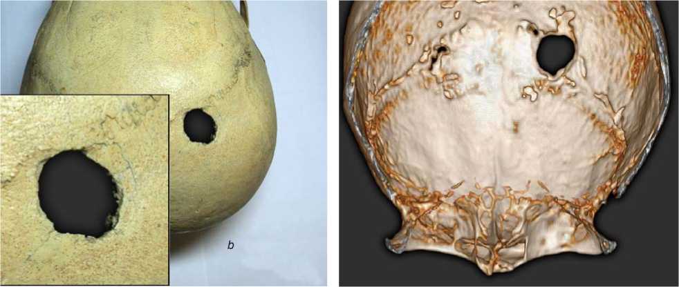
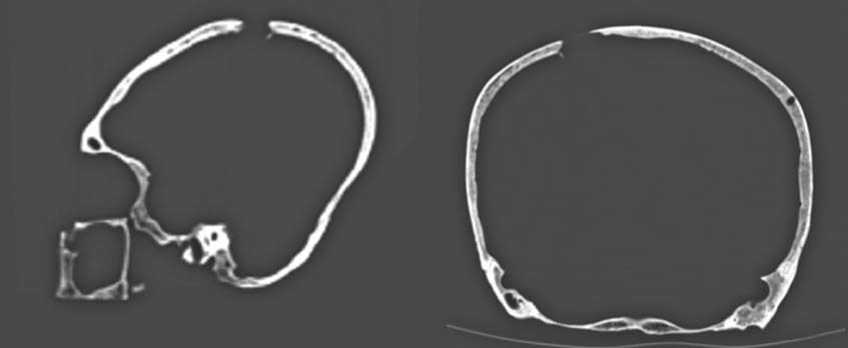
5148-8. Male, maturus . An oval traumatic penetration is visible in the right parietal bone, at the interface with the coronal suture, and 2 cm from the sagittal suture (Fig. 4). This trauma was caused by a weapon with a sharpened striking part. The external size of the lesion is 20 × 15 mm, internal is slightly larger. A bone-crushing and a semicircular fissure that mark the point of the initial application of force are observable on the right side of the penetration. A 1 cm long oblique fracture radiates from the left side of the perforation toward the sagittal suture. No manifestation of healing was detected, so the individual died shortly after the injury.
An extensive cleavage accompanied by the introduction of a bone fragment into the cranial cavity is visible at the endocranial surface in the left part of the lesion (Fig. 5). Depending on the depth of penetration into the cranial cavity of the weapon that caused the injury, the cause of death could have been either an interception of the brain, complicated by intracerebral and meningeal hematomas, or direct damage to vital centers of the brain stem, with respiratory and cardiac arrest.
5148-11. Female, senilis . A crushing injury 20 × 15 mm in size was detected in the right parietal
tubercle. This is a healed trace of a blow caused by an item with a convex impact surface. No signs of trauma are present in the internal compact layer. Thus, the brain likely was not damaged. A blow to this area might have led to contusion of the right parietal lobe of the brain and, as a consequence, provoked symptoms of irritation, paresthesia, loss over time, partial seizures, and concussion.
5148-20. Male, adultus . A post-traumatic deformation of the right nasal bone is present. This deformation occurred long before the death of the individual. A crushing injury of an irregular shape
аc
Fig. 4. General view and details of the lesion of the parietal bone of skull No. 5148-8.
a – bone lesions at the margins of the perforation; b – general view of the trauma from the outside; c – general view of the trauma on the endocranial surface.
b
Fig. 5. Computed tomography scan of skull No. 5148-8. a – lateral norm; b – occipital norm.
without signs of inflammation is visible in the center of the lower margin of the lesion.
Discussion
Traumatic lesions were detected in 5 out of the 20 skulls (25 %) from Pucará de Tilcara. In one case, a perimortem trauma of the cranial vault was accompanied by an earlier injury of the nose. Most of the lesions were antemortem (4 cases, 80 % of the total number of traumas), and in only two cases (40 %) were perimortem wounds that might have been the cause of death detected.
The antemortem traumas include nasal bone fractures (three cases, 50 % of the total number of traumas), and parietal bones injuries, which were struck with relatively little force, from the back, perhaps without the use of military weapons but with an object to hand. This allows the assumption of domestic conflicts with no relation to warfare. Two of the antemortem traumas were detected in male skulls, while one was observed in a female skull, and one in an adolescent individual of undetermined sex.
Both perimortem lesions were found in males older than 35 years. It is difficult to determine precisely the tools by which their traumas were caused, since some of the various types of weapons known from the archaeological complexes of the Quebrada de Humahuaca valley can leave wounds of similar shape. Stone or bone arrowheads and spearheads, stone or bronze club-heads, stone axes, balls for boleadoras or slingshots are all typical of the sites of the Omaguaca Indians (Handbook…, 1946: 627–628). The weaponry of the Inca was analogous, differing only in the variety of shapes and skill of making throwing-balls and pommels of battle-clubs (Marino, Gonzales-Portillo, 2000: 944). Stone six-pointed or round tops of war clubs (Zaburlín, Otero, 2014: 171, 200), bone arrowheads (Ibid.: 195), and stone weights for boleadoras (MAE, No. 1800-57, 58) are represented in the archaeological complex of Pucará de Tilcara.
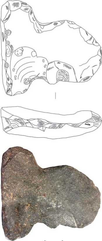
The trauma detected in specimen No. 5148-3 definitely could not have been caused by any item leaving penetrating wounds of the head, e.g. arrows, spears, sharp-pointed maces, etc. This injury was struck by a weapon with a flat impact surface, such as bolas, boleadoras (Fig. 6), or stone axes (Fig. 7).
The traumatic perforation observed in specimen No. 5148-8 is similar to wounds caused by a pickaxe (Borodovsky et al., 2010: 41, 63), but the irregular margins of the defect and the presence of short radiating fissures suggest that the striking surface was fairly flat and rather blunt. It is highly likely that the injury was caused by a war-club with a pointed stone pommel or some similar weapon.
The comparison of the cranial collection from Pucará de Tilcara with the aggregate sample from the Quebrada de Humahuaca valley, including the skulls from the sites belonging to the Regional Development Period (Yakoraite, Los Amarillos, and La Huerta), has shown that the proportion
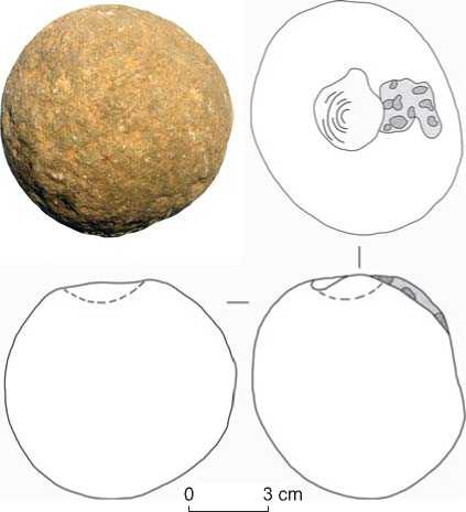
Fig. 6. Bolas stone cannonball (MAE, No. 1800-57) from the excavations of the fifth expedition of the Faculty of Philosophy and Literature at Pucará de Tilcara in 1909 (according to the General Catalogue of the Ambrosetti Ethnographic Museum).
of skulls with traumas differs little between the two samples: 25 % (including the subadult skull; subadult individuals were included in the reference samples as well) vs. 34.6 % (Table 1). The value of the χ2 criterion is 0.74; thus, the difference is not significant.
The distribution of the lesions among parts of the skull is, nevertheless, specific in the Pucará de Tilcara collection as compared to the Regional Development Period samples (Table 2). First, the prevalence of nasal bone injuries is increased at Pucará de Tilcara: three cases (50 % of the total number of traumas) vs. one case at Yakoraite (4 %) and no cases at Los
3 cm
Fig. 7. Stone double-faced axe (MAE, No. 1800-55), probably fastened in the central part by clamping in a split wooden handle. From the excavations of the sixth expedition of the Faculty of Philosophy and Literature at Pucará de Tilcara in 1910 (according to the General Catalogue of the Ambrosetti Ethnographic Museum).
Table 1. Distribution of cranial traumatic lesions in the samples from the Quebrada de Humahuaca valley
|
Sample |
Total number of skulls |
Skulls with traumas |
Skulls without traumas |
Trophy skulls |
|||
|
Number |
% |
Number |
% |
Number |
% |
||
|
Los Amarillos * |
60 |
13 |
21.7 |
42 |
70 |
4 |
6.7 |
|
La Huerta * |
49 |
15 |
30.6 |
23 |
46.9 |
11 |
22.4 |
|
Yakoraite * |
44 |
25 |
56.8 |
18 |
36.7 |
2 |
4.5 |
|
Aggregate * |
153 |
53 |
34.6 |
83 |
54.2 |
17 |
11.1 |
|
Pucará de Tilcara |
20 |
5 |
25 |
15 |
75 |
0 |
0 |
*Here and in Tables 2 and 3 data are according to (Seldes, Botta, 2014).
Table 2. Distribution of various types of cranial traumas in the samples from the Quebrada de Humahuaca valley
|
Type of trauma |
Los Amarillos |
La Huerta |
Yakoraite |
Aggregate |
Pucará de Tilcara |
|
Antemortem |
12 (92.3) |
9 (56.2) |
22 (78.6) |
43 (75.4) |
4 (66.7) |
|
Perimortem |
1 (7.7) |
7 (43.7) |
6 (21.4) |
14 (24.6) |
2 (25) |
|
Fractures |
11 (84.6) |
15 (100) |
19 (70.4) |
45 (81.8) |
6 (100) |
|
Cutting wounds |
2 (15.4) |
0 |
8 (29.6) |
10 (18.2) |
0 |
|
Traumas of the bones: |
|||||
|
frontal |
4 (33.3) |
11 (52.4) |
4 (16) |
19 (32.8) |
1 (16.7) |
|
parietal |
7 (58.3) |
5 (23.8) |
13 (52) |
25 (43.1) |
2 (33.3) |
|
occipital |
1 (8.3) |
4 (19) |
3 (12) |
8 (13.8) |
0 |
|
maxilla |
0 |
1 (4.75) |
0 |
1 (1.73) |
0 |
|
zygoma |
0 |
0 |
1 (4.0) |
1 (1.73) |
0 |
|
nasal |
0 |
0 |
1 (4.0) |
1 (1.73) |
3 (50) |
|
temporal |
0 |
0 |
3 (12) |
3 (5.18) |
0 |
Note. In parentheses, the percentage from the total number of skulls with traumas is given.
Table 3 . Sex-specific distribution of cranial traumas in the sample from Pucará de Tilcara and the aggregate sample from Los Amarillos, La Huerta, and Yakoraite
|
Sample |
Males |
Females |
Undetermined (including children and adolescents ) |
|||
|
Total |
With traumas |
Total |
With traumas |
Total |
With traumas |
|
|
Pucará de Tilcara |
11 |
3 (27.3) |
7 |
1 (14.3) |
2 |
1 (50) |
|
Aggregate |
88 |
28 (31.8) |
43 |
16 (37.2) |
24 |
9 (37.5) |
Note. In parentheses, the percentage from the total number of the corresponding skulls is given.
Amarillos and La Huerta. Second, injuries to the occipital bone, which are typical of the Yakoraite, Los Amarillos, and La Huerta samples, were not found at Pucará de Tilcara.
Some differences are also observed in the number of female skulls with traumas: their percentage in the Pucará de Tilcara collection is almost 2.5 times lower than in the aggregate sample (Table 3). Though this difference is not statistically significant, this may indicate less involvement of women in armed clashes. Notably, the percentage of male skulls with traumas is roughly equal in both samples. It is also of note that trophy skulls, which are present in all the other samples, are absent in the Pucará de Tilcara collection (see Table 1).
Judging by the distribution of traumatic lesions in the studied sample and the pattern of differences with the cranial samples from the sites belonging to the Regional Development Period, it seems possible that the arrival of the Inca to Pucará de Tilcara had led to some social changes that directly affected the pattern of military and domestic traumatism in this group. The general level of violence remained high; however, in the later stages of the existence of the fortress, the number of mass military affairs involving the whole population decreased, while the prevalence of interpersonal violence, not associated with warfare, increased. This is suggested by the absence of trophy skulls in the sample, a decreased (as compared to the aggregate sample) percentage of injured female skulls, a lower number of cranial traumas in the general profile of traumatization of the population, and the prevalence of antemortem lesions of the nasal bones. Indirect evidence is also the absence of occipital bone traumas typically received when fleeing from an armed enemy.
Irrespective of the colonizing policy of the Inca, the number of combat weapons suitable for inter-tribal warfare had not decreased in Inca’s archaeological complexes as compared to Omaguaca sites (Pérez Pieroni, Becerra, 2018). The materials from the Pucará de Tilcara fortress, nevertheless, suggest a social character of the colonization related to the interest of the Incas in the extraction of mining raw materials and the production of luxury goods by local craftsmen. The advent and long-term presence of the Incas at Pucará de Tilcara led to the emergence of a new social layer of specialized craftsmen working at administrative centers and providing luxury goods to the elite (Zaburlín, 2009: 99–100). This is suggested by the architecture and archaeological complexes excavated by J.B. Ambrosetti named “House of the Jeweler” and “Copper House” (“La Casa del Joyero” and “La Casa de los Cobres”) (Ibid.: 94–95). Representatives of this social group were likely emancipated from participation in mass warfare, which might have affected the pattern and prevalence of the traumatic lesions of the skulls from the fortress.
Conclusions
The results of our analysis of markers of violence in the cranial sample from the Pucará de Tilcara fortress suggest that at the late stages of its history, i.e. the time of the Incas’ expansion in Argentina, the pattern of interpersonal violence in the Quebrada de Humahuaca valley has changed as compared to the previous Regional Development Period. In the Pucará de Tilcara cranial sample, only two traumas that can be considered combat-related were detected, trophy skulls are absent, and the prevalence of nasal bone fractures is increased as compared to the samples belonging to the Regional Development Period. These observations may evince a decrease in the number of mass warfare encounters aimed at establishing control over resources, and a shift toward domestic conflicts.
The observed picture can indirectly suggest that the arrival of the Incas to the Quebrada de Humahuaca region eased social tension and contributed to some stabilization of the political situation. These were achieved through establishing control over trade routes and fertile areas, which were previously the main cause of local armed conflicts (Seldes, Botta, 2014). This hypothesis requires further testing employing more numerous materials, since the skulls used in the present study do not represent a proper sample from the Pucará de Tilcara population. However, as a first approximation, anthropological data support this view.
References Cranial traumas in a sample from the Pucar'a de Tilcara fortress (Jujuy province, Argentina)
- Borodovsky A.P., Zubova A.V., Pozdnyakov D.V., Tabarev A.V., Cheremisin D.V. 2010 Arkheologiya nasiliya (interpretatsiya materialov arkheologicheskikh, antropologicheskikh i izobrazitelnykh kompleksov): Ucheb.-metod. posobiye. Novosibirsk: Novosib. Gos. Univ.
- Dmitrenko L.M., Zubova A.V. 2020 Collection related to the Omaguaca Indians from the Pucará de Tilcara Fortress, Northwestern Argentina, at the Museum of Anthropology and Ethnography RAS, St. Petersburg: Tentative Findings. Archaeology, Ethnology and Anthropology of Eurasia, vol. 48 (1): 149-157.
- Greco C., Otero C. 2015 The chronology of settlements with pre-inca and inca occupations superimposed: The case of Pucará de Tilcara (Humahuaca Gorge, Argentina). Archaeometry, vol. 58 (5): 848-862.
- Handbook of South American Indians. 1946 Vol. 2: The Andean Civilizations. Washington: Government Printing Office, 1946. (Smithsonian Institution Bureau of American Ethnology; bull. 143).
- Mahinda H., Murty O.P. 2009 Variability in thickness of human skull bones and sternum- an autopsy experience. Journal of Forensic Medicine, vol. 26 (2): 26-31.
- Marino R., Gonzales-Portillo M. 2000 Pre-conquest Peruvian neurosurgeons: A study of Inca and pre-Columbian trephination and the art of medicine in ancient Peru. Neurosurgery, iss. 47: 940-950.
- Otero C. 2013 La arqueología en el relato ofi cial del estado nacional: El caso Del Pucará de Tilcara (Jujuy, Argentina). Arqueología Suramericana/Arqueología Sul-Americana, vol. 6 (1/2): 87-111.
- Pererva E.V. 2015 Rentgenologicheskoye issledovaniye deformirovannykh cherepov zolotoordynskogo vremeni s territorii Nizhnego Povolzhya (paleoliticheskiy aspect). Vestnik arkheologii, antropologii i etnografi i, No. 2: 98-114.
- Pérez Pieroni M.J., Becerra M.F. 2018 Archaeology of Northwestern Argentinean Puna. In Encyclopedia of Global Archaeology, C. Smith (ed.). New York: Springer.
- Plitnichenko B.G., Telkov V.E. 2011 Anatomicheskiye osobennosti tolshchiny kostey cherepa u zhiteley Tulskoy oblasti i vliyaniye ikh na obrazovaniye perelomov. Vestnik novykh meditsinskikh tekhnologiy, vol. XVIII (3): 314-316.
- Seldes V., Botta F.N. 2014 Violence indicators in Quebrada de Humahuaca, Jujuy, Argentina: The Regional Development Period from a regional perspective. Anthropological Review, vol. 77 (1): 87-109.
- Sprovieri M. 2013 El mundo en movimiento: Circulación de bienes, recursos e ideas en el valle Calchaquí, Salta (Noroeste Argentino): Una vision desde La Paya. Oxford: Archaeopress. (BAR Int. Ser.; No. 2487).
- Thulung S., Ranabhat K., Bishokarma S., Gongal D.N. 2019 Morphometric measurement of cranial vault thickness: A Tertiary hospital based study. Journal of Nepal Medical Association, vol. 57 (215): 29-32.
- Zaburlín M.A. 2009 Historia de ocupación del Pucará de Tilcara (Jujuy, Argentina). Intersecciones en Antropología, vol. 10 (1): 89-103.
- Zaburlín M.A., Otero C. 2014 Un manuscrito olvidado de J.B. Ambrosetti: “Exploraciones arqueológicas en la Antigua ciudad del Pukará de Tilcara”. In Investigaciones del Instituto Interdisciplinario Tilcara. Buenos Aires: Editorial de la Facultad de Filosofia y Letras, pp. 161-220.
- Zubova A.V., Ananyeva N.I., Moiseyev V.G., Stulov I.K., Dmitrenko L.M., Obodovsky A.V., Potrakhov N.N., Kulkov A.M., Andreev E.V. 2020 The use of computed tomography for the study of chronic maxillary sinusitis: Based on crania from the Pucará De Tilcara Fortress, Argentina. Archaeology, Ethnology and Anthropology of Eurasia, vol. 48 (3): 143-153.
- Zubova A.V., Pikhur O.L., Obodovskiy A.V., Malyutina A.A., Dmitrenko L.M., Chugunova K.S., Pozdnyakov D.V., Bessonov V.B. 2020 A case of surgical extraction of the lower third molars in a cranial series from the Pucará de Tilcara Fortress (Jujuy Province, Argentina). Archaeology, Ethnology and Anthropology of Eurasia, vol. 48 (2): 149-156.
