
Cyclophosphamide-induced nephrotoxicity: a mini-review
Journal: Journal of Stress Physiology and Biochemistry @jspb
Article in issue: 2 т.22, 2026.
Free access
Cyclophosphamide (CP) is a widely used alkylating chemotherapeutic and immunosuppressive drug for treating malignancies, autoimmune disorders, and renal diseases. However, its clinical use is limited by nephrotoxicity caused by toxic metabolites such as phosphoramide mustard and acrolein. These metabolites induce oxidative stress, inflammation, mitochondrial dysfunction, and apoptosis in renal tissues. CP administration results in elevated renal biomarkers including serum creatinine, blood urea nitrogen, cystatin-C, KIM-1, and NGAL, along with severe histopathological damage. Activation of inflammatory cytokines and NF-κB pathways further aggravates renal injury. Various antioxidants and pharmacological agents have shown potential renoprotective effects.
Short address: https://sciup.org/143186138
IDS: 143186138
Text of the scientific article Cyclophosphamide-induced nephrotoxicity: a mini-review
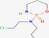
Cyclophosphamide (CP) is a widely used alkylating agent belonging to the oxazaphosphorine class and is extensively employed in the treatment of malignant disorders as well as autoimmune and inflammatory diseases due to its potent cytotoxic and immunosuppressive properties. ( hosh et al., 1999; Abraham and Isaac, 2011). CP is a prodrug that undergoes hepatic biotransformation by cytochrome P450 enzymes to generate active metabolites, including phosphoramide mustard and acrolein, which are responsible for its antineoplastic efficacy as well as systemic toxicity ( hosh et al., 1999; Haque et al., 2003). Despite its therapeutic importance, CP administration is associated with significant dose-limiting adverse effects, among which nephrotoxicity represents a clinically relevant complication affecting both glomerular and tubular structures (Hauritz et al., 2001; Abraham et al., 2007). Experimental studies have demonstrated that CP-induced renal injury is closely linked to oxidative stress, characterized by increased lipid peroxidation, depletion of endogenous antioxidants, and disruption of redox homeostasis within renal tissues (Ayhanci et al., 2009; Abraham and Rabi, 2009). Elevation of biochemical markers such as malondialdehyde and alterations in antioxidant enzyme activities have been consistently reported following CP exposure, indicating enhanced reactive oxygen species generation (Abraham and Sugumar, 2008; unes et al., 2017). In addition to oxidative damage, CP-induced nephrotoxicity involves inflammatory responses, leukocyte infiltration, and enzymatic leakage, contributing to progressive renal dysfunction ( ashlan et al., 2013; Kocahan et al., 2017). Ultrastructural investigations have further revealed that CP causes profound subcellular alterations, including mitochondrial deformation, lysosomal depletion, peroxisome proliferation, and endoplasmic reticulum dilation, underscoring the organelle-specific nature of CP-mediated renal injury (Abraham and Isaac, 2011). Collectively, these findings establish cyclophosphamideinduced nephrotoxicity as a multifactorial process involving oxidative stress, inflammation, and organelle dysfunction, thereby justifying continued investigation into protective and therapeutic strategies (Adikwu et al., 2019; Alghamdi et al., 2024) (Fig. 1).
EFFECTS OF CYCLOPHOSPHAMIDE ONKIDNEY
Cyclophosphamide-induced nephrotoxicity is multifactorial, involving biochemical, structural, inflammatory, and mitochondrial disturbances. Rodent studies consistently show elevated serum creatinine, BUN, uric acid, and cystatin-C after CP exposure, indicating sharp impairment in renal filtration ( unes et al., 2016). These functional changes correlate with increased oxidative stress, as evidenced by elevated MDA levels and suppression of endogenous antioxidants including SH, SOD, CAT, and Px (Ayhanci et al., 2010).
Histopathological studies reveal clear renal damage such as tubular necrosis, loss of brush border, glomerular shrinkage, epithelial desquamation, and interstitial inflammation (Alabi et al., 2022). Some investigations also report mesangial proliferation, Bowman’s space widening, and glomerulonephritis-like changes (Zonozi et al., 2021). Inflammatory activation is a major factor in CP nephrotoxicity. CP upregulates pro-inflammatory cytokines such as TNF-α, IL-1β, and IL-6, and activates NF-κB signaling, resulting in prolonged renal inflammation (Dobrek et al., 2017) CP further induces apoptosis, indicated by increased Bax, p53, and caspase-3 expression and decreased Bcl-2 levels, contributing to tubular cell loss (Tohamy et al., 2021). Recent mechanistic insights highlight early mitochondrial membrane damage, ROS overproduction, ATP depletion, and autophagy– apoptosis imbalance as critical contributors to kidney deterioration (Jiang et al., 2020).
EARLY BIOMARKERS OFNEPHROTOXICITY
Traditional markers like serum creatinine rise only after major nephron loss. Clinical proteomic approaches using N AL, KIM-1, clusterin, and urinary protein fragments allow early and more accurate detection of CP-induced renal damage, improving patient monitoring and intervention timing (Kim et al., 2015).
METHODOLOGY
The author followed PRISMA, the Preferred Reporting Items for systematic Reviews and Meta-Analyses.
Recommendations are a minimal collection of things based on evidences. The author focused on both experimental and non-experimental studies, using four internet databases, including oogle scholar, PubMed, and Science Direct Research articles published between and were searched for using search engines. Research articles published between were searched for using search engines. Research articles published between 2001 and 2025 were searched for using search engines.
BIOCHEMICAL CHANGES
Cyclophosphamide (CP) administration has been consistently associated with marked biochemical disturbances reflecting renal dysfunction and oxidative stress. Experimental studies in rodents demonstrated significant elevations in serum creatinine, blood urea nitrogen (BUN), and urea levels following CP exposure, indicating impaired glomerular filtration and renal clearance (Bhat et al., 2018; Adikwu et al., 2019; Al-Salih et al., 2019; Yadav et al., 2021). CP treatment also induced pronounced oxidative stress, evidenced by increased malondialdehyde (MDA) levels and reactive oxygen species (ROS), accompanied by depletion of endogenous antioxidant defenses such as superoxide dismutase, catalase, glutathione, and glutathione peroxidase (Elwakeel et al., 2021; Tohamy et al., 2021; Ahmad et al., 2021). Furthermore, elevated levels of pro-inflammatory cytokines including TNF-α, IL-1β, and IL-6, along with activation of apoptotic markers such as caspase-3, were reported, highlighting the role of inflammation and apoptosis in CP-induced nephrotoxicity (Jiang et al., 2020; Alghamdi et al., 2024). Similar biochemical impairments have also been observed in clinical settings, where prolonged CP therapy was associated with progressive renal functional decline (Cortazar et al., 2017; Sedhain et al., 2018).
HISTOLOGICAL CHANGES
Histopathological evaluation of kidney tissues following cyclophosphamide exposure revealed extensive structural damage affecting both tubular and glomerular compartments. Common histological alterations included tubular epithelial degeneration, tubular necrosis, loss of brush border, epithelial desquamation, and disruption of normal tubular architecture (Bhat et al., 2018; Olukole et al., 2020; Tohamy et al., 2021). lomerular changes such as shrinkage, congestion, distortion of Bowman’s capsule, and glomerular atrophy were frequently observed, along with interstitial edema and vascular congestion (Elwakeel et al., 2021; Yadav et al., 2021). Severe inflammatory cell infiltration and apoptotic features, including nuclear pyknosis and cellular fragmentation, were evident in high-dose or prolonged CP exposure models, confirming progressive renal injury (Jiang et al., 2020; Ahmad et al., 2021; Eldien and Alenzi, 2023; Kabil et al., 2023; Seker et al., 2024). These histological findings strongly correlate with the observed biochemical abnormalities and collectively confirm that cyclophosphamide induces nephrotoxicity through oxidative stress–mediated inflammation and apoptosis, leading to structural and functional deterioration of the kidney.
PROTECTIVE TREATMENTS AGAINSTCYCLOPHOSPHAMIDE NEPHROTOXICITY
A wide range of natural antioxidants and pharmacological agents have shown promising Reno protective effects in experimental models. Melatonin, quercetin, gallic acid, Selen methionine, caffeic acid phenethyl ester, royal jelly, hesperidin, vitamins C and E, and several plant-derived extracts (e.g., Ocimum gratissimum , Phoenix dactylifera seed extract) demonstrated significant reversal of oxidative injury, restoration of antioxidant enzymes, and improvement in tubular architecture (Ayhanci et al., 2010).
Compounds such as NAC (N-acetylcysteine), diosmin, zinc oxide nanoparticles, sesamin, baicalein, and bioactive saponins showed dual antioxidant and anti-inflammatory effects (Tohamy et al., 2021). Advanced regenerative strategies, including mesenchymal stem cells (MSCs), have been particularly effective in repairing renal injury by suppressing inflammation and promoting tissue regeneration (Eldien and Alenzi 2023). L-carnitine has also been shown to stabilize mitochondria and reduce renal and neural toxicity during CP or combination chemotherapy (Kocahan et al., 2016)
Major Clinical Limitation: Dose-Limiting Toxicities
-
1. Myelosuppression
-
2. Hemorrhagic Cystitis
Caused by acrolein accumulation in the bladder; prevented with MESNA and hydration (Sedhain et al., 2018).
-
3. Nephrotoxicity
-
4. Cardiotoxicity (High Dose)
The most common dose-limiting toxicity causing neutropenia, anemia, and thrombocytopenia (Hauritz et al., 2001).
Nephrotoxicity Leads to oxidative stress, tubular necrosis, inflammation, and renal (Morid et al., 2023).
High-dose CP can cause acute cardiomyopathy and arrhythmias (Hauritz et al., 2001).
Therefore, the present review aims to provide a comprehensive analysis of the cyclophosphamideinduced nephrotoxicity effects and renal risks of this widely used anticancer drug.
Cl
Figure 1. Cyclophosphamide. (Source:
Table 1 . Comparative analysis of biochemical changes
|
Model organism |
Dose of CP |
Duration |
Major biochemical changes observed |
References |
|
Rats ( Rattus norvegicus ) |
200 mg/kg |
10–14 days |
Increased oxidative stress markers (increase MDA), decreased antioxidant enzymes (decrease SOD, CAT, SH) |
Bhat et al., (2018) |
|
Rats ( Rattus norvegicus ) |
150 mg/kg |
10 days |
Oxidative stress–mediated kidney damage, altered renal biomarkers |
Adikwu et al., (2019) |
|
Rats ( Rattus norvegicus ) |
150 mg/kg |
7–14 days |
Altered renal biochemistry (increase serum urea, creatinine) |
Al-Salih et al., (2019) |
|
Rats ( Rattus norvegicus ) |
200 mg/kg |
14 days |
Elevated inflammatory markers and oxidative stress parameters |
Olukole et al., (2020) |
|
Mice ( Mus musculus ) |
200 mg/kg |
24–72 hours |
Acute oxidative stress, depletion of antioxidant defense |
Jiang et al., (2020) |
|
Rats ( Rattus norvegicus ) |
150 mg/kg |
14 days |
Increased renal inflammatory cytokines and oxidative damage |
Elwakeel et al., (2021) |
|
Rats ( Rattus norvegicus ) |
150 mg/kg |
7–10 days |
Oxidative stress–mediated nephrotoxicity |
Yadav et al., (2021) |
|
Rats ( Rattus norvegicus ) |
200 mg/kg |
14 days |
Activation of apoptotic pathways and inflammatory mediators |
Ahmad et al., (2021) |
|
Rats ( Rattus norvegicus ) |
50 mg/kg |
21 days |
Persistent oxidative stress and altered renal enzymes |
Alaqeela and Al-Hariri, (2023) |
|
Rats (Sprague Dawley) |
150 mg/kg |
19 days |
Oxidative stress, inflammation, and apoptosis-related biochemical alterations |
Alghamdi et al., (2024) |
Table 2. Comparative analysis of histological damage
|
Model organism |
Dose of CP |
Duration |
Major histopathological changes observed |
References |
|
Rats ( Rattus norvegicus ) |
200 mg/kg |
10–14 days |
Renal structural damage, tubular degeneration |
Bhat et al., (2018) |
|
Rats ( Rattus norvegicus ) |
150 mg/kg |
7–14 days |
Tubular necrosis and glomerular damage |
Al-Salih et al., (2019) |
|
Rats ( Rattus norvegicus ) |
200 mg/kg |
14 days |
Tubular injury and inflammatory cell infiltration |
Olukole et al., (2020) |
|
Mice ( Mus musculus ) |
200 mg/kg |
24–72 hours |
Early tubular necrosis and epithelial damage |
Jiang et al., (2020) |
|
Rats ( Rattus norvegicus ) |
200 mg/kg |
10 days |
Tubular injury and renal tissue degeneration |
Tohamy et al., (2021) |
|
Rats ( Rattus norvegicus ) |
200 mg/kg |
14 days |
Apoptosis, inflammation, and renal architectural distortion |
Ahmad et al., (2021) |
|
Rats ( Rattus norvegicus ) |
70 mg/kg (single dose) |
4–10 days |
Severe renal injury with inflammatory infiltration and tissue damage |
Eldien and Alenzi, (2023) |
|
Mice ( Mus musculus ) |
175 mg/kg |
30 days |
Marked kidney injury and deterioration of renal histoarchitecture |
Kabil et al., (2023) |
|
Rats ( Rattus norvegicus ) |
50 mg/kg |
21 days |
Tubular damage and loss of normal renal morphology |
Alaqeela and Al-Hariri, (2023) |
|
Mice ( Mus musculus ) |
200 mg/kg |
14 days |
Renal dysfunction with histopathological alterations |
Seker et al., (2024) |
Table 3. Comparative analysis of Ameliorative effects
|
Protective Agent / Strategy |
Nature of Agent |
Major Protective Mechanisms |
Observed Reno protective Effects |
References |
|
Melatonin |
Endogenous hormone / Antioxidant |
Free radical scavenging, enhancement of antioxidant enzymes |
Reduced lipid peroxidation, restoration of renal antioxidant status, improved tubular histology |
Ayhanci et al., (2010) |
|
Quercetin |
Flavonoid (Natural antioxidant) |
Antioxidant, antiinflammatory |
Decreased oxidative stress, improved renal architecture |
Ayhanci et al., 2010 |
|
allic acid |
Phenolic compound |
ROS scavenging, inhibition of oxidative injury |
Protection of renal tubular cells, normalization of biochemical markers |
Ayhanci et al., 2010 |
|
Selen methionine |
Organic selenium compound |
Boosts glutathione peroxidase, antioxidant defense |
Reduced renal oxidative damage, improved kidney function |
Ayhanci et al., 2010 |
|
Caffeic Acid Phenethyl Ester (CAPE) |
Polyphenolic compound |
Anti-inflammatory, antioxidant |
Preservation of renal structure, reduction in inflammatory damage |
Ayhanci et al., 2010 |
|
Royal Jelly |
Natural bee product |
Antioxidant, immunomodulatory |
Attenuation of tubular degeneration and oxidative stress |
Ayhanci et al., 2010 |
|
Hesperidin |
Citrus flavonoid |
Antioxidant, antiinflammatory |
Restoration of antioxidant enzymes, reduced nephron injury |
Ayhanci et al., 2010 |
|
Vitamin C |
Water-soluble antioxidant |
ROS neutralization |
Reduced CP-induced oxidative renal injury |
Ayhanci et al., 2010 |
|
Vitamin E |
Lipid-soluble antioxidant |
Prevention of lipid peroxidation |
Protection of renal membranes and tubular integrity |
Ayhanci et al., 2010 |
|
Ocimum gratissimum extract |
Medicinal plant extract |
Antioxidant, antiinflammatory |
Improved renal histology and antioxidant status |
Ayhanci et al., 2010 |
|
Phoenix dactylifera seed extract |
Plant-derived antioxidant |
Free radical scavenging |
Reduction in biochemical and histopathological damage |
Ayhanci et al., 2010 |
|
N-Acetylcysteine (NAC) |
Synthetic antioxidant |
lutathione replenishment, antiinflammatory |
Reduced oxidative stress and inflammation in renal tissue |
Tohamy et al., 2021 |
|
Diosmin |
Flavonoid glycoside |
Antioxidant, antiinflammatory |
Improved renal biochemical parameters |
Tohamy et al., 2021 |
|
Zinc oxide nanoparticles |
Nanoparticlebased antioxidant |
Reduction of oxidative and inflammatory responses |
Protection against CP-induced renal damage |
Tohamy et al., 2021 |
|
Sesamin |
Lignan (Sesame-derived) |
Antioxidant, antiinflammatory |
Attenuation of renal oxidative stress |
Tohamy et al., 2021 |
|
Baicalein |
Flavone (Herbal origin) |
Suppression of inflammatory mediators |
Reduced tubular injury and oxidative stress |
Tohamy et al., 2021 |
|
Bioactive saponins |
Plant secondary metabolites |
Antioxidant, antiinflammatory |
Improved renal function and histology |
Tohamy et al., 2021 |
|
Mesenchymal Stem Cells (MSCs) |
Regenerative therapy |
Anti-inflammatory, tissue regeneration |
Repair of renal tissue, suppression of CP-induced inflammation |
Eldien and Alenzi, 2023 |
|
L-Carnitine |
Mitochondrial stabilizer |
Mitochondrial protection, antioxidant |
Reduced renal and neural toxicity, improved energy metabolism |
Kocahan et al., 2016 |
CONCLUSION
From the available literature, it is evident that cyclophosphamide induces significant nephrotoxicity. Although cyclophosphamide is a highly effective anticancer and immunosuppressive agent, its clinical use is limited by dose-dependent renal toxicity. The severity of kidney injury varies with species, dosage, duration of exposure, metabolic capacity, and route of administration. Cyclophosphamide-induced nephrotoxicity is characterized by oxidative stress, inflammation, tubular and glomerular damage, and impaired renal function. Overall, the combination of cyclophosphamide therapy with appropriate nephroprotective agents or optimized dosing strategies may significantly improve renal safety and reduce kidney damage. Further investigations are essential to elucidate the long-term renal effects of cyclophosphamide and to develop effective therapeutic interventions for minimising its nephrotoxic potential.
CONFLICTS OF INTEREST
The author declares no conflicts of interest.