Decision support system in radiology for fast diagnostics of thoracic diseases under COVID-19 pandemic conditions

Author: Borodyansky I.M.
Journal: Cardiometry @cardiometry
Section: Short report
Article in issue: 21, 2022.
Free access
In the present article the relevance of using DSS under the current conditions for image recognition and, as a more specific application, for the purpose of additional assistance rendered to medical experts (radiologists) in their decision-making and preparing findings upon assessment of X-ray images is considered. The paper analyzes the requirements for some expert DSS and their main characteristics that they should have; considered and selected is the necessary software for making rapid diagnoses of diseases of the thorax. All these modern requirements and characteristics are met by the Deep Learning Studio (DLS) software, which allows using deep convolutional neural network Inception V3 to teach this network and further obtain optimal results in the recognition and diagnosis of diseases of the thorax by assessing X-ray images. As a result of this study, a ready-made DSS intended for use by medical institutions for additional assistance to radiologists to prepare findings according to X-ray images has been obtained.
Dss, dls, transfer learning, inception v3 neural network, x-ray images, pneumonia, myocarditis
Short address: https://sciup.org/148324179
IDR: 148324179
Text of the scientific article Decision support system in radiology for fast diagnostics of thoracic diseases under COVID-19 pandemic conditions
Imprint
Ilya M. Borodyansky. Decision support system in radiology for fast diagnostics of thoracic diseases under COVID-19 pandemic conditions. Cardiometry; Issue 21; February 2022; p. 50-54; DOI: 10.18137/ cardiometry.2022.21.5054; Available from: http://www.cardiometry. net/issues/no21-february-2022/decision-support-system
At present, information technologies have been rapidly introducing into the broadest areas of human activity, and in this sense the impact made by digitaliza- tion in the spheres of society related to natural science, education, medical care, etc. is especially noticeable. We all have witnessed a huge push towards the growth of digitalization, “a global process that is subjugating the planet and even the space beyond its borders more and more every day” [1] during the global Covid-19 pandemic. The worldwide pandemic of coronavirus infection has revealed some deep-seated problems in society that have already been experienced by the society, but have been not so acute. For example, it is a well-known fact that there is a problem of providing primary care by qualified medical experts. Similar difficulties in finding narrow-field experts also affect the secondary link of municipal medical institutions (advisory centers, hospitals, etc.). The scarce specialties include, in particular, radiology.
The basic function of the lungs is to saturate the blood with oxygen, and during illness this function is significantly weakened. Therefore, the heart has to work in an extremely abnormal mode. Operating in this mode leads to rapid fatigue of the heart muscle.
The SARS-CoV-2 virus increases coagulation, resulting in the formation of blood clots. Damage to the walls of blood vessels can also provoke their inflammation and detachment of blood thrombi, which can lead to a heart attack, stroke or pulmonary embolism. Viral myocarditis (inflammation of the heart muscle fibers of an infectious nature) is also a very dangerous complication after the Covid-19 disease. The diagnosis of myocarditis is based on patient complaints and instrumental examinations, the most informative of which are CT and MRI of the heart, which are capable of showing pathological structural changes and the presence of an inflammatory process.
Recently, medical consultation systems based on the “second opinion” principle have found a widespread application: they allow properly assessing images and interpret the results [2].
An important task is to design medical decision support systems (DSS), which are information systems functioning autonomously or as a part of medical information systems (MIS). To reduce the severity of the problem, it is necessary to apply an approach to the development and implementation of the above information systems and DSS from the standpoint of the system analysis, one of the stages of which is mathematical modeling, including a construction and an analysis of deterministic models [3]. The decision support system allows you to use the data obtained, on the basis of which it helps the doctor in decision-making and also provides information support for the decision being made [4-7].
Radiography is the most common tool for review and further analysis of images, necessary for screening, diagnostics, and treatment of diseases, including pneumonia. However, it is estimated that two thirds of the world’s population do not have access to radiological diagnostics. With the use of automated radiography systems at the expert level, this technology can also improve rendering of the medical care service and increase access to high-quality medical imaging expertise in those parts of the world where access to qualified radiologists is limited.
However, in order to implement full automation of the X-ray imaging in medicine, it is required to solve a set of complex tasks.
For example, it will be necessary to standardize the format and quality of the output of images produced by various X-ray equipment types. The absence of a standard regulating the parameters and quality of images will not guarantee the stability of the model and the reliability of the results upon data transfer from one device to another.
Next, you will need to add regular quality check of the model by submitting a reference test sample verified by doctors. At the same time, the quality of the model will be constantly tested. And finally, confidence thresholds should be built into the model, and, if exceeded, the images are to be submitted to the doctor for classification. Based on the current state of our primary and secondary health care, we are not yet ready to fully automate the process.
By DSS, we will understand an interactive automated system that helps the user (the decision maker (DM)) using data and models to identify and solve problems and make decisions. The DSS should be capable to work with interactive queries with a query language that is easy enough to learn.
According to D. Power [8, 9], the DSS has the following four main features:
-
1) DSS uses both data and models;
-
2) DSS is designed to help DM in decision-making for poorly structured and unstructured tasks;
-
3) DSS supports, but does not replace, decision-making by experts;
-
4) the purpose of using the DSS is to increase the efficiency in decision-making.
The typical features of an intelligent decision support system are the following:
-
– a clearly defined limitation of the subject area;
-
– availability of a knowledge base;
– separation of declarative and procedural knowledge (facts and decision-making mechanisms);
– capability to make decisions in unique problem situations for which the algorithm is not known in advance and is formed from the source data in the form of chains of decision-making rules from the knowledge base;
– capability to solve the problem under conditions of incompleteness, unreliability, ambiguity of the initial data and the lack of quantitative estimates of alternatives;
– capability to deduce decision rules well in time and answer specific user questions;
– using the interface that is most acceptable for the user of the given specialty [10].
There are some interesting features which an intelligent system may have and which are related to knowledge bases (KB), and one of them is such a property of machine learning as a modification of its own KB in the process of an intelligent system performance, an adaptation to a problem area. It is similar to a human ability to “gain experience”.
Machine Learning is an extensive subsection of artificial intelligence that studies methods for constructing algorithms capable of learning. There are two types of teaching: 1. Case-based learning, or inductive learning, is based on identifying patterns in empirical data. 2. Deductive learning involves the formalization of experts’ knowledge and their transfer to a computer in the form of a knowledge base. Deductive learning is usually attributed to the field of expert systems, so the terms machine learning and case-based learning can be considered synonymous [11, 12].
There is one area of AI that has achieved the greatest success in recent years, namely the field of image recognition and convolutional neural networks. In some tests, AI algorithms outperform humans in image recognition. Here are two examples: Large Scale Visual Recognition Challenge and German Traffic Sign Recognition Benchmark.
Therefore, our idea is to apply AI to the field of image recognition in the processes, where doctors are engaged, namely, to the analysis of X-ray images.
Issue 21. February 2022 | Cardiometry | 51
In the differential diagnosis of respiratory diseases, it is extremely important to identify the key radiological syndrome that allows the doctor to outline the potential list of diseases and apply additional examination methods [13–15].
The most informative method of the X-ray examination in respiratory diseases and diagnosis of viral pneumonia (including Covid-19) is a computed tomography (CT). This method is the most relevant under the current conditions [13–15].
To teach our neural network model, a pre-marked data set will be needed and, most importantly, the presence of a validation sample will also be a good advantage, however if it is not available, it will be possible to take a small part of the marked training sample, remove it from the set before teaching our model, and after training, use it as a validation one.
There are a lot of datasets containing X-ray images of the lungs, both of a healthy and pathological nature. After studying several examples of suitable datasets, our preference was given to a dataset freely hosted on Kaggle Chest X-Ray Images (Pneumonia), since it fits several parameters such as given below:
-
1) the dataset is already marked up;
-
2) large training sample size (5216 images);
-
3) availability of a validation sample;
-
4) it’s an authoritative source.
All these parameters allow us evaluating this dataset as the most convenient to use.
The initial data for verification were taken from the Russian official website of State Budgetary Institution “Scientific Clinical Center for Diagnostics and Telemedical Technologies of the Department of Healthcare”, , Moscow, and the Society of Radiologists, Moscow.
For our analysis of radiographs, all lung images were checked for image quality, and images of poor quality or poorly digitized were removed.
When trying to teach our model, we face three serious problems:
-
1) Insufficient data: as we have already said, it is difficult to find eligible datasets for each task, and it was a great luck for us to find them.
-
2) Lack of capacity: a system capacity is a big problem, it is almost impossible to teach a serious model at home; renting computers with powerful GPUs or virtual machine in the cloud is not always possible; the platforms with free access often have
low capacities, and the huge disadvantage of renting a computer is the lack of knowledge necessary to configure this machine, when an extra worker is needed for this purpose.
-
3) Lack of time: much time is needed to teach the model.
Thus, we use the transfer training, which helps us retrain the final layer of the already trained Inception V3 model to new categories from scratch.
The main problem in training a neural network is a computing capacity, but in addition to that, besides the theoretical knowledge in the field of artificial intelligence, programming skills at a sufficiently high level may be required. Deep Learning Studio (DLS), on the other hand, simplifies the creating of architecture, training and retraining of the model, followed by the subsequent analysis of training outcomes as much as possible.
Since it was decided to use the Deep Learning Studio software in our research, it was also necessary to convert the data in the dataset into the form that the program could process [16]. For that aim the special Python3 program was developed, the task of which was to quickly and conveniently make a table, in which each image would contain a record to classify it. The program successfully produced such tables, and, after that, the archive with the tables and images was uploaded to DLS.
After uploading the data, the network training process was carried out several times, until it was possible to achieve a verification accuracy approximately of 74 percent (see Figure 1 herein).
With an increase in the number of the training epochs, the accuracy of the DSS will be significantly higher.
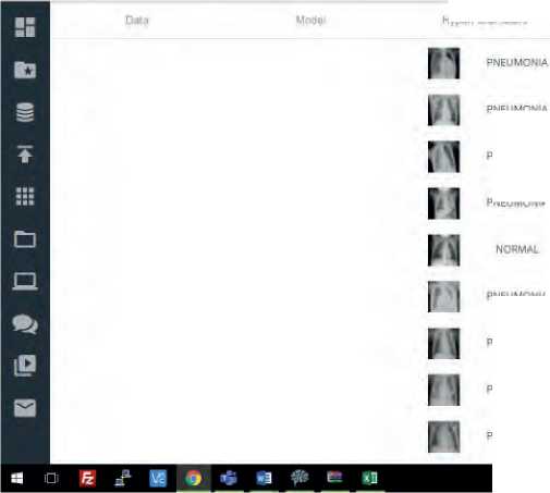
And the already trained network was considered as a DSS. We managed to process the X-ray images from the hospital, and the findings were obtained as indicated herein (see Figure 2).
Our DLS has a convenient interface, which allows you to upload images for processing via a tab in the browser, and, in our opinion, it greatly simplifies the work by a doctor in making a diagnosis. It makes the system user-friendly for applications by a medical institution, since every expert who has an access thereto can use one and the same trained network, and there is no need for deep knowledge in the field of AI, so that all this allows the doctor to better concentrate on making a diagnosis.
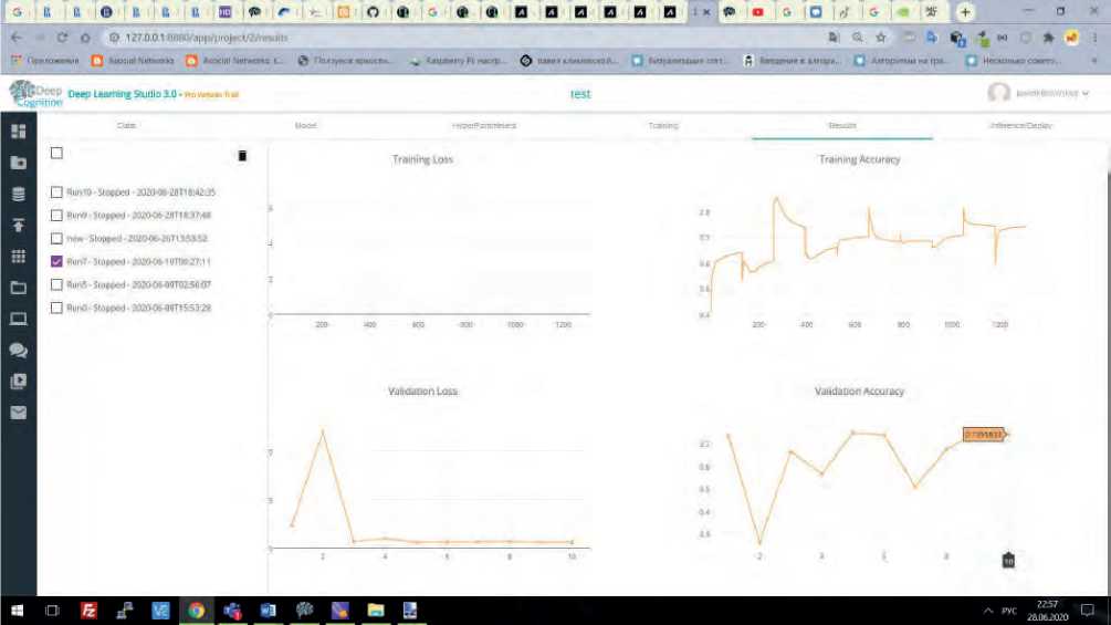
Figure 1. Training accuracy verification
3 ■ю о I,', 1Хоч> ■ 8 Ипзсе © ^ 8 type С 15 Поаез- 3 орея: 3 Pltho И кл : й ' «не fl йнлг« □ О ^ - х fl Хидек; ^ Нпдг- +

pavelklimovsKoy v test
1 )
4yper Pa takers
PNEUMONIA
PNEUMONIA
PNEUMONIA
PNEUMONIA
PNEUMONIA
PNEUMONIA
PNEUMONIA
PNEUMONIA
PNEUMONIA
PNEUMONIA
PNEUMONIA
PNEUMONIA
PNEUMONIA
PNEUMONIA
Figure 2. Validation sample test
PNEUMONIA
PNEUMONIA
Training
ReMiits
Infetenne/DopJoy
2.5601
2.55*97
2.5595
2.5598
2.5596
a
Statement on ethical issues
Research involving people and/or animals is in full compliance with current national and international ethical standards.
Conflict of interest
None declared.
Author contributions
The authors read the ICMJE criteria for authorship and approved the final manuscript.
References Decision support system in radiology for fast diagnostics of thoracic diseases under COVID-19 pandemic conditions
- Development of the digital economy: theoretical and practical significance for the agro-industrial complex: Proceedings of the International Scientific and Practical Conference. / Ed. by Sharikova IV. Saratov: LLC “TseSain”, 2019. 361 p. [in Russian]
- COVID-19 Resources. NIH. National Cancer Institute. https://www.cancer.gov/publications/dictionaries/cancer-terms/def/second-opinion
- Gavrilova, et al. Knowledge bases of intelligent systems. Textbook for universities. SPb.: Piter, 2000. [in Russian]
- Kravchenko TK. Expert decision support system. Open education. 2010;6:147-56 [in Russian]
- Suslova EV. Intelligent decision support systems. Young scientist. 2017;3(137):171-4. [in Russian]
- Castiglioni I, et al. AI applications to medical images: From machine learning to deep learning. Physica Medica. 2021;83:9-24.
- Haick H, Tang N. Artificial Intelligence in Medical Sensors for Clinical Decisions. ACS Nano. 2021;15(3):3557-67.
- P ower DJ. A brief history of decision support systems. DSSResources.com. Ver. 2.8,31 May 2003. URL: http://DSSResources.COM/history/dsshistory.html
- Power DJ. Supporting decision-makers: An expanded framework. e-Proceedings Informing Science Conference. Krakow, Poland, June 19-22, 2001. P. 431-436.
- Sechopoulos I, Teuwen J, Mann R. Artificial intelligence for breast cancer detection in mammography and digital breast tomosynthesis: State of the art. Seminars in Cancer Biology. 2021;72:214-25.
- Tran KA, et al. Deep learning in cancer diagnosis, prognosis and treatment selection. Genome Medicine. 2021;13(1):152. DOI: 10.1186/s13073-021-00968-x.
- Borges AFS, et al. The strategic use of artificial intelligence in the digital era: Systematic literature review and future research directions. International Journal of Information Management. 2021;57:102225
- Varela C, et al. Use of prior mammograms in the classification of benign and malignant masses. European Journal of Radiology. 2005;56(2):248-55.
- El-Rashidy N, et al. Mobile health in remote patient monitoring for chronic diseases: Principles, trends, and challenges. Diagnostics. 2021;11(4):607.
- Gelig Thurfjell M, Vitak B, Azavedo E, Svane G, Thurfjell E. Effect on sensitivity and specificity of mammography screening with or without comparison of old mammograms. Acta Radiologica. 2000;41(1):52-6.
- Borodyanskaya SE. Decision support system in radiology for the rapid diagnosis of pneumonia based on machine learning. Dissertation. Master’s Degree. Southern Federal Universitty. July, 9, 2020. [in Russian]
