
Diagnostic Uncertainty in Palaeopathology: An Epistemological Analysis of the Zeytinli Ada Cranial Lesions
Журнал: Материалы по археологии и истории античного и средневекового Причерноморья @maiask
Рубрика: Археология
Статья в выпуске: 22, 2026 года.
Бесплатный доступ
Palaeopathological diagnosis moves from a macroscopic lesion to a disease label through several inferential steps, each carrying its own uncertainty. This study examines eleven previously unstudied crania from the Byzantine assemblage of Zeytinli Ada, a monastic and healing complex on an island off the Erdek peninsula, Marmara Sea, Türkiye. The commingled material, which precludes postcranial association for any individual, is treated not as a limitation but as a condition exposing the epistemic structure of palaeopathological reasoning itself. A four-layer inferential model, the Layered Palaeopathological Inference Framework (LPIF), is applied: macroscopic observation, differential diagnosis, osteobiographic inference, and population-level interpretation. Rhinomaxillary destruction, basicranial lytic activity, and anaemia indicators recur across the adult series and support a treponemal reading in most of it (7 of 10 adult individuals scoring 2 or above on a Hackett-based matrix); with a series of this size the count is descriptive and carries no prevalence estimate. It aligns with a previously published tertiary syphilis case from the same site (ZK-61). One subadult cranium (CR-9) is reclassified from probable treponemal disease to infantile scurvy, based on a differential matrix and radiographic exclusion of thalassaemia and congenital syphilis. The results argue that diagnostic confidence depends less on the number of criteria met than on how explicitly inference is marked as it gives way to speculation, particularly in commingled series where anatomical completeness cannot be assumed.
Короткий адрес: https://sciup.org/14139204
IDS: 14139204 | DOI: 10.53737/2713-2021.2026.93.36.014
Диагностическая неопределённость в палеопатологии: эпистемологический анализ краниальных поражений с острова Зейтинли-Ада
Палеопатологический диагноз проходит путь от макроскопического поражения до нозологической категории через ряд этапов вывода, каждый из которых несёт свою неопределённость. В работе исследуются одиннадцать ранее не изученных черепов из византийского комплекса Зейтинли-Ада — монастырского и лечебного центра на острове у полуострова Эрдек, Мраморное море, Турция. Смешанный характер материала, исключающий посткраниальное сопоставление, рассматривается не как ограничение, а как условие, раскрывающее эпистемическую структуру палеопатологического рассуждения. Применена четырёхуровневая модель, обозначенная как Layered Palaeopathological Inference Framework (LPIF): макроскопическое наблюдение, дифференциальная диагностика, остеобиографическая и популяционно-историческая интерпретация. Риномаксиллярная деструкция, базикраниальная литическая активность и признаки анемии повторяются в серии взрослых и поддерживают трепонемную интерпретацию для большей её части (7 из 10 взрослых получили оценку 2 балла и выше по матрице на основе критериев Хакетта); при таком объёме выборки этот показатель носит описательный характер и не является оценкой распространённости. Он согласуется с ранее опубликованным случаем третичного сифилиса с того же памятника (ZK-61). Череп субадульта (CR-9) переклассифицирован из вероятного трепонемного случая в инфантильную цингу на основании дифференциальной матрицы и рентгенологического исключения талассемии и врождённого сифилиса. Результаты показывают, что уверенность в диагнозе зависит не столько от числа критериев, сколько от того, насколько явно обозначена граница перехода от вывода к предположению, особенно в смешанных сериях, где анатомическая полнота не гарантирована.
Текст научной статьи Diagnostic Uncertainty in Palaeopathology: An Epistemological Analysis of the Zeytinli Ada Cranial Lesions
МАИАСП № 22. 2026
palaeopathology at a persistent epistemic fault line, between the diagnostic certainty expected of clinical medicine and the interpretive uncertainty built into archaeological material (Mays 2010; Aufderheide, Rodriguez-Martin 1998).
Two structural problems recur across the field. The first is what Wood et al. (1992) termed the osteological paradox. Its core claim is that a skeletal sample is not a random draw from a once-living population but a selectively filtered one: only those who died are available for study, and among them, only certain trajectories of disease leave a durable skeletal signature. Selective mortality means the sample systematically over-represents individuals who survived long enough for bone to remodel in response to disease, and under-represents those who died acutely, before any lesion could form. Hidden heterogeneity in risk means that an apparently healthy skeleton may reflect either genuine resistance or simply a shorter exposure to whatever killed the individual, and the bone alone cannot distinguish between the two. Taken together, these mechanisms invert the intuitive reading of skeletal evidence: a heavily lesioned individual, such as CR-11, may represent someone whose body fought a long, survivable battle, while a lesion-free individual, such as CR-2, may equally represent someone who died before the disease had time to register on bone, rather than someone who was never exposed (DeWitte, Stojanowski 2015). The paradox has no methodological fix; it can only be made explicit.
The second problem is equifinality, a term with origins outside palaeopathology. In systems theory, it describes how a system can reach the same final state from many different initial conditions and through many different pathways; in geomorphology, it describes how visually identical landforms can result from entirely different combinations of erosion, deposition, and uplift (Roberts, Manchester 2010). Applied to skeletal lesions, equifinality means that aetiologically distinct disease processes converge on the same macroscopic morphology. Destructive rhinomaxillary lesions, central to much of the differential diagnosis in this paper, are produced convergently by treponemal disease, leprosy, and other chronic granulomatous infections, and no amount of macroscopic description can, by itself, decide among them. Neither the osteological paradox nor equifinality is solved by technical refinement: higher-resolution observation, more detailed photography, more sophisticated radiography, makes the underlying ontological uncertainty more visible, not less, because the problem is not a lack of resolution but a genuine many-to-one relationship between cause and effect. Any inferential framework applied to this material has to be built to accommodate that relationship instead of eliminating it, which is the starting point for the layered model introduced below.
This study examines eleven crania, and several associated mandibles, from the Byzantine layers of Zeytinli Ada, none of which has been previously analysed or published. The postcranial skeletons could not be matched to the crania, a direct consequence of the commingled deposition. Rhinomaxillary destruction, palatal porosity, basicranial lytic activity, and anaemia indicators are all present and diagnostically suggestive, but each carries its own interpretive uncertainty. Rather than treating this as an obstacle, the study uses it as a test case for the limits of palaeopathological diagnosis in commingled assemblages.
To manage this uncertainty, the Layered Palaeopathological Inference Framework (LPIF), a four-layer inferential model, is applied throughout: (1) macroscopic observation of the lesion itself, (2) differential diagnostic interpretation linking the lesion to clinical categories, (3) osteobiographic and archaeological inference about the individual, and (4) population-level historical interpretation. Each layer adds explanatory reach and, at the same time, compounds uncertainty. Marking clear boundaries between these layers is what converts palaeopathology’s methodological limitations from a weakness into a controlled analytic procedure. The central question is not whether treponemal disease was present at Zeytinli Ada, but where, and how, the line between observed lesion and diagnosed disease is actually drawn. This paper therefore pursues two aims jointly: to propose the LPIF as a transferable heuristic for structuring palaeopathological reasoning, and not as a validated diagnostic algorithm, then to apply it in sufficient depth to the Zeytinli Ada material for the case itself to stand as a contribution to Byzantine palaeopathology.
Tolga Köroğlu, Burak Atik, Fadime Suata, Frank Rühli, Sercan Acar, Onur Alpaslan, Gülfirde Akgül, Ayşe Yeşim Demir
МАИАСП № 22. 2026
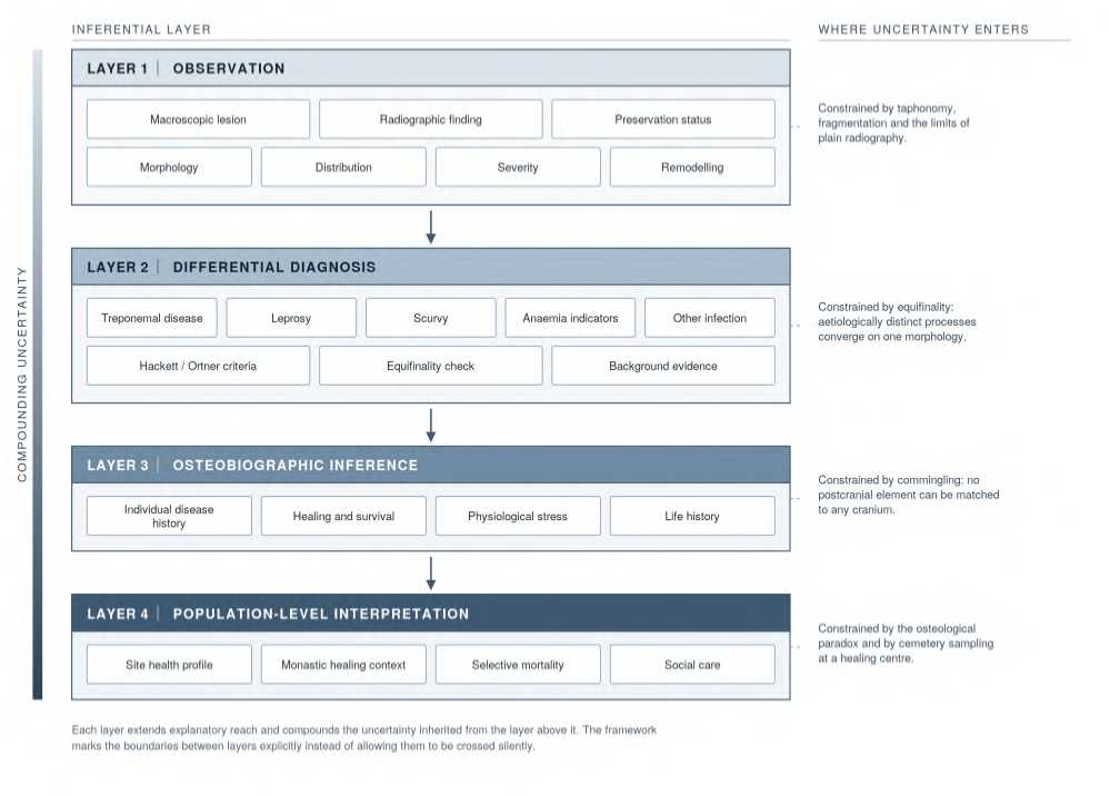
Fig. 1 The Layered Palaeopathological Inference Framework (LPIF).
Archaeologicaland HistoricalContext
Zeytinli Ada is a small island (170 × 50 m) of Erdek, Balıkesir province, in the south-western Marmara Sea, with occupation layers spanning the Roman, Byzantine, and Ottoman periods (Öztürk, Kavaz 2012). The Church of Panagia, a baptistery, a chapel, monastic cells, holy springs (ayazma), a bath, and harbour structures form a cluster typical of Byzantine monastic-healing complexes. In 1250, the Byzantine monk Maximos Planoudes, passing through Erdek (Artake), recorded in his fifty-fifth letter that a church dedicated to the Panagia stood on the island alongside a thermal healing spring (Leone 1991: Ep. 55). This letter is the clearest textual evidence for the site’s dual religious and therapeutic function.
Excavation has recovered dense tile-grave cemetery areas, in keeping with high mortality during the island’s use as a healing centre; Takunyacıoğlu (2019) links this density to a population of sick visitors. This historical profile bears directly on the sampling dynamics of the skeletal series analysed here: individuals recovered from the cemetery are more likely to represent a self-selected, unwell population than a cross-section of the regional Byzantine community, a point that matters for population-level interpretation later in this paper.
The assemblage is commingled. Centuries of superimposed burial in a spatially constrained cemetery, combined with taphonomic disturbance, have separated crania from postcranial elements, and no technique available to this study could restore that association. The eleven crania analysed here are previously unpublished. A prior study by Suata Alpaslan and Bekmez (2015) examined the overall commingled assemblage, and reported a single articulated case, ZK-61, in which cranial and long-bone (tibia, fibula) lesions together supported a secure diagnosis of tertiary syphilis. ZK-61 provides an
№ 22. 2026 An Epistemological Analysis of the Zeytinli Ada Cranial Lesions independent postcranial anchor for treponemal disease at the site and informs the population-level interpretation offered in Section 5, but it is not part of the cranial series analysed here.
The commingled structure has a direct epistemic consequence. Without anatomical completeness, individual-level diagnosis loses the corroborating evidence that a full skeleton would provide, and the equifinality problem described in Section 1 becomes harder to resolve. Several diagnoses in this study are therefore expressed as population-level probability instead of individual certainty. One subadult cranium, CR-9, illustrates this directly: metabolic stress markers and infection-consistent lesions co-occur in a case where the absence of long bones removes the evidence that would otherwise settle the differential diagnosis (see Section 4.5).
Materialsand Methods
Sample
The sample comprises eleven crania, excavated 2007—2011 under the direction of N. Öztürk. The series includes ten adults, nine male and one female, together with one subadult (CR-9, 2—4 years) whose sex was left undetermined. Sex was estimated from cranial morphological traits (nuchal crest, mastoid process, supraorbital margin, glabella, and mental eminence) following Buikstra and Ubelaker (1994); no sex estimate was attempted for CR-9, since these traits are not reliably expressed before adulthood. Age atdeath wasestimatedfrom cranialsuture closure in the adultsand, for CR-9,from dental development and cranial size following the same standards. These eleven crania were selected because they constitute newly available material: none had been described, scored, or published prior to this study, in contrast to other skeletalelements from the same cemetery already examined in prior dental and jaw pathology work (Bıçak, Suata Alpaslan 2015). No postcranial elements could be matched to any cranium. Cranial morphological characteristics are summarised in Table 1.
Table 1. Sample composition and cranial preservation (n = 11). AMTL = antemortem tooth loss.
|
ID |
Sex |
Age |
Preservation |
Mandible |
Dental status |
|
CR-1 |
M |
Adult |
Frontal, ethmoid, partial zygomatic, maxilla, palate, bilateral orbits |
Absent |
Severe AMTL |
|
CR-2 |
F |
Adult |
Right frontal (4/5), temporal, bilateral zygomatic/maxilla/palate/orbits/nasals |
Absent |
Moderate AMTL, no caries |
|
CR-3 |
M |
Adult |
Bilateral, partial |
Absent |
Moderate-severe abscessing, caries |
|
CR-4 |
M |
Older adult |
Bilateral, near-complete |
Present |
Total AMTL |
|
CR-5 |
M |
Adult |
Partial bilateral; calvaria not preserved |
Absent |
Multiple abscesses |
|
CR-6 |
M |
Adult |
Near-complete (left zygomatic process absent) |
Absent |
Moderate |
|
CR-7 |
M |
Adult |
Complete cranium |
Absent |
Moderate; caries + abscess |
|
CR-8 |
M |
Older adult (45—60) |
Near-complete |
Present (left ramus fractured) |
Severe abscessing + AMTL |
|
CR-9 |
Un det. |
Subadult (2-4 yr) |
Frontal, bilateral parietal, temporal (right squama absent), occipital, partial sphenoid |
Absent |
Deciduous, none erupted |
|
CR-10 |
M |
Middle adult |
Bilateral, present |
Absent |
Multiple abscesses |
|
CR-11 |
M |
Adult |
Bilateral, near-complete |
Present |
Severe AMTL |
Tolga Köroğlu, Burak Atik, Fadime Suata, Frank Rühli, Sercan Acar, Onur Alpaslan, Gülfirde Akgül, Ayşe Yeşim Demir
МАИАСП № 22. 2026
Methods
Lesions were assessed macroscopically, following standard descriptive protocols for surface bone pathology (location, distribution, margin morphology, and evidence of activity versus healing). Examination and scoring were carried out collaboratively among the authors: primary assessments for each specimen were cross-checked by a second author, and any disagreement in scoring was resolved through joint re-examination and discussion, reflecting a consensus procedure and not independent parallel assessment. Lesions were recorded on standardised forms adapted from Buikstra and Ubelaker (1994), and macroscopic photographs of every scored lesion were reviewed jointly during the consensus sessions. Because scoring was not independent, and because eleven crania cannot support a meaningful interobserver statistic such as Cohen’s kappa, no such measure is reported here; formal interobserver testing on a larger series is left to future work (Section 5.5). Treponemal evaluation followed the criteria of Hackett (1976), Ortner (2003), and Harper et al. (2011) (Table 2).
The subadult case (CR-9) was evaluated against Lewis (2007), Brickley and Ives (2008), Snoddy et al. (2018), and Ortner (2003) (Table 3).
Differential diagnosis relied on lesion combinations and contextual data rather than any single finding, following the four-layer model outlined in Section 1: observation, diagnostic interpretation, probability-based inference.
This study does not aim to establish, independently, that treponemal disease was present at Zeytinli Ada; ZK-61 (Suata Alpaslan, Bekmez 2015) already provides secure postcranial evidence for this. The aim instead is to examine the epistemic structure of the diagnostic process applied to the cranial series. Other infections capable of producing similar lesions (tuberculosis, leprosy, brucellosis, other granulomatous disease) were not systematically excluded but were retained in the differential diagnosis wherever equifinality applied; quantifying their prevalence falls outside the scope of this study.
Digital radiographs were obtained for three crania (CR-4, CR-9, CR-11), assessed for diploic structure, endosteal change, and the extent of lytic areas. Radiography is treated here not as an additional, independent evidentiary layer, but as a mediating technique with its own blind spots, capable of narrowing or sustaining the probability space without resolving it outright.
МАИАСП № 22. 2026
An Epistemological Analysis of the Zeytinli Ada Cranial Lesions
Table 2. Treponemal diagnostic criteria by anatomical region.
Sources: Hackett (1976); Ortner (2003: 257—291); Harper et al. (2011).
Tolga Köroğlu, Burak Atik, Fadime Suata, Frank Rühli, Sercan Acar, Onur Alpaslan, Gülfirde Akgül, Ayşe Yeşim Demir
МАИАСП № 22. 2026
Table 3. Paediatric skeletal lesions under the Lewis (2007) framework: diagnostic specificity and associated conditions (infant and child).
Sources: Lewis (2007); Ortner (2003); Brickley & Ives (2008); Snoddy et al. (201 8).
|
Anatomical Region |
Lesion Type |
Morphological Description |
Specificity |
Associated Condition |
Age at Risk |
Differential Diagnosis |
|
Orbital roof (internal surface) |
Cribra orbitalia (active) |
Fine, sponge-like new bone formation on the endocranial surface of the orbital roof; small, sharp-edged channels |
High (for scurvy) |
Infantile scurvy (vitamin C deficiency) |
0-4 yr (peak 0-2) |
Haemolytic anaemia; tuberculosis; may co-occur with rickets |
|
Sphenoid (greater wing) |
Subperiosteal porosity |
Dense, irregular porosity on the greater wing of the sphenoid; subperiosteal haemorrhage traces |
High (for scurvy) |
Infantile scurvy |
0-4 yr |
Highly specific to scurvy; not expected in thalassaemia |
|
Cranial vault (general) |
Porotic hyperostosis |
Dense, irregular porosity on the outer |
Moderate (non- |
Iron-deficiency |
Throughout |
Diploic widening is |
|
(active) |
table; may accompany diploic widening; active phase shows new bone deposition |
specific) |
anaemia, chronic infection, scurvy |
childhood |
pronounced in thalassaemia; minimal in scurvy |
|
|
Diploe (cranial table) |
Diploic widening |
Marked widening between inner and |
High (for |
Thalassaemia, |
Throughout |
Not expected in scurvy; its |
|
(“hair-on-end”) |
outer tables; comb-like trabecular pattern on radiograph |
thalassaemia) |
haemolytic anaemia |
childhood |
absence in CR-9 excludes thalassaemia |
|
|
Long bone metaphysis/diaphysis |
Subperiosteal new bone formation |
New bone layer beneath the periosteum on the diaphysis and metaphysis; bilateral; smooth or irregular |
High (for scurvy) |
Infantile scurvy, congenital syphilis |
0-4 yr |
Scurvy: widespread and bilateral. Congenital syphilis: more focal, metaphysis-weighted |
|
Long bone metaphysis |
Wegener’s bands (radiographic) |
Radiopaque band at the metaphyseal-diaphyseal junction; radiolucent Trummerfeld zone |
High (congenital syphilis) |
Congenital syphilis (early) |
0-2 yr |
Not seen in scurvy; may be confused with rickets |
|
Cranial morphology |
Caput natiforme (frontal bossing) |
Marked outward expansion of the frontal and parietal bones; “square head” appearance |
High (congenital syphilis) |
Congenital syphilis (late, 2+ yr) |
2-5 yr |
Also seen in rickets; its absence in CR-9 weakens the congenital syphilis reading |
|
Facial skeleton |
Saddle nose |
Collapse of the nasal bones and narrowing of the piriform aperture |
High (congenital syphilis) |
Congenital syphilis (late) |
2+ yr |
Leprosy; not assessable in CR-9 due to absence of the facial skeleton |
|
Maxilla (Hutchinson's |
Dental morphology |
Barrel-shaped upper central incisors |
Moderate |
Congenital syphilis |
6+ yr |
Non-specific; fluorosis, |
|
teeth) |
defect |
with notched incisal edge and posteriorly tapering crown |
(congenital syphilis) |
(late) |
(permanent dentition) |
other developmental defects; deciduous dentition not present in CR-9 |
|
Rib (internal surface) |
Visceral surface new bone formation |
Irregular new bone layer on the internal rib surface; active and healed forms |
Moderate |
Tuberculosis (pulmonary), chronic infection |
Throughout childhood |
Non-specific; respiratory infections; may co-occur with scurvy |
Table 3. Paediatric skeletal lesions under the Lewis (2007) framework: diagnostic specificity and associated conditions (infant and child) (continued).
|
Anatomical Region |
Lesion Type |
Morphological Description |
Specificity |
Associated Condition |
Age at Risk |
Differential Diagnosis |
|
Cranial vault (endocranial) |
Endocranial new bone or porosity |
Fine new bone formation or porosity on the internal table surface; granular appearance in active phase |
Moderate |
Tuberculosis (meningeal), scurvy, congenital syphilis |
Throughout childhood |
Non-specific; meningitis, endocranial inflammation |
|
Long bones (all) |
Haematogenous osteomyelitis |
Focal or multifocal lytic lesion; cloacae; sequestrum; reactive new bone |
Moderate (focal) |
Pyogenic bacterial infection |
Throughout childhood |
Unlike syphilis, asymmetric and focal; bilateral distribution not expected |
|
Dental enamel |
Enamel hypoplasia (LEH) |
Horizontal line or pitting on the enamel surface; timing of the stress episode is chronologically estimable |
Non-specific |
Nutritional stress, chronic illness |
Tooth development periods |
Specific cause not distinguishable; indicates presence of stress only |
|
Long bones (metaphysis) |
Harris lines (radiographic) |
Growth-arrest lines; transverse radiopaque bands on radiograph |
Non-specific |
Nutritional stress, chronic illness |
Active growth periods |
Indicates a stress episode during growth; cause unspecified |
|
General growth |
Growth stunting |
Deviation from age-expected bone length and morphology |
Non-specific |
Nutritional insufficiency, chronic infection |
Throughout childhood |
Genetic factors; requires detailed demographic data |
МАИАСП № 22. 2026
An Epistemological Analysis of the Zeytinli Ada Cranial Lesions
Tolga Köroğlu, Burak Atik, Fadime Suata, Frank Rühli, Sercan Acar, Onur Alpaslan, Gülfirde Akgül, Ayşe Yeşim Demir
МАИАСП № 22. 2026
Results:craniallesions
Table 4 summarises macroscopic findings for all eleven individuals, scored 0 —3 for treponemal suspicion (0 = absent, 1 = limited/non-specific, 2 = probable, 3 = high probability). Radiographic findings for CR-4, CR-9, and CR-11 are given in Table 5. The account below follows the four cases that carry the most diagnostic and epistemic weight, with the remaining individuals summarised at the end of each subsection.
Rhinomaxillary destruction and mid-facial lesions
Rhinomaxillary syndrome, resorptive widening of the piriform aperture, destruction of the nasal septum and vomer, and palatal perforation with loss of the anterior nasal spine, is considered by Hackett (1976) one of the most characteristic cranial signatures of treponemal disease, particularly tertiary syphilis. Leprosy produces overlapping facial destruction, though typically more limited, and palatal perforation with basicranial extension is unusual for leprosy (Ortner 2003); several Zeytinli Ada individuals nonetheless show morphology close enough to this overlap zone to require full differential treatment.
CR-4 (score 3; Figure 2a, Figure 3a) presents the clearest case in the series. The nasal aperture margins are resorbed and widened, and most of the nasal bones and vomer are lost. Smoothed, sclerotic margins point to a long-standing chronic process and rule out a peri-mortem origin. Radiography supports the macroscopic pattern but does not resolve it: whether the palatal defect is a true perforation or a superimposed air space from projection angle could not be determined from the image, nor could density differences in the parietal and occipital regions be separated from superimposed auditory structures. This combination of macroscopic and radiographic evidence places CR-4 among the highest-confidence cases in the series, though the unresolved radiographic ambiguity noted above means the diagnosis is best read as a high probability, not a certainty (see Section 5.3).
CR-10 (score 3) shows a different pattern: multifocal lytic activity extending beyond the face into the frontal, parietal, and zygomatic bones. This wider distribution, compared with CR-4’s more localised lesion, suggests systemic spread and indicates that individuals scoring identically on the matrix can nonetheless represent different stages or intensities of the same disease process; scoring narrows probability without fixing a diagnosis.
CR-3 (score 3; Figure 2c) is the clearest instance of unresolved equifinality in the series. Palatal perforation and alveolar lytic lesions (alveolar cloacae) initially favour a treponemal reading, but Ortner (2003) notes that leprosy can produce similar findings; syphilitic alveolar destruction tends to be confluent across the palate, while leprosy produces more localised, anteriorly concentrated erosion (Aufderheide, Rodriguez-Martin 1998). CR-3’s partial preservation (Table 1) does not allow this distinction to be made, and the case is retained at “moderate confidence” within the high-probability band. Set against CR-4, whose pattern aligns more consistently with Hackett’s criteria, CR-3 shows how the qualitative interpretation of lesion distribution, not the raw score, ultimately governs diagnostic confidence.
Basicranial involvement
The basicranium and foramen magnum region, well protected from taphonomic damage and a key crossing point for systemic infection spread, shows lytic activity in three individuals: CR-4, CR-5, and CR-11.
CR-5 (score 2) combines basicranial erosion with diploic thickening on the internal occipital surface. Roberts and Manchester (2010) note that this combination also admits tuberculosis, which is normally confirmed through vertebral collapse (Pott’s disease) and rib lesions, evidence unavailable here due to the absence of postcranial material. CR-5 is retained at “probable,” with tuberculosis kept open as an alternative.
МАИАСП № 22. 2026
CR-11 (score 3; Figure 2b, Figure 3c) shows the most extensive and chronologically active basicranial destruction in the series, with dense porosity across the sphenoid and basal occipital regions. Sclerotic zones indicate bone remodelling alongside active lytic areas, suggesting different stages of the same disease process co-existing at the time of death. This pattern is consistent with tertiary syphilis, but Table 5’s radiographic findings show it fits tuberculosis equally well; pronounced sutural lines visible on radiograph may reflect antemortem remodelling or postmortem desiccation, and the two cannot be distinguished at this resolution. Compared with CR-4, CR-11’s basicranial score is identical (3), but its wider anatomical distribution suggests a more advanced stage of infection; in both cases, tuberculosis remains an open differential, meaning that even a score of 3 should be read as “high probability,” not certainty.
Anaemia indicators
Cribra orbitalia and porotic hyperostosis, present in CR-6 and CR-7, are not disease-specific; they reflect general physiological stress from iron deficiency, parasitic load, gastrointestinal disturbance, chronic infection, or malnutrition, and cannot on their own support an aetiological diagnosis (Walker et al. 2009).
CR-7 (score 1) shows healed porotic lesions with no evidence of active infection or ongoing metabolic disturbance, and no independent sign of treponemal disease. A healed childhood metabolic crisis in an individual who survived to adulthood is a direct illustration of the osteological paradox (Wood et al. 1992): bone that shows healing marks resistance, not disease burden, in a way that a lesion-free bone cannot demonstrate.
CR-6 (score 2) combines porotic hyperostosis with palatal porosity and early-stage rhinomaxillary involvement, moving the case out of the non-specific category and into the probable treponemal group; the early stage of the facial lesion keeps a localised infection plausible, so the case remains at moderate confidence. The co-occurrence of cribra orbitalia with palatal porosity may indicate treponemal disease triggering an anaemic process, but the non-specific nature of anaemia markers prevents establishing that causal link (Ortner 2003). CR-7’s healed profile functions as a population-level baseline against which CR-6’s active lesions can be read.
Dental sepsis
Dental sepsis, untreated caries, advanced periodontal disease, or pulpitis spreading from the tooth into surrounding bone, produces cloacae, confluent bone loss, and localised osteomyelitis of the jaw (Dias et al. 2007; Waldron 2009). Beyond poor oral hygiene, it signals restricted access to treatment in pre-antibiotic communities and, when it spreads intracranially, carries systemic mortality risk (Hillson 2005).
CR-8 (Figure 2e), a male aged 45—60, shows the most severe dental sepsis in the series: confluent maxillary alveolar destruction, multiple cloacae, and dense palatal abscessing, with secondary porotic reaction on the internal cranial surface. No independent treponemal indicator is present, and the case (basicranial porosity scored 1) is consistent with periapical infection spreading internally. CR-8 is classified as chronic odontogenic sepsis, not treponemal disease, and represents the assemblage’s clearest evidence that the site’s pathological profile is not reducible to a single disease category.
Paediatric case
CR-9 (Figure 2d, Figure 3b), the series’ only subadult (2—4 years), demonstrates how the absence of postcranial material can leave a diagnosis genuinely unresolved rather than merely uncertain. Table 4 removes CR-9 from the treponemal category and classifies it as probable scurvy. The lesion combination, bilateral cribra orbitalia, sphenoid greater-wing porosity, and normal
Tolga Köroğlu, Burak Atik, Fadime Suata, Frank Rühli, Sercan Acar, Onur Alpaslan, Gülfirde Akgül, Ayşe Yeşim Demir
МАИАСП № 22. 2026
diploic architecture, matches the infantile scurvy profile described by Ortner (2003) and Brickley and Ives (2008). Radiography (Table 5) supports this: no diploic widening or “hair-on-end” pattern consistent with thalassaemia or haemolytic anaemia, and no “caput natiforme” pattern associated with congenital syphilis. Here radiography works by elimination and not by positive diagnosis: it rules out competing aetiologies without confirming one, and that asymmetry illustrates the central epistemological argument of this paper (Table 3).
The age range (2—4 years) supports a scurvy diagnosis on historical grounds; this corresponds to the post-weaning period in Byzantine populations, when children face peak risk of vitamin C deficiency and nutritional stress (Lewis 2007). Congenital syphilis cannot be fully excluded, since its late clinical presentation can theoretically overlap this age range, but its typical cranial markers, caput natiforme and saddle nose, are absent, and the single potentially diagnostic right parietal lesion does not carry enough weight on its own. Had postcranial elements survived, periosteal reaction or bowed (“saber”) tibiae would have supported congenital syphilis, and subperiosteal haemorrhage would have supported scurvy; without them, neither possibility can be settled, and CR-9 stands as a direct illustration of how commingled assemblage conditions turn diagnostic uncertainty into a structural feature of the data rather than a research failure.
Diagnostic confidence synthesis
Table 6 applies a seven-criterion Hackett/Ortner matrix across all adult individuals. CR-4 and CR-11 both score 3 (high probability) but for different reasons: CR-4 combines caries sicca with rhinomaxillary syndrome, a single-region but highly specific signature, while CR-11 shows multifocal involvement across four anatomical regions without rhinomaxillary destruction, a broader but less specific pattern. Identical scores conceal different diagnostic bases. CR-3, also scored 3, is comparatively fragile: caries sicca is equivocal and the facial destruction pattern sits at the treponemal-leprosy intersection described in Section 4.1. Among the score-2 individuals, CR-1’s facial lesions cannot yet be assigned to antemortem or postmortem origin, CR-5 keeps tuberculosis open pending postcranial evidence that does not exist, and CR-6 lacks caries sicca and endocranial involvement altogether, consistent with early, localised disease. CR-2 returns a fully negative profile and serves as the series’ pathology-free reference point. Across the ten adult crania, seven score 2 or above; with a sample of this size, this count is best read as a description of the series and not as a population-level statistic.
The 0—3 scale used in Table 6 is a summary device, not a direct transcription of Hackett’s (1976) original protocol. Hackett’s criteria are hierarchical rather than additive: certain lesion combinations, caries sicca together with rhinomaxillary destruction among them, carry categorically more diagnostic weight than the same numerical count built from lower-weight findings. Table 6 collapses this hierarchy into a single ordinal score for ease of comparison across the series, but the underlying criteria in Table 2 retain their original qualitative weighting (pathognomonic, diagnostic, supportive, non-specific). A score of 3 built from one pathognomonic finding, as in CR-4, is therefore not equivalent, in evidentiary terms, to a score of 3 built from several supportive findings distributed across multiple regions, as in CR-11; Table 6 should be read alongside Table 2, not as a substitute for it.
Caries sicca is classified as pathognomonic for treponemal disease in Table 2, following Hackett (1976), and CR-4, CR-10, and CR-11 all score positively for it. Under a strict reading of Hackett’s criteria, this would justify certainty for these three individuals instead of probability. This paper nonetheless keeps them within the high-probability band and stops short of certainty, for a reason specific to this assemblage: pathognomonicity is normally established on complete, individually attributed skeletons, whereas the calvarial fragments considered here carry no matched postcranial confirmation (Section 2) and cannot be fully excluded from taphonomic surface alteration or, in principle, from an advanced non-specific osteomyelitic process of the kind documented in CR-8. A pathognomonic criterion observed on a commingled, postcranially unconfirmed specimen carries less evidentiary weight than the same criterion observed on an articulated skeleton such as ZK-61. High
№ 22. 2026 An Epistemological Analysis of the Zeytinli Ada Cranial Lesions confidence, rather than certainty, is retained as the appropriate designation throughout this paper for exactly this reason.
Table 4. Treponemal suspicion scores by individua
(0 = absent, 1 = limited/non-specific, 2 = probable, 3 = high probability).
|
ID |
Score |
Key findings |
Note |
|
CR-1 |
2 |
Palatal porosity; right infraorbital lesion; equivocal antemortem remodelling at piriform margins |
Conditional; would benefit from microscopic confirmation |
|
CR-2 |
0 |
No active lesion; limited midline palatal porosity; normal nasal aperture |
Pathology-free reference individual |
|
CR-3 |
3 |
Palatal perforation threshold; rhinomaxillary widening; infraorbital and frontal endocranial lytic activity; multifocal alveolar destruction |
Leprosy retained in differential |
|
CR-4 |
3 |
Classic rhinomaxillary syndrome; dense basicranial lytic activity; sphenoid porosity; endosteal mandibular involvement |
Strongest treponemal case in the series |
|
CR-5 |
2 |
Occipital diploic thickening with patchy lytic activity; dense basicranial involvement; moderate rhinomaxillary change |
Basicranial tuberculosis is the primary differential |
|
CR-6 |
2 |
Cribra orbitalia and porotic hyperostosis; palatal porosity; early rhinomaxillary change; basicranial porosity |
Retained within probable treponemal group |
|
CR-7 |
1 |
Healed porotic hyperostosis; localised infraorbital remodelling; no active lesion |
Adult profile consistent with resolved childhood metabolic crisis |
|
CR-8 |
1 |
Severe chronic odontogenic sepsis; confluent maxillary alveolar destruction; secondary endocranial porosity |
No corroborating treponemal criteria |
|
CR-9 |
Probabl e scurvy |
Bilateral cribra orbitalia; sphenoid greater-wing porosity; normal diploic architecture; right parietal lesion equivocal |
Reclassified as infantile scurvy; removed from treponemal category |
|
CR-10 |
3 |
Generalised palatal porosity; dense basicranial porosity and lytic activity; infraorbital and zygomatic lytic lesions |
Classic active treponemal presentation |
|
CR-11 |
3 |
Chronic-active basicranial honeycombing; foramen magnum lytic activity; frontal lytic/porotic change; dense endocranial involvement |
Consistent with advanced, active tertiary-stage disease |
Table 5. Radiographic findings for CR-4, CR-9, and CR-11, and their epistemic function within the differential diagnosis.
|
ID |
Radiographic findings |
Diploic status |
Epistemic function |
|
CR-4 |
Piriform margin obscured; midline alveolar radiolucency; patchy parietal/occipital density change; basicranial density increase |
Normal |
Supports macroscopic rhinomaxillary pattern; palatal and basicranial margins remain unresolved |
|
CR-9 |
Suture patency age-consistent; orbital roof porosity confirmed; sphenoid greater-wing porosity weakly resolved; sharp sutural lines; no caput natiforme configuration |
Preserved, not thinned; no hair-on-end pattern |
Excludes thalassaemia/haemolytic anaemia; weakens congenital syphilis reading; works by elimination |
|
CR-1 |
Sutural lines unusually pronounced; frontal |
Diploic widening |
Supports chronic-active tertiary- |
|
1 |
lytic/porotic areas confirmed; dense, irregular radiodensity in parietal/occipital regions; radiolucent defects at foramen magnum margin |
visible in preserved vault segment |
stage reading; does not resolve tuberculosis differential |
Tolga Köroğlu, Burak Atik, Fadime Suata, Frank Rühli, Sercan Acar, Onur Alpaslan, Gülfirde Akgül, Ayşe Yeşim Demir
МАИАСП № 22. 2026
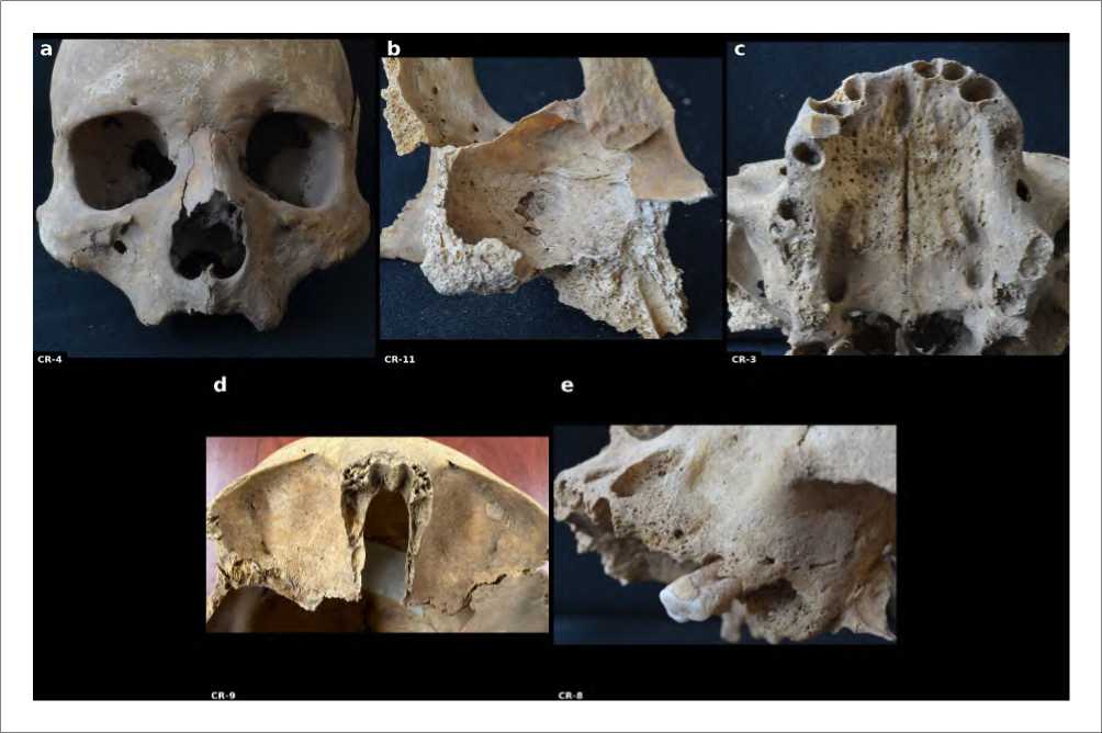
Fig. 2 Representative macroscopic lesions from the five individuals central to the differentia diagnosis discussed in Section 4. a — CR-4, rhinomaxillary lesion; b — CR-11, basicranial lytic lesion; c — CR-3, palatal destruction; d — CR-9, cribra orbitalia; e — CR-8, maxillary alveolar sepsis.
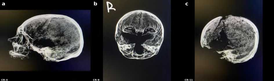
Figure 3 Radiographic findings for CR-4, CR-9, and CR-11 (see Table 5).
№ 22. 2026 An Epistemological Analysis of the Zeytinli Ada Cranial Lesions
Table 6. Hackett (1976) / Ortner (2003) treponemal criteria matrix. + present, - absent/not assessable, ? Equivocal.
|
ID |
Caries sicca |
Rhinomax. |
Endocranial |
Basicranial |
Calvarial |
Mandibular |
Score |
|
CR-1 |
? |
?+ |
- |
- |
- |
- |
2 |
|
CR-2 |
- |
- |
- |
- |
- |
- |
0 |
|
CR-3 |
? |
+ |
+ |
+ |
+ |
- |
3 |
|
CR-4 |
+ |
+ |
+ |
+ |
? |
+ |
3 |
|
CR-5 |
- |
+ |
? |
+ |
+ |
- |
2 |
|
CR-6 |
- |
+ |
- |
+ |
- |
- |
2 |
|
CR-7 |
- |
- |
- |
+ (healed) |
- |
- |
1 |
|
CR-8 |
- |
- |
+ |
+ |
- |
+ |
1 |
|
CR-9 |
n/a |
n/a |
n/a |
n/a |
n/a |
n/a |
Probable scurvy |
|
CR-10 |
+ |
+ |
- |
+ |
+ |
- |
3 |
|
CR-11 |
+ |
- |
+ |
+ |
+ |
+ |
3 |
Discussion
ZK-61 as contextual evidence
Suata Alpaslan and Bekmez’s (2015) case ZK-61, an articulated skeleton of a woman around 30 years old with periosteal reaction on the tibia and fibula alongside cranial destruction consistent with tertiary syphilis, sits outside the cranial series analysed here but provides its most important contextual anchor. It confirms, independently and at the postcranial level, that treponemal disease circulated in this population. The reasoning applied here is Bayesian only in a qualitative sense: ZK-61 functions as background evidence that raises confidence in a treponemal reading of the cranial lesions described in this paper, not as an input to a formal probabilistic calculation. In palaeopathology, this kind of background evidence is typically drawn from historical, epidemiological, or previously documented material independent of the specimens under study (Mays 2012; Chamberlain 2006). ZK-61 raises confidence that the cranial lesions described here reflect treponemal disease. It does not, however, resolve individual cases: CR-5’s tuberculosis differential and CR-3’s leprosy overlap are unaffected by ZK-61’s existence, since population-level background evidence does not transfer automatically to individual osteobiography.
Since the original macroscopic and radiographic description of ZK-61, an attempt has been made to confirm the diagnosis molecularly. Ancient DNA analysis of the ZK-61 skeleton (laboratory sample name Sivas_merged_data, an internal identifier assigned by the sequencing facility and unrelated to the provenance of the specimen) was conducted at the Institute of Evolutionary Medicine, University of Zurich, and the results have been made available to the authors with permission (Rühli, Akgül 2018). Authentic ancient DNA was recovered from the sample, confirmed by a 15% cytosine-to-thymine misincorporation pattern typical of degraded ancient material, but endogenous human DNA represented only 2.747% of reads, with a mean genomic coverage of 0.0061X. Shotgun sequencing mapped only nine reads to the Treponema pallidum pallidum genome; a subsequent targeted enrichment capture, using biotinylated baits against four treponemal-specific regions (tprB, tprH, tprK, tpp15), recovered only 32 mapped reads, none showing the damage pattern expected of authentic ancient pathogen DNA. The report concludes that treponemal DNA could not be detected in the sample, and the project was closed on that basis (Rühli, Akgül 2018).
It should also be acknowledged that the molecular investigation of ZK-61 was undertaken approximately a decade ago, within the methodological framework available at that time. The reported negative result therefore reflects the analytical sensitivity and technical possibilities of the original ancient DNA workflow rather than constituting a definitive exclusion of treponemal infection. Since ancient pathogen genomics has continued to develop substantially, with improvements in library preparation, enrichment strategies, sequencing depth, and bioinformatic authentication, re-analysis of the same material using contemporary approaches may potentially yield different results. Accordingly, the previous molecular study is best interpreted as an important but
Tolga Köroğlu, Burak Atik, Fadime Suata, Frank Rühli, Sercan Acar, Onur Alpaslan, Gülfirde Akgül, Ayşe Yeşim Demir
МАИАСП № 22. 2026
methodologically time-bound line of evidence whose negative outcome should not be treated as equivalent to biological absence.
This negative result does not, by itself, overturn the treponemal diagnosis established for ZK-61 on morphological and postcranial grounds (Suata Alpaslan, Bekmez 2015); a failure to detect pathogen DNA is not equivalent to evidence that the pathogen was absent. Low endogenous DNA content and poor pathogen-DNA preservation are common in the burial environments found across Anatolia, and the low proportion of human DNA recovered here indicates a sample already close to the practical limit of what shotgun and capture-based methods can resolve, well before pathogenlevel detection is considered. The absence of a damage pattern in the 32 reads that did map to the treponemal genome is, on current evidence, more consistent with background noise than with authentic ancient pathogen DNA, and the original report treats the result accordingly. What this attempt demonstrates in concrete terms is a point raised elsewhere in this paper (Section 5.5): aDNA is not a straightforward adjudicator for the equifinality problems affecting CR-3 and CR-5, since a negative molecular result in commingled, poorly preserved cranial material is uninformative, not disconfirming. ZK-61’s diagnosis therefore continues to rest on the same converging morphological and postcranial evidence as before this analysis was undertaken.
The underlying logic is worth making explicit, because it recurs at several points in this paper. In formal Bayesian inference, a prior probability, representing what is known before new data are considered, is updated by a likelihood function derived from the data themselves, producing a posterior probability. Applying this apparatus rigorously would require, at minimum, a specified base rate of treponemal disease in comparable Byzantine populations and a quantified likelihood for each lesion pattern given each candidate diagnosis; neither exists in the palaeopathological literature at a resolution fine enough to support this material, and manufacturing such figures for a series of eleven crania would produce spurious precision instead of genuine inference. What ZK-61 supplies is a much weaker, but still useful, version of the same logic: independent evidence, external to the cranial series itself, that shifts the analyst’s confidence in one direction before the cranial lesions are even examined. This is the sense in which the reasoning here is Bayesian, an orientation rather than a calculation, and the distinction matters, because collapsing it, treating a raised confidence as though it were a computed posterior, would misrepresent the epistemic status of the conclusion.
Epistemic tension in the diagnostic method
Hackett (1976) was the first to systematise treponemal skeletal criteria, working from clinical case files, museum specimens, and well-preserved dissection material, all with anatomical completeness intact. None of these reference conditions hold for the Zeytinli Ada material: subsurface taphonomy, postmortem disturbance, commingling, and the absence of postcranial elements all work against Hackett’s original criteria. Mays (2012) argued that later researchers hardened Hackett’s originally probabilistic framework into something closer to deterministic rule (“this lesion means this disease”), collapsing the distinction between diagnostic confidence and diagnostic certainty. This study does not attempt to resolve the tension between Hackett’s criteria and Mays’s critique, but keeps both explicit: Hackett’s matrix functions here as the working diagnostic tool, and Mays’s caution is applied at every inferential step to keep the residual uncertainty visible. CR-1’s rhinomaxillary pattern, CR-5’s basicranial activity, and CR-3’s palatal perforation all look persuasive under Hackett’s framework; under Mays’s framework, that same persuasiveness reflects unresolved alternatives and not their exclusion, which is a difference in the limits of knowledge, not merely a terminological one.
МАИАСП № 22. 2026
The epistemic role of radiography
Radiography performed differently across the three cases examined (CR-4, CR-9, CR-11), and that unevenness is informative about its evidentiary status in bioarchaeology. In CR-9, radiography worked by elimination, ruling out thalassaemia, haemolytic anaemia, and congenital syphilis, and thereby strengthening the macroscopic scurvy interpretation without establishing a new positive finding on its own. In CR-4, radiography tracked the macroscopic pattern but widened rather than closed the probability space. In CR-11, it deepened understanding of lesion severity and internal bone structure without resolving the differential. Andersen and Manchester (1992) documented comparable radiographic limits for leprosy-related rhinomaxillary syndrome, and Ortner and Ericksen (1997) found that paediatric cranial porosity on radiograph functioned mainly to eliminate alternatives instead of confirming one, a pattern consistent with what is observed here. Radiography, in this material, is not an independent evidentiary layer stacked on top of macroscopic observation; it is an instrument with its own resolution limits, angle-dependent artefacts, and blind spots. A conventional two-dimensional projection radiograph compresses a three-dimensional structure onto a single plane, so that overlapping anatomical layers, such as the petrous pyramid superimposed on the basicranium in CR-4, cannot always be separated from genuine pathological change; this projection limitation, rather than any failure of the observers, is a primary reason why the radiographic findings reported here narrow or sustain a probability without settling it.
Interpreting Zeytinli Ada archaeologically
The fourth inferential layer, that the sick were brought to this island for healing or in search of a holy spring, rests on Maximos Planoudes’s letter and not on skeletal data, and the skeletal evidence can only support it indirectly. What the skeletal data do show is a pathological profile too heterogeneous for a single-disease narrative: CR-4, CR-10, and CR-11 present advanced treponemal signs; CR-8 presents severe, localised dental sepsis unrelated to treponemal disease; CR-9 presents probable scurvy; CR-7 presents a healed lesion profile in an individual who survived to adulthood. Miller (1985) and Horden (2005) describe Byzantine monastic healing institutions as admitting patients according to geography, denomination, and bed capacity, a pattern the Zeytinli Ada material broadly fits, since the site’s architecture could plausibly have housed resident monks and staff alongside temporary visiting patients. Some cemetery individuals may represent long-term island residents who died of age or local infection and not visiting patients, a possibility that adds a layer to the “healing pilgrims” hypothesis without replacing it.
At the population level, the finding that seven of ten adult crania score 2 or above is inflated by sampling bias in one direction and deflated by the osteological paradox in the other; with a sample this size, no percentage should be read as a population parameter. Zeytinli Ada was not a demographically representative cemetery; if the island functioned as a healing centre, its dead are disproportionately drawn from an already unwell population, and this count describes that selected pool, not regional Byzantine disease prevalence. In the opposite direction, individuals who died in the acute phase of infection, before bone remodelling had time to register the disease, are systematically invisible in this kind of analysis (Wood et al. 1992). Read together with ZK-61, this pattern is compatible with an endemic treponemal presence, but extending that biological reading into the social claim that these individuals were brought to the island specifically for healing goes beyond what skeletal data alone can support; that claim rests on the textual record, not the bone.
Tolga Köroğlu, Burak Atik, Fadime Suata, Frank Rühli, Sercan Acar, Onur Alpaslan, Gülfirde Akgül, Ayşe Yeşim Demir
МАИАСП № 22. 2026
Limitations and future directions
This study relies on macroscopic observation and conventional radiography for the cranial series itself; no micro-computed tomography was performed, and molecular confirmation was attempted only for ZK-61, the external contextual case discussed in Section 5.1, not for the crania analysed here. That attempt is itself informative about the limits of the method for this material: despite authentic ancient DNA being recovered from the ZK-61 sample, endogenous human DNA content was low, and neither shotgun sequencing nor a targeted treponemal enrichment capture recovered enough authentic pathogen reads to confirm or exclude treponemal infection (Rühli, Akgül 2018). This suggests that aDNA-based pathogen confirmation, while in principle capable of resolving the equifinality problems affecting CR-3 and CR-5, may not be straightforwardly achievable across this assemblage even where it is attempted, given the preservation conditions typical of the site. Microcomputed tomography remains untried here and could still convert CR-9’s “normal diploic architecture” criterion from a qualitative judgement into a measurable one, along with clarifying whether CR-4’s palatal defect is a genuine antemortem perforation; access to this technique continues to be limited by cost and logistics for most anthropology laboratories in Turkiye. The absence, and in the case of aDNA the attempted but inconclusive use, of these techniques is reported here as a methodological constraint on what this material can currently support, not as an oversight. A formal interobserver reliability protocol, with lesions scored independently by multiple observers and agreement quantified using a statistic such as Cohen’s kappa, was likewise not applied here, for the reasons given in Section 3.2; this remains an appropriate target for future work on larger, noncommingled series where independent parallel scoring is logistically feasible.
Three interpretive constraints in practice
Three interpretive constraints run through this paper and are worth drawing together explicitly, since each surfaces repeatedly under a different name across the case material. Equifinality is the constraint that governs individual diagnosis: CR-3’s position at the treponemal-leprosy intersection, and CR-5’s and CR-11’s unresolved tuberculosis differentials, are not failures of observation but direct expressions of the fact that different diseases converge on the same bony response. The osteological paradox is the constraint that governs population-level reading: the series’ apparently high burden of treponemal disease, and CR-7’s healed lesions in an individual who survived to adulthood, only make sense once it is accepted that the skeletons available for study are not a neutral sample of who once lived, but a biased record of who died and how. Background evidence, weighed qualitatively in the sense set out in Section 5.1, is the constraint that governs how findings external to the sample, such as ZK-61, are allowed to shift confidence without dictating individual conclusions; the inconclusive aDNA result reported for ZK-61 (Section 5.1) illustrates the same point from the opposite direction: a negative molecular finding does not erase the background evidence a case already provides, just as a positive one would not have transferred automatically to individual cranial diagnoses in the present series. None of these three constraints is specific to Zeytinli Ada, and none is resolved by better technique alone; a higher-resolution photograph of CR-3 would not settle whether it is treponemal disease or leprosy, and a larger sample would not, by itself, remove the selective mortality built into any cemetery population. What the LPIF offers is not a solution to any of the three constraints but a discipline for keeping them visible: by separating observation from differential diagnosis, differential diagnosis from osteobiographic inference, and osteobiographic inference from population-level interpretation, the model ensures that equifinality, the osteological paradox, and background evidence are confronted at the specific layer where each actually operates, instead of blurring together into an undifferentiated sense of uncertainty.
МАИАСП № 22. 2026
Conclusion
The eleven Zeytinli Ada crania were analysed to examine the epistemic structure of the diagnostic process, not to establish a single confirmed diagnosis. Macroscopic lesions document morphological reality; radiography narrowed some probability spaces while widening others, exposing the unevenness of imaging technology as an evidentiary tool without resolving it. Applying Hackett (1976) and Ortner’s (2003) criteria sorts individuals into high- and moderate-confidence groups, but CR-3’s equifinality with leprosy and CR-5’s open tuberculosis differential show that these groupings narrow probability without fixing a diagnosis. CR-8’s severe dental sepsis, excluded from the treponemal category, confirms that the site’s pathological profile is not reducible to one disease. CR-9, lacking postcranial evidence, shows that congenital syphilis and infantile scurvy cannot always be separated on morphology alone, a direct limit on what commingled cranial material can answer.
The finding that seven of ten adult crania score 2 or above, together with the previously documented ZK-61 case, is consistent with an endemic presence at the site, but this reading must be held against both the selection bias built into a healing-centre cemetery and the osteological paradox that hides lesion-free deaths from view. The biological heterogeneity of the sample confirms that Zeytinli Ada was not a uniform syphilis-burial ground, but is compatible with a mixed Byzantine healing complex population, resident monks and desperate outside visitors together, as described by Miller (1985) and Horden (2005).
The Layered Palaeopathological Inference Framework proposed here treats diagnosis not as a single determination but as a cumulative process, where each successive layer adds explanatory value while also compounding uncertainty. Palaeopathology studies the traces disease leaves, not disease itself. Making the distinction between diagnostic confidence and diagnostic certainty visible, instead of collapsing it, is what allows this material’s methodological limitations to become an honest tool for interpreting the past instead of an obstacle to it.