Diagnostics of coronary blood flow: advantages of using the cardiometric method

Author: Rudenko Mikhail Y., Berseneva Irina А., Bersenev Evgeniy Y., Deberdeev Marat P., Chepiga Artem O.
Journal: Cardiometry @cardiometry
Section: Editorial
Article in issue: 25, 2022.
Free access
The article discusses the features of the diagnosis of coronary blood flow using the cardiometric method. Diagnosis of coronary blood flow is of great importance in cardiology. The causes of incomplete filling of the coronary arteries with blood are considered, criteria for rechecking the correctness of the diagnosis of the functioning of the coronary blood flow, including amount of lactate, stress index and type of adaptation reaction, are identified. A possible therapy for the early stage of reducing the blood filling of the coronary arteries is given.
Cardiometry, coronary, blood flow, lactate, stress index, adaptation reaction
Short address: https://sciup.org/148326335
IDR: 148326335 | DOI: 10.18137/cardiometry.2022.25.510
Text of the scientific article Diagnostics of coronary blood flow: advantages of using the cardiometric method
Imprint
Mikhail Y. Rudenko, Irina А. Berseneva, Evgeniy Y. Bersenev, Marat P. Deberdeev, Artem O. Chepiga. Diagnostics of coronary blood flow: advantages of using the cardiometric method. Cardiometry; Special issue No. 25; December 2022; p. 5-10; DOI: 10.18137/cardiometry.2022.25.510; Available from: ber-2022/diagnostics-coronary-blood-flow
Accurate diagnostics of coronary blood flow is of great importance in medicine, and sports medicine in particular. The cardiometric method for diagnosing coronary blood flow was tested, including on athletes from the Southern Federal University. The cardiometric method evaluates the general state of the hemodynamics of the coronary arteries and does not indicate the location of the problem of narrowing of the coronary arteries. The starting position in diagnostics is based on the assessment of the uniformity of the rate of filling of the coronary arteries with blood at a strictly defined time in the initial phase of early diastole, namely in Tk- Uн. This process is a criterion for normal arterial hemodynamics. The slowing of the blood flow velocity is the earliest sign for predicting the development of the situation. Change can happen for so many reasons, so assessing it at an early stage is extremely important. This will effectively correct the situation and prevent complications in the future, and for preventive purposes - to prevent critical situations.
In the diagnostics of coronary blood flow, one cannot make a mistake, as this can lead to serious consequences. Its consideration requires the analysis of both the ECG and the Rheogram in the Tk-Uн segment [1-3].
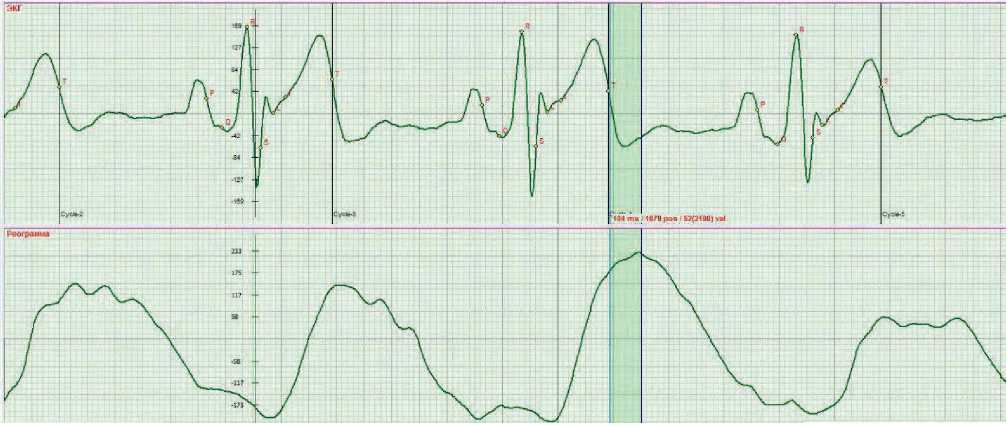
The coronary blood flow is filled with blood at the beginning of the early diastole phase, namely in the segment from the Tk point to the beginning of the Uн wave. From the Tk point, the closure of the aortic valve begins and, at the same time, relaxation of the myocardial muscles begins. This process promotes the absorption of blood into the coronary arteries, because when the muscles are contracted, blood cannot enter the arteries. When the valve closes, the orifices of the coronary arteries open and the filling of the coronary arteries begins. At the beginning of wave U, the process of complete closing of the valve ends. Simultaneously, the muscles of the ventricles relax, which contributes to the filling of the coronary arteries with blood. The rheogram reflects the process of blood filling and it is by it that the quality of the filling of the coronary arteries is assessed - by a uniform decline in the rheogram in the Tk-Uн segment.
Fig. 1 shows the norm of a decline in Rheo.
The method allows only a qualitative assessment of the filling of the coronary arteries, but this is enough to understand the state of the coronary arteries and make an effective decision.
Cause of incomplete filling of the coronary arteries with blood
With a decrease in lung function, an imbalance in the functioning of the systemic and pulmonary circulation will occur. Subjectively, it manifests itself in
Issue 25. December 2022 | Cardiometry | 5
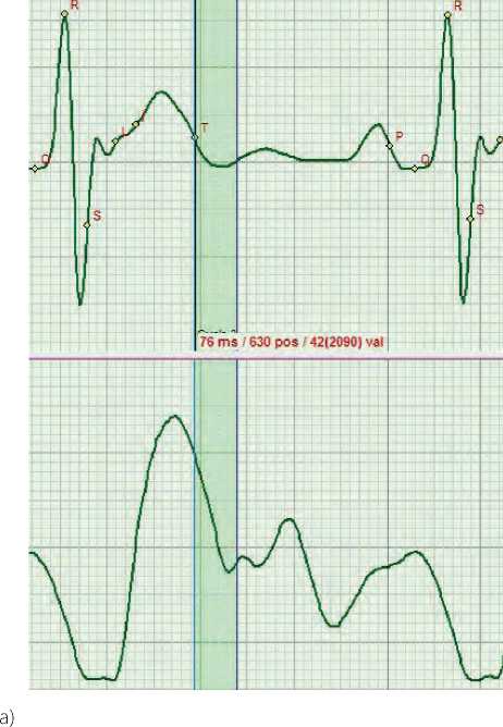
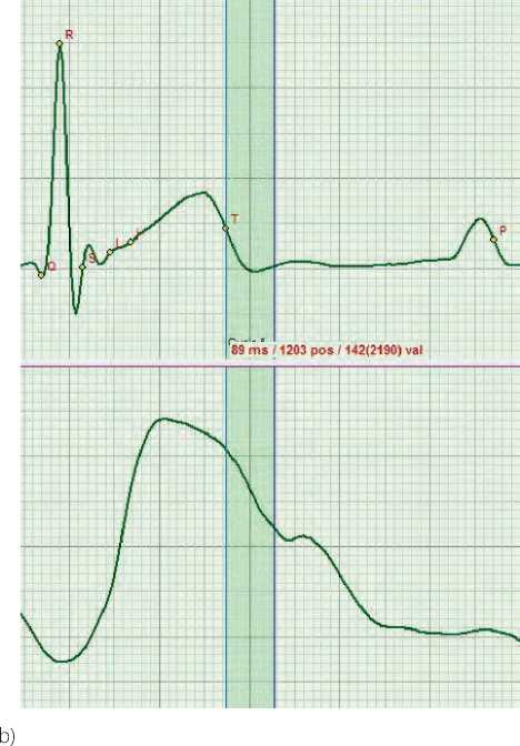
Fig. 1. The norm of a uniform decrease in Rheo in the Tk-Uн segment: a) Close to the ideal shape of Rheo b) The most common shape of Rheo
non-synchronous breathing in relation to heart contractions. As a result, systolic pressure does not reach the norm, which often becomes a habit for a person.
Insufficient systolic pressure is not able to provide normal blood volumes to fill the coronary arteries (Fig. 2). This begins the natural process of narrowing
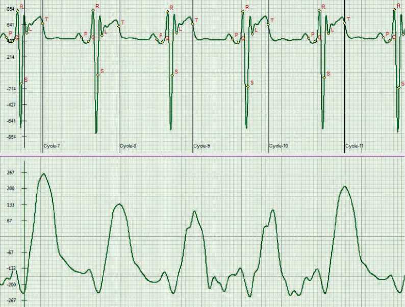
Fig. 2. Rheo shape with full exhalation. The top of the Rheo in the Tk-Uн segment changes shape.
of the coronary arteries, which subsequently leads to their pathological narrowing.
Aortic calcification also leads to insufficient filling of the coronary arteries with blood. In both cases, the result will be narrowing of the coronary arteries (Fig. 3).
Criteria for rechecking the correctness of the diagnostics of the coronary blood flow functioning
Once again, we note the importance of assessing coronary blood flow. To make a decision, it is necessary to be confident in the correct diagnosis and choice of therapy. No instructions can achieve this. Confidence is formed on the basis of the principle of evidence existing in the natural sciences, namely: an observed phenomenon is considered to be true if it is connected by three cause-and-effect relationships with other observed effects. For ECG and Rheo, this is a manifestation of several changes in any of the phases, for example, the amplitude of the ECG, the metabolic parameter and the form of Rheo, and it can also be supported by a change in the compensation mechanism - the influ-
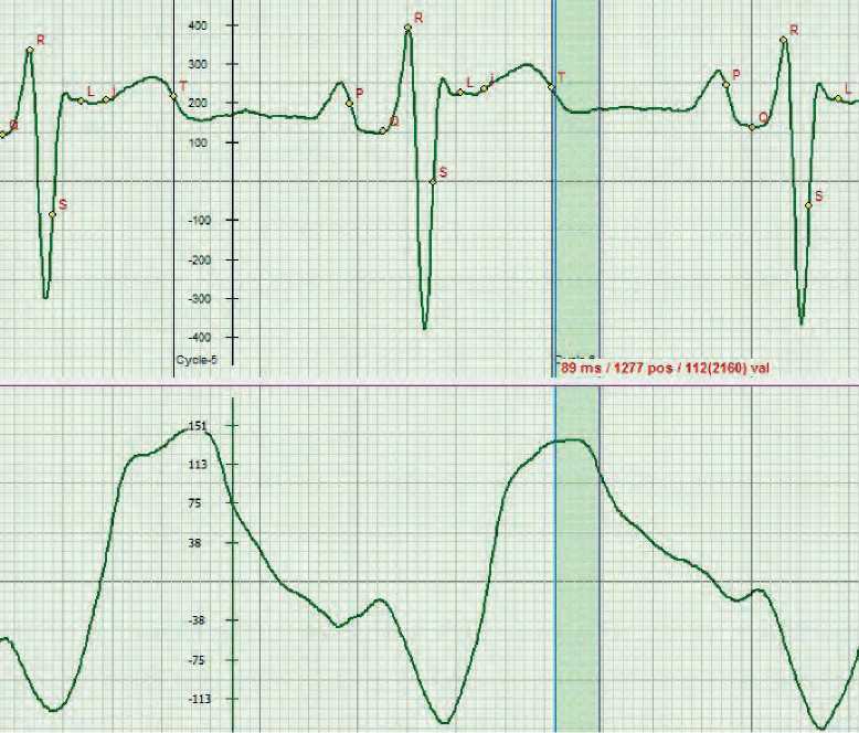
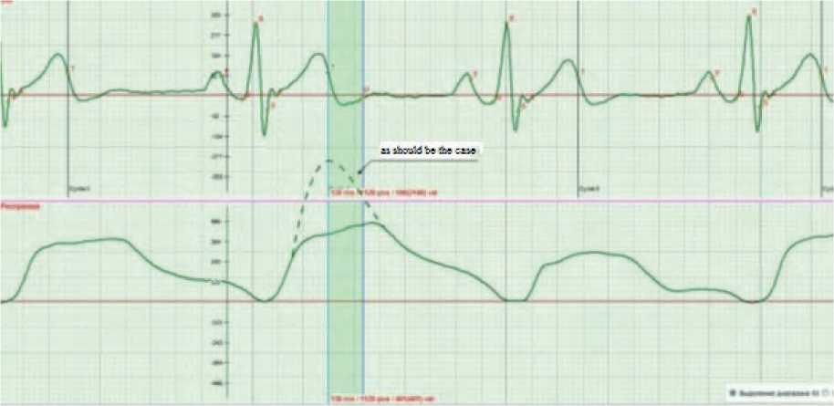
Fig. 3. Shape of Rheo in aortic calcification
ence of one phase on another, or a systemic parameter, the stress index.
Figure 3 shows examples of assessing the quality of blood supply to the coronary arteries. The shape of the Rheogram in the Tk-Uн segment is the main criterion for a qualitative assessment of the state of coronary blood flow. The criterion of the norm is a uniform negative decline in the Rheogram in the Tk-Un segment of the early diastole Tk-Rн phase.
The first additional criterion confirming the presence of a problem in the blood filling of the coronary arteries is the amount of lactate (lactic acid) in the heart muscles.
The second additional criterion is the stress index (SI) . Normally, its value should be at the level of 250 c.u. [4]
The third additional criterion is the type of adaptation reaction . We can talk about an increase in the power of the spectrum at lower frequencies in the spectral characteristic of the adaptation reaction type.
Taking into account the importance and information content of the noted additional criteria, let us consider it in more details.
1. Criteria for the quantitative assessment of lactate in the heart muscles
Causes of changes in the amount of lactate in the heart muscles
Lactate is a metabolic product of anaerobic reactions. Its concentration in the muscles characterizes fatigue. The cardiac muscle, like smooth muscles, also accumulates lactate during rhythmic contractions. With a normal rhythm and a sufficient supply of oxygen, the accumulated lactate is restored to glycogen due to a short rest. If the load was significant or prolonged, then this process requires additional methods to reduce lactate levels. In the case of coronary blood flow problems, the heart muscles will constantly have elevated lactate levels due to the increased workload [3].
Increased lactate in the heart muscles
The decrease in oxygen in the muscles with increasing physical activity increases the amount of lactate. Due to coronary blood flow problems, this level will be significantly higher than normal, as the muscles will receive less oxygen. Figure 4 illustrates such a case. The lactate value is 23.25 c.u. This is well above the normal range of 3-7 c.u., indicating an oxygen de- 8 | Cardiometry | Issue 25. December 2022
ficiency due to coronary blood flow problems and a large accumulation of lactate. In this case, the amount of lactate is modulated by the rhythm of respiration, which also indicates the problem of oxygen deficiency.
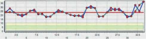
Fig. 4. Increased content of lactate in the heart muscles (c.u.)
Decreased lactate in the heart muscles
A decrease in lactate indicates a decrease in the intensity of muscle contraction: it simply does not accumulate; in this case, attention should be paid to the PV2 parameter, which characterizes the atrial systole. In the event of failure of the myocardium, a compensation mechanism is triggered and the atria take on the entire load. PV2 will increase. In the fig. 5 example, the average value of lactate is 1.88 c.u. This is a very low indicator and can be attributed to a critical level.
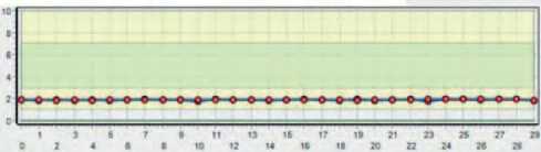
Fig. 5. Decreased lactate in the heart muscles (c.u.)
2. The value of the stress index (SI)
Taking into account the importance of diagnosing coronary arteries, it is necessary to obtain comprehensive information about their condition. It is important that it duplicates the main one, thereby confirming it. This will allow you to be more confident in making a diagnosis.
The second additional criterion is the magnitude of the stress index (SI). Let us consider the information content of SI as an example.
Coronary angiography performed on a patient whose ECG and Rheogram in Figure 6 showed the following:
LAD stenosis (medium 1/3) up to 90%;
DV stenosis (mouth) up to 80%
OB stenosis (medium 1/3) on the verge of occlusion
RCA stenosis (proximal 1/3) on the verge of occlusion.
With simultaneous cardiometric examination, the following data were obtained, presented in Table 1.
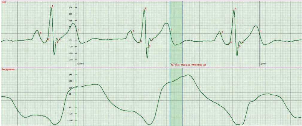
a) in a lying position
b) in a sitting position
Fig. 6. Qualitative assessment of the filling of coronary arteries with blood at the initial stage of the early diastole phase in segment Тк-Uн
Table 1
Cardiometric indicators in case of changes in the blood flow of the coronary arteries
|
Position of the body |
SV-stroke volume, ml. |
MV-minute volume, l/min. |
RV1 (62) – ejection fraction, % |
SI (150 … 300) stress index by R.M. Baevsky, c.u. |
|
lying |
54.8 |
3.3 |
68 |
381 |
|
sitting |
59 |
4.0 |
67 |
5556 |
It can be seen that the parameters of hemodynamics and metabolism are normal. The only parameter that draws attention to itself is the stress index SI. When moving to a vertical position, it reaches 5556 c.u. The reason for such a high index is the low filling of the coronary arteries, which was confirmed by both car-diometry and coronary angiography. The patient underwent coronary bypass surgery.
The use of SI as an additional criterion for assessing the state of coronary blood flow is a reliable indicator. It is used in addition to the criterion described above, thereby enhancing the certainty of the diagnosis.
3. Adaptation reaction type.
The third additional criterion confirming the presence of a problem in the blood filling of the
Issue 25. December 2022 | Cardiometry | 9
coronary arteries is the adaption reaction type. An increase in the power of the spectrum at lower frequencies in the spectral characteristic of the adaption reaction type. Coronary blood flow problems will affect the energy of the heart. An indicator of such an influence is the type of the adaption reaction. In this case, the important point is the amplitude of the spectrum at lower frequencies for the type of reaction characteristic of stress (Fig. 7b). A rise at frequencies of 0.2 Hz indicates general physical fatigue.
Note that this criterion for assessing general physical fatigue serves as an additional one, in order to clarify the diagnosis of coronary blood flow pathology.
It is important to know that this criterion characterizes general physical fatigue even with normal coronary blood flow, so it is used as an additional one to the main one.
Having, along with the main criterion, three more additional confirming criteria, one can be highly confident in the accuracy of the diagnosis. It is important to assess the patient’s subjective symptoms. In this case, it remains to choose the right therapy and monitor its effectiveness.
The elasticity of blood vessels depends to a large extent on the level of CO2 in the blood. Consider an example showing the effectiveness of vascular treatment with hypoxia.
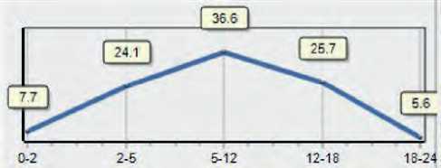
а) Adaptation reaction: calm activation
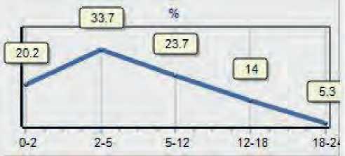
b) Adaptation reaction: stress
Fig. 7. Energy state of the heart tor: the state of the coronary arteries, and, first of all, their elasticity. The basis of treatment and prevention is correct breathing.
-
1. Use a breathing simulator of the Samozdrav type to achieve a balance of O2 and CO2. This will greatly help in restoring the elasticity of blood vessels.
-
2. Use ethylmethylhydroxypyridine succinate (Mexicor) to maintain myocardial energy.
-
3. Apply the drug “Ginkgotropil” to improve microcirculation.
-
4. Take L-carnitine (“Elkar” (solution)) to improve the energy of mitochondria.
-
5. In emergency cases, use phenobarbital + ethyl ester of α-bromisovaleric acid (Valocordin preparation). All preparations based on valerian are reliable and effective.
-
6. Drink Nuxen VI tincture (black walnut) containing the following components: black walnut, hawthorn flowers, gingko biloba, rose hips, lemon balm herb, oregano herb, hop cones, Caucasian rhododendron, purified water, alcohol up to 3% . The effectiveness of the tincture has been tested in practice.
-
7. You can drink cedar balm (cedar needles), which are a source of dehydroquercetin - a substance that stimulates the synthesis and stabilizes the structure of collagen fibers.
-
8. In all cases, acupuncture is indicated in combination with other therapeutic methods and means.
References Diagnostics of coronary blood flow: advantages of using the cardiometric method
- Rudenko MY, Voronova OK, Zernov VA. Theoretical Principles of Heart Cycle Phase Analysis. Munchen, London, New York: Fouque Literaturverlag; 2009.
- Rudenko MY, Zernov VA, Voronova OK. Study of Hemodynamic Parameters Using Phase Analysis of the Cardiac Cycle. Biomedical Engineering. July 2009;43(4): 151-5.
- Rudenko MY, et al. Adrenaline heart. Cardiometry; August 2022; 23:106-107;.
- Baevsky RM, Berseneva AP. Evaluation of adaptation abilities of organism and pathology development risk. Moscow: Medicina; 1998.
