
Investigation of HeLa cell culture lipid peroxidation level induced by core-shell Fe/Fe3O4 nanoparticles
Journal: Siberian Journal of Oncology @siboncoj
Section: Лабораторные и экспериментальные исследования
Article in issue: 4 т.25, 2026.
Free access
Introduction. The application of iron and iron oxide nanoparticles for cancer therapy is a promising strategy due to their high surface area, chemical stability, low toxicity and high biological activity against cancer cells. The mechanism of iron oxide-induced toxicity against cells is the production of reactive oxygen species. the investigation of the molecular mechanisms of the effect of particles on cells and the assessment of the influence of the mass ratio of iron to iron oxide on the lipid peroxidation parameters is of great importance for their application. Goals and Objectives: to synthesize core-shell Fe/Fe3O4 nanoparticles to study the effect of iron oxide and iron mass fraction on Hela cell viability and the content of malondialdehyde, bityrosine, catalase, superoxide dismutase and cathepsin D activity and analyze the oxidative modification of proteins in HeLa cancer cell line. Material and Methods. Core-shell Fe/Fe3O4 nanoparticles with an iron oxide mass ratio from 5 to 90 wt. % were synthesized using the electrical explosion of wires in an oxygen-containing atmosphere. Results. The resulting nanomaterials contained a phase composition of Fe, Fe3O4 and FeO, and their biological impact was evaluated by measuring lipid peroxidation levels through standard biochemical assays. The average particle size was approximately 60−80 nm, and the zeta potential of the particles increased from 0.47 to 6.37 mV with increasing mass fraction of iron oxide. The cytotoxicity of the core-shell Fe/Fe3O4 nanoparticles towards HeLa cancer cells was evaluated by an MTT assay and lipid peroxidation parameter determination. The Fe/Fe3O4 nanoparticles with Fe3O4 mass ratio of 40% was found to have a significant effect on the lipid peroxidation intensity, however a maximal protein oxidative modifications accumulation was observed. The correlations obtained need to be taken into account when obtaining iron and iron oxide nanoparticles for cancer treatment. Conclusion. We identified changes in lipid peroxidation and cathepsin D activity in HeLa cancer cell cultures induced by Fe/Fe3O4 nanoparticles. Based on these results, we concluded that Fe/Fe3O4 nanoparticles can be used as an antitumor additive, however their activity depended on Fe3O4 mass ratio. These findings are essential for producing iron and iron oxide nanoparticles for cancer cell targeting.
Short address: https://sciup.org/140316527
IDS: 140316527 | UDC: 577.115:576.5:544.77 | DOI: 10.21294/1814-4861-2026-25-4-89-99
Исследование параметров процессов перекисного окисления липидов в культуре клеток HeLa, индуцированного наночастицами Fe/Fe3O4 с морфологией «ядро-оболочка»
Введение. Благодаря развитой поверхности, химической стабильности, низкой токсичности и высокой биологической активности в отношении раковых клеток применение наночастиц железа и оксида железа является перспективной стратегией противораковой терапии. Механизм токсического действия на клетки железа и оксида железа заключается в образовании активных форм кислорода. Изучение молекулярных механизмов воздействия частиц на клетки и оценка влияния соотношения железа и оксида железа на параметры процессов перекисного окисления липидов имеют важное значение для их применения. Целью исследования является синтез наночастиц Fe/Fe3O4 типа «ядро-оболочка» для изучения влияния массовой доли оксида железа на жизнеспособность клеток HeLa и содержание малонового диальдегида, битирозина, активность каталазы, супероксиддисмутазы и катепсина D и анализ окислительной модификации белков в линии раковых клеток HeLa. Материал и методы. Наночастицы Fe/Fe3O4 типа «ядро-оболочка» с массовым соотношением оксида железа от 5 до 90 мас. % были синтезированы физическим методом – электрическим взрывом проводников в кислородсодержащей атмосфере. Определение параметров перекисного окисления липидов проводили при помощи стандартных биохимических тестов. Результаты. Получены частицы Fe/Fe3O4, содержащие Fe, Fe/Fe3O4 и FeO. Массовая доля Fe/Fe3O4 варьировалась от 1 до 91 % мас. cредний размер частиц составлял около 80 нм, дзета-потенциал частиц увеличивался с 0,47 до 6,37 мВ с увеличением массовой доли оксида железа. Цитотоксичность наночастиц Fe/Fe3O4 по отношению к раковым клеткам HeLa оценивали при помощи МТТ-теста и определения параметров перекисного окисления липидов. Установлено, что наночастицы Fe/Fe3O4 с массовой долей Fe3O4 40 % оказывали значительное влияние на интенсивность перекисного окисления липидов, однако наблюдалось максимальное накопление окислительно модифицированных белков. Заключение. Выявлены особенности изменения активности перекисного окисления липидов и активности катепсина D в культуре раковых клеток HeLa для наночастиц Fe/Fe3O4. На основании полученных результатов сделан вывод, что наночастицы Fe/Fe3O4 могут быть использованы в качестве противоопухолевой добавки, однако их активность зависит от массовой доли Fe3O4. Полученные корреляции необходимо учитывать при получении наночастиц железа и оксида железа для воздействия на раковые клетки.
Text of the scientific article Investigation of HeLa cell culture lipid peroxidation level induced by core-shell Fe/Fe3O4 nanoparticles
Iron oxide nanoparticles (NPs) have been used in clinical practice for decades [1, 2] as powerful platforms for targeted drug delivery [3], contrast agents for magnetic resonance imaging and cancer treatment [4]. Magnetite (Fe3O4) NPs are core superparamagnetic iron oxide nanomaterials due to their physicochemical properties, such as high surface area, small size, optimum crystallization, magnetism, adsorption capacity and biocompatibility [5]. The Food and Drug Administration (FDA) has approved Fe3O4 NPs for clinical use, including diagnostic imaging contrast and localized magnetic hyperthermia therapy [5]. The development of Fe3O4 nanomaterials with anticancer properties is believed to be an important area of activity in targeted theranostics [6].
Ferropotosis is a type of programmed cell death during iron accumulation associated with activation of lipid peroxidation [7]. Activation of free-radical oxidation in tissues is able to modulate the antitumor effect [8]. ROS generation can cause protein carbonylation, DNA breakage, lipid peroxidation, proteolysis and apoptosis through the activation of proteins of the caspase family [9]. Studies by Y. Wang et al. [10] demonstrated that Fe3O4 nanoparticles could induce cytotoxicity and genotoxicity in MCF-7 cells via oxidative stress. L. Magtanong et al. [11] reported the strong oxidative effect of these materials, as well as properties related to the modulation of tumor microenvironment.
The morphology and physicochemical properties of Fe3O4 NPs are known can influence the anticancer action including an inducing a pro-oxidant effect [12]. Multiple studies [13] show increased toxicity from smaller Fe3O4 particles compared to larger particles. X. Zhou et al. [14] examined the role of different modes of Fe3O4 NP synthesis and their influence on the antitumor properties of these NPs. The anticancer effects of Fe3O4 NPs with various morphologies were evaluated by M.A. Siddiqui et al. [15] against human A549 lung cancer cells. The cell viability in the presence of octahedrons, cubes, rods and wires was slightly lower than in the sample without NPs, but still exceeded 75 %, indicating low anticancer activity of these particles. It was also reported that the surface charge of Fe3O4 NPs could produce genotoxicity and cytotoxicity [16]. Nevertheless, the most of the studies were conducted on normal cells [17].
Recent studies indicate that zero-valent iron nanoparticles exhibit cancer-selective cytotoxicity, but their efficacy in cancer therapy is limited due to the chemical instability of nanoscales [18]. On the one hand, iron is a critical component for many cellular functions, including DNA replication and repair, and it is essential for cell viability. On the other hand, iron accumulation leads to the production of a large quantity of free radicals, which can increase the risk of fatal outcomes of the disease and excess iron may lead to oxidative damage to DNA. H. Liang et al. [19] reported a new strategy to stabilize Fe(0) via a porous yolk shell Fe/Fe3O4 nanostructure for enhanced cancer therapy. These NPs displayed superior inhibition cancer HepG2 cell growth compared to the iron oxide NPs as a control. The results of the study by B.H. Domac et al. [20] indicated that intratumoral administration of Fe/Fe3O4 core/shell nanoparticles could attenuate mouse melanoma after alternating magnetic field exposure. Iron species are known to display a dual effect on cancer cells.
Universal indicators of the lipid peroxidation are malondialdehyde content, defense antioxidant enzyme activity, and the content of oxidized forms of proteins which can regulate many intracellular processes [21]. The development of oxidative stress with the subsequent accumulation of its products is believed to contribute to the initiation of programmed cell death, activation of lysosomal enzymes, in particular cathepsin D [22]. Therefore, the aim of the present study was to synthesize core-shell Fe/Fe3O4 NPs to investigate the effect of iron oxide/iron mass ratio on the HeLa cell viability and content of malonic dialdehyde, protein oxidative modifications, bityrosine, as well as catalase, superoxide dismutase and cathepsin D activities in cancer cell line.
Goals and objectives: to synthesize core-shell Fe/ Fe3O4 nanoparticles to study the effect of iron oxide and iron mass fraction on HeLa cell viability and the content of malondialdehyde, bityrosine, catalase, superoxide dismutase and cathepsin D activity and analyze the oxidative modification of proteins in HeLa cancer cell line.
Material and Methods
Preparation of Fe/Fe3O4 nanoparticles
To prepare NPs, the electric explosion of iron wire (EEW) of 0.3 mm in diameter, 65 mm long in argon or oxygen-containing atmosphere (argon + oxygen) was used. The EEW parameters were as follow: the capacitor capacity was 1.2 μF; the voltage was 16-29 kV.
The capacitive energy storage device provided the fast electrical explosion mode that described in detail by Bakina at al. [23]. Immediately after synthesis, the particles were passivated by slow air passing. The nanoparticle EEW synthesis parameters are summarized in Table 1.
Table 1/Таблица 1
Parameters of Fe/Fe3O4 nanoparticles production
Параметры синтеза наночастиц Fe/Fe3O4
|
Sample/Образец |
Gas media/Газовая среда |
Volumetric oxygen content in the gas media, wt%/ Содержание кислорода в газовой смеси, % об. |
U0 ,kV/ U0 кВ/ |
|
Fe |
Ar |
0.00 |
1 , 6 |
|
Fe/Fe3O4(5) |
Ar + O2 |
1.00 |
16 |
|
Fe/Fe3O4(20) |
Ar + O2 |
1.30 |
19 |
|
Fe/Fe3O4(40) |
Ar + O2 |
1.75 |
22 |
|
Fe/Fe3O4(60) |
Ar + O2 |
3.40 |
26 |
|
Fe/Fe3O4(90) |
Ar + O2 |
5.00 |
29 |
Note: created by the authors.
Примечание: таблица составлена авторами.
Nanoparticles characterization
The morphology of the NPs was investigated by transmission electron microscopy using a JEM 2100 microscope (JEOL, Japan). The NP phase composition was investigated by X-ray phase analysis using XRD-6000 diffractometer (Shimadzu, Japan) using CuKα radiation with a step width of 0.02 θ and exposure of 1 s. The surface area of the nanoparticles was measured by nitrogen adsorption/desorption using a Sorbtometer M (Katakon, Russia) automatic analyzer in the relative pressure range of 0.05-1.00. The size of nanoparticle agglomerates was determined using a CPS DS24000 disc centrifuge (CPS Instruments, USA). Zeta potential measurements were performed in deionized water at 25 °С and pH 7 value using Zetasizer Nano ZS instrument (Malvern, GB).
Cell culture
The study was carried out on cell culture HeLa provided the Cancer Research Institute (Russia). Cells were grown and cultured as a monolayer in Eagle MEM nutrient medium (Biolot, Russia) with addition of 10% fetal calf serum (Biolot, Russia) and 1% penicillin/streptomycin solution (Biolot, Russia). The cells were cultured in a 25 mL culture plate at 37 °C in a CO2 atmosphere.
MTT assay
The MTT test is a high-throughput screening test for cell viability. Briefly it can be described as follows. The HeLa cells were grown in 96-well microplate at 37±1 ºC in CO2 incubator (Sanyo, Japan). The initial cell concentration was 5 × 10³ cells per well. After 24 h incubation spent media was removed and fresh media with NP extracts (1–20% wt.) was added. The cells were incubated with NP extracts for 24 h at 37±1 ºC in CO2. Then MTT test was performed using (3-(4.5-dimethyl thiazol-2-yl)-2.5-diphenyl-tetrazolium bromide solution and dimethyl sulfoxide (Biolot, Russia). The optical density was determined using the Multiscan FC microplate spectrophotometer (Termo Scientific, Germany) at a wavelength of 570 nm. The cell viability was normalized to control sample (cell media only).
Cell homogenates preparation
To prepare HeLa cell homogenates we selected 5 × 10⁴ cells and replaced them to a culture plate containing nutrient medium and NPs water suspension (100 μg/mL). The cells were cultured for 24 hours and then treated with ultrasound using a conventional 100 W ultrasonic bath for 10 min to obtain a homogenate. The homogenate was centrifuged for 60 minutes at 10000 g and 37 °C. The supernatant was collected and used in the following experiments.
The level of protein assay
The level of protein in homogenate sample was determined by the Lowry method. This method was based on the interaction of protein nitrogen with copper ions in an alkaline medium and the reduction of the product with phosphomolybdic phosphotungstic acid. In typical procedure, 0.8 ml of distilled water and 4.5 ml of a working reagent containing 48 ml of a Na2CO3 (2 % wt.) solution in NaOH (0,1 M), 1 ml of an aqueous solution of sodium tartrate tetrahydrate (1 % wt.), and 1 ml of an aqueous solution of CuSO4 (0.5 % wt.) were added to 0.2 ml of homogenate. The sample was incubated at a room temperature for 30 min. The optical density of the resulting mixture was measured spectrophotometrically at a wavelength of 660 nm.
Malondialdehyde level assay
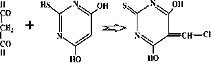
The assay method was based on spectrophotometric determination of the optical density of a colored complex formed by the reaction of malondialdehyde (MDA) and thiobarbituric acid (TBA) according to the reaction:
но
ТВА MDA Colored complex
To carry out this reaction, 1 ml of trichloroacetic acid (20 % by weight) and 1.0 ml of TBA (0.8% by weight) were added to 0.1 ml of the homogenate sample prepared above. The mixture was heated to 100 °C and held for 10 min. After centrifugation, the optical density of the supernatant was determined using a Specol 1500 UV-Vis spectrophotometer (Analytik Jena, Germany) at a wavelength of 540 nm and a cuvette optical path length of 10 mm.
Spontaneous and metal-dependent modification of protein assay
To determine spontaneous oxidative modification of proteins (sPOM) in vitro , 0.05 ml of homogenate and 0.95 ml of 0.05 M phosphate buffer (pH 7.4) were added to the blank and test samples. The obtained samples were incubated at 37 °C for 15 min. After this, 1.0 ml of a 2,4-dinitrophenylhydrazine solution (0.01 M) in hydrochloric acid (2 M) and 1.0 ml of a trichloroacetic acid solution (20 %wt.) were added to the samples. The samples were incubated at a room temperature for 1 h. After incubation time, the samples were centrifuged at 3000 rpm for 15 min and the supernatant was removed. For extraction of lipids and removal of unreacted dinitrophenylhydrazine, the precipitate was washed twice with 3 ml of a mixture of ethanol and ethyl acetate (1:1). Between washes, the sample was centrifuged again. The precipitate was then air-dried to constant weight. 3 ml of urea (8 M) and 20 µl of hydrochloric acid (2 M) were added.
To determine metal-dependent oxidative modification of proteins (iPOM) in vitro , Fenton’s medium containing ferrous sulfate (4 mM) and EDTA (1 mM) was used. All procedures described above for sPOM) were then carried out.
Catalase activity assay
To determine catalase activity, 0.1 ml of homogenate and 2.0 ml of hydrogen peroxide water solution
(0.03 %wt.) were added and mixed thoroughly. The control sample contained only 2.0 mL of distilled water. Control and test samples were incubated at 37 ºC for 10 min. After incubation time, 10 mL of ammonium molybdate solution (4% wt.) was added and mixture was centrifuged at 4000 rpm for 10 min. The optical density of supernatant was measured using spectrophotometer at a wavelength of 410 nm.
Activity of superoxide dismutase assay
The method was based on reaction of autoxidation of adrenaline at pH 10.65. To determine superoxide dismutase (SOD) activity, 10 μL of homogenate to 100 μL of adrenaline solution (0.1 %) was added and replaced to the spectrophotometric cuvette (10 mm) with 2 mL of the carbonate buffer and mixed. The optical density of solution was measured at λ=347 nm every 30 sec for 5 min. No adrenaline was added to the blank sample.
Bityrosine level assay
Bityrosine is a marker of oxidative damage to proteins. For bityrosine determination, 50 μL of homogenate was mixed with 0.95 ml phosphate buffer (pH 7.4) and incubated for 2 h. After incubation, the test sample was diluted 5-fold with distilled water. The fluorescence was measured using a spectrofluorimeter Agilent (USA) at a wavelength of 415 nm, excitation wavelength was 325 nm.
Free and total cathepsin D activity assay
Free cathepsin D activity was determined as follows. First, we prepared a hemoglobin solution (1 % by weight) in acetate buffer (0.1 M, pH 5). 0.15 ml of the hemoglobin solution was added to 0.3 ml of the homogenate. The obtained solution was mixed and incubated at 37°C for 30 min with constant stirring. After incubation time, 0.6 ml of 10 % trichloroacetic acid solution and 2.1 ml of distilled water were added to the test sample. The sample was then centrifuged at 4000 rpm for 10 min, and the optical density of the supernatant was measured at 280 nm in 10 mm quartz cuvettes. Distilled water was used as a control. To determine total cathepsin D activity, 0.1 ml of 1 % Triton X-100 solution was added to 0.3 ml of the homogenate. The mixture was incubated at 37°C for 10 min. After incubation time, 0.15 ml of hemoglobin solution was added to the sample and the mixture was incubated again for 30 min at 37 °C. After incubation, 0.6 ml of 10% TCA solution and 2 ml of distilled water (with Triton X-100) were added to the mixture. The sample was also centrifuged and the optical density was determined at a wavelength of 280 nm.
Statistical processing
All tests were performed in triplicate, and biochemical tests were performed in ten replicates. STA-TISTICA 10.0 was used for statistical processing of the results. All biochemical test data were analyzed for normality using the Kolmogorov–Smirnov criterion. The Mann–Whitney U-test was also used to assess significant differences between two independent groups. Nonparametric analysis of variance using the
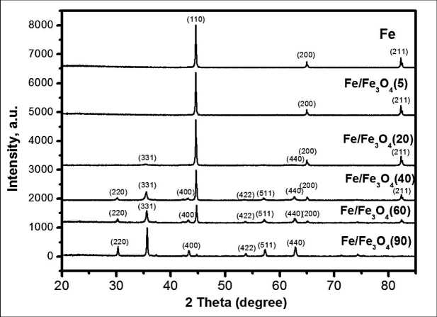
Fig. 1. X-ray diffraction pattern of Fe/Fe3O4nanoparticles. Note: created by the authors
Рис. 1. Дифрактограммы наночастиц Fe/Fe3O4 Примечание: рисунок выполнен авторами
Kruskal–Wallis test was used to compare two independent groups. Experimental data for determining lipid oxidation markers are presented as Me (Q1; Q3).
Results
Fe/Fe3O4 core-shell NPs of complex shape, predominantly spherical and faceted in shape, with different Fe3O4 mass ratio were formed during the EEW of iron wire in oxygen-containing atmosphere. Immediately after synthesis, the particles were passivated by a slow injection of air. XRD pattern of Fe/Fe3O4 NPs showed Fe, Fe3O4 peaks and FeO peaks with weak intensity (Fig. 1). According to quantitative X-ray phase analysis dates the Fe3O4 mass ratio depended on the oxygen content in a gas mixture (Table 2).
Typical TEM images of the synthesized NPs are presented in Fig. 2a. Fe and Fe/Fe3O4(5) NPs have a spherical or almost spherical shape. As the oxygen content in an explosive media increases, the number of nanoparticles with non-spherical shape increases significantly. Hexagonal and square-shaped nanoparticles with core-shell structure are observed in the samples Fe/Fe3O4(20) and Fe/Fe3O4(40). At oxygen content of 3.4 and 5 vol. % the Fe/Fe3O4(60) and Fe/Fe3O4(90) NPs are mostly spherical in shape, the number of nanoparticles with deviation from spherical shape is too small. The nanoparticles formed at EEW demonstrate a separation of components (Fig. 2b) with core enriched in iron and shell enriched in iron oxide observed according to EDX elemental mapping data.
All NPs have the average NP size less than 100 nm. The NP size was depended on the iron oxide mass ratio (Fig. 3a). The Fe/Fe3O4 NPs had a normal-logarithmic particle size distribution and an average NP size of less than 100 nm (Fig. 3b). The average NP size depended on the Fe3O4 mass ratio.
The prepared NPs had similar values of specific surface area (SSA) from 8 to 11 m2/g (Table 2). All Fe/Fe3O4 samples are characterized by positive zeta
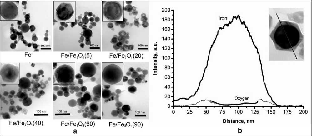
Fig. 2. TEM images (a) and EDS line-scan analysis (b) of Fe/Fe3O4 NPs. Note: created by the authors Рис. 2. ПЭМ изображения (а) и ЭДС анализ (b) наночастиц Fe/Fe3O4. Примечание: рисунок выполнен авторами
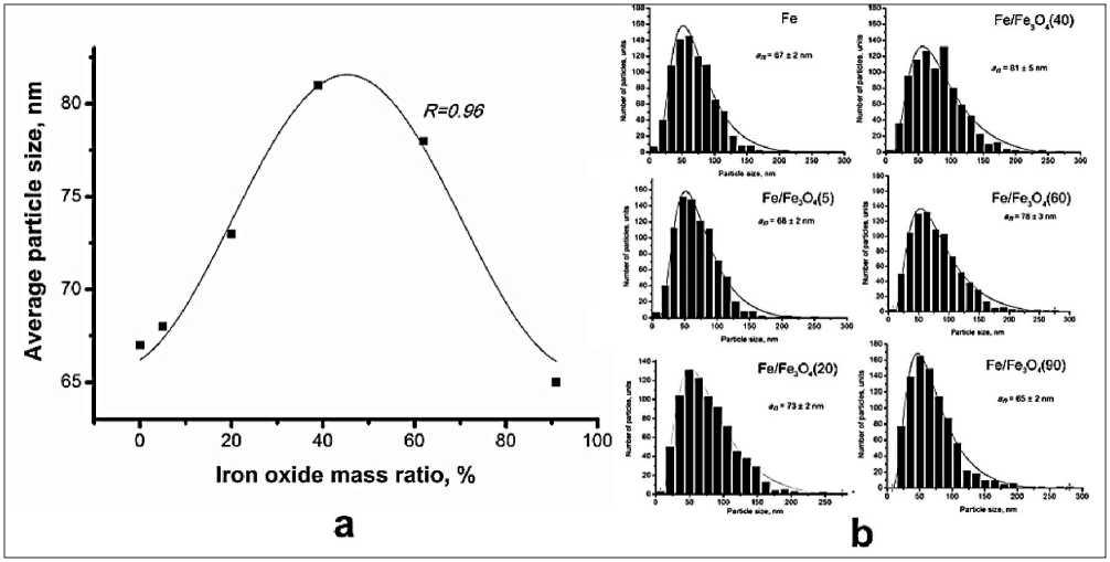
Fig. 3. The dependence of average particle size on Fe3O4 mass ratio (a) and NP size distribution (b). Note: created by the authors Рис. 3. Зависимость размера частиц от массовой доли Fe3O4 (а) и распределение частиц по размерам (b).
Примечание: рисунок выполнен авторами
Table 2/Òàблицà 2
Properties of nanoparticles
Физиêî-õимичåñêиå ñвîéñтвà нàнîчàñтиц
|
NPs/Наночастицы |
Mass ratio/Массовая доля |
SSA, m2/g/ Удельная поверхность, м2/г |
Zeta potential, mV/ Дзета потенциал, мВ/ |
||
|
a Fe |
Fe 3 O 4 |
FeO |
|||
|
Fe |
99.0 % |
1.0 % |
- |
8 ± 0.2 |
-3.36 ± 0.2 |
|
Fe/Fe3O4(5) |
95.1 % |
4.9 % |
- |
11 ± 0.2 |
0.37 ± 0.0 |
|
Fe/Fe3O4(20) |
81.0 % |
17.0 % |
2.0 % |
11 ± 0.2 |
2.31 ± 0.2 |
|
Fe/Fe3O4(40) |
60.0 % |
35.0 % |
5.0 % |
10 ± 0.2 |
3.52 ± 0.3 |
|
Fe/Fe3O4(60) |
37.0 % |
61.0 % |
2.0 % |
11 ± 0.2 |
5.62 ± 0.4 |
|
Fe/Fe3O4(90) |
7.0 % |
91.0 % |
2.0 % |
11 ± 0.2 |
5.51 ± 0.3 |
Note: created by the authors.
Примечание: таблица составлена авторами.
potential in 0,9% NaCl solution at pH 7.2 and temperature 25 °C. The zeta potential values vary among different NPs, as indicated by the results presented in Tab. 1. The presence of Fe3O4 on the NP surface leads to an increase in zeta potential from -3.4 to 5.6 mV. As reported in the literature, the zeta potential for neat Fe3O4 is about 20 mV [24-26]. Iron NPs have a negative zeta potential [27]. The zeta potential value of Fe/ Fe3O4 NPs becomes more positive with increasing iron oxide phase due to the increasing amount of it on the NP surface. A positive zeta potential value improves the interaction of nanoparticles with the surface of the bacterial cell, which is negatively charged.
The MTT test was used to determine toxicity of Fe/ Fe3O4 NPs against HeLa cells. The phosphate buffer saline (pH 7.2) was used as a control sample. The NPs showed dose-dependent cytotoxicity in HeLa cells (Fig. 4 ) . At concentrations from 10 to 100 μl, Fe and Fe/Fe3O4(5) NPs had almost no effect on cell viability. For Fe and Fe/Fe3O4(5) NP-treated cells, the cell viability was about 90 % at 1 wt.%, and 50-60 % at 20 wt.%. However, there was decrease in cell viability of HeLa at all concentration of Fe/Fe3O4 NPs with Fe3O4 at 20-90 wt. % (less than 70% vs. control). The Fe/Fe3O4(40) NP-treated cells showed a significant (p<0.05) increase in cytotoxicity compared to the control group. At 10 μg/ml of Fe/Fe3O4(40) NPs, 14.18 % decrease in cell viability of HeLa was observed. These results provide substantial evidence of the cytotoxic nature of the synthesized NPs against HeLa cells, which can be further explored for anti-cancerous activity.
In order to assess the effect of Fe/Fe3O4 on the oxidative signaling, the current study has evaluated the levels of cardinal components of the anti-oxidant
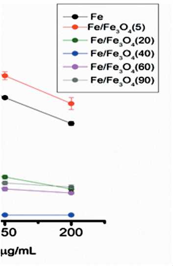
Fig. 4. Analysis of HeLa cell viability with NPs by MTT test. Note: created by the authors
Рис. 4. Жизнеспособность клеточной линии HeLa, определенная при помощи МТТ-теста.
Примечание: рисунок выполнен авторами system: MDA concentration, catalase activity, content of spontaneous (sPOM) and induced (iPOM) protein oxidative modifications, content of bityrosine and SOD activity (Fig. 5).
After exposure to Fe/Fe3O4 for 24 hours, the obtained catalase activity was found. As the observed result showed, an increase of IO mass ratio for 40 % caused a statistically significant (p<0.05) increase in catalase activity (Fig. 5). A 1.4-6.0-fold decrease in MDA content was found in HeLa supernatants incubated with Fe/Fe3O4 NPs with 0-60 wt. % Fe3O4 mass
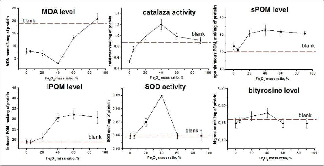
Fig. 5. Fe3O4 mass ratio effect of Fe/Fe3O4 on the oxidative parameters. Data are presented as the means ± SD.
Note: created by the authors
Рис. 5. Влияние содержания Fe3O4 в наночастицах Fe/Fe3O4 на параметры перекисного окисления. Данные представлены в виде: значение ± стандартное отклонение. Примечание: рисунок выполнен авторами
Table 3/Òàблицà 3
Indices of lipid peroxidation in HeLa culture under the effect of NPs compared to the blank samples Пîêàзàтåли пåðåêиñнîгî îêиñлåния липидîв в êóльтóðå HeLa пîд дåéñтвиåм нàнîчàñтиц
|
Parameter/Параметр |
Blank/ Контроль |
Fe NPs/ Наночастицы Fe/ |
Fe/Fe3O4(40) |
Fe/Fe3O4(90) |
|
Free cathepsin D nmol/mg of protein/ |
41.83 |
24.83 |
43.31 |
34.39 |
|
Свободный катепсин D |
(36.96; 47.67) |
(23.14; 32.67)* |
(26.21; 100.68) |
(31.03; 37.45) |
|
Total cathepsin D nmol/mg of protein/ |
152.86 |
106.60 |
264.26 |
160.24 |
|
Общий катепсин D |
(113.65; 169.48) |
(102.20; 130.26)* |
(185.34; 447.46)* |
(158.20; 183.63)* |
Kruskal–Wallis test:
H (1, N=20)=3.89
p=0,0485
Notes: * – significance of differences compared to blank sample (p<0.05); created by the authors.
Примечания: * – различия статистически значимы по сравнению с контрольной группой (p<0,05); таблица составлена авторами.
ratio compared with the blank samples. A decrease in catalase activity was detected in cell culture incubated with Fe and Fe/Fe3O4(5) NPs compared to blank sample. However, when incubated with other NPs, increase in the activity of the studied enzyme was observed.
It is known that protein oxidative modifications (sPOM and iPOM) can be caused by chemical interaction between amino acid residues of protein and reactive oxygen or nitrogen species [28]. The cell cultures incubated with NPs showed an increase in the content of sPOM and iPOM (iPOM) compared to the blank samples.
Low SOD activity was detected for the cell culture incubated with Fe/Fe3O4 NPs. At the same time, an increase in SOD activity was observed only for HeLa cell culture incubated with Fe/Fe3O4(20) and Fe/Fe3O4(40) NPs. The change of intracellular SOD activity can result from the increase of molecular activity induced by directly interacting with iron oxide NPs and ROS-inhibition of activity [29].
Bityrosine is a by-product resultant from the exposition of proteins to •OH radicals, often present and a marker or intra- or intermolecular crosslinking? No changes in the bityrosine content in the studied cell cultures were detected under the influence of nanoparticles. Significant intergroup differences have been obtained for the total cathepsin D activities. Incubating cells or samples with Fe NPs causes a 1.4-fold decrease in total cathepsin D activity compared to the blank control. Conversely, incubating with Fe/Fe3O4(40), composite sample causes a 1.7-fold increase in cathepsin D activity relative to the blank (Table 3).
Discussion
Previous in vitro studies have reported that Fe3O4 and Fe NPs induce cellular cytotoxicity through the generation of free radicals in HeLa cell lines [30]. As-synthesized Fe/Fe3O4NPs due to their positive charge are strongly attracted to the cell membrane, where they can generate reactive oxygen species (ROS) and lead to outer membrane lipid peroxidation. The results of this study provide data on the differences in cancer response to incubation with NPs of different Fe3O4 mass ratio. A pronounced antioxidant effect for Fe/ Fe3O4(40) NPs was observed. In this case, there was a decrease not only in MDA level, but also an increase in the antioxidant enzymes levels such as catalase and SOD. One of the causes of lipid peroxidation is an imbalance in the level of antioxidant enzymes. The revealed changes were accompanied by an increase in cathepsin D activity, indicating the involving of lysosomes in the formation of the molecular pattern of programmed cell death. At the same time, for Fe/ Fe3O4(90) NP sample showed an effect on the POM content, both spontaneous and induced, without an increase in the activity of antioxidant enzymes and cathepsin D. For sample Fe and Fe/Fe3O4(5) NPs, the minimum amount of spontaneous and induced POM was detected. The effect of iron and iron species on the activity of lipid peroxidation processes and the development of programmed cell death is well known. Oxidative stress is one of the main pathogenetic mechanisms in the initiation of oncogenesis. Decrease in MDA content is observed for all studied samples. An important step in the activation of ferroptosis, programmed cell death influenced by the accumulation of iron and iron species is known to be the alteration of POL activity, the formation of free radicals, which are able to trigger cell death mechanisms [12]. It is important to recognize that the powerful pro-oxidant effect of iron nanoparticles can be associated with high antioxidant activity, activation of ferroptosis [7].
Conclusion
The changes in lipid peroxidation and cathepsin D activities were revealed for iron oxide nanoparticles incubated in the culture of HeLa cancer cells. Based on our results, we conclude that Fe/Fe3O4nanoparticles can be used as an antitumor supplement. However, pre-clinical and clinical studies of NPs are needed from for their validation. The present data also suggest that it is important to control the iron oxide content to avoid any adverse effects of NPs and regulate their anticancer activity.