Магнитно-резонансная томография как инструмент дифференциальной диагностики при поражении ствола головного мозга у детей

Автор: Марченко Наталья Викторовна, Войтенков Владислав Борисович, Скрипченко Наталья Викторовна, Бедова Мария Алексеевна, Курзанцева Ольга Олеговна
Журнал: Клиническая практика @clinpractice
Рубрика: Обзоры
Статья в выпуске: 1 т.11, 2020 года.
Бесплатный доступ
В данном обзоре литературных данных мы рассматриваем магнитно-резонансную томографию как инструмент дифференциальной диагностики поражения ствола головного мозга у детей. Показано, что поражение ствола мозга, как изолированное, так и связанное с супратенториальными изменениями, возникает при широком спектре патологических состояний - при острых нарушениях мозгового кровообращения, дисметаболических нарушениях, воспалительных процессах как инфекционной, так и неинфекционной (аутоиммунной, паранеопластической) этиологии, нейродегенеративных заболеваниях, опухолях. Небольшой размер ствола, а также менее четкое различие между серым и белым веществом и определенное ограничение мультипараметрической магнитно-резонансной томографии в случае исследования стволовых структур часто приводят к затруднениям в проведении дифференциальной диагностики. Иногда большое диагностическое значение приобретают топография очага и интенсивность сигнала от него. Для целей дифференциальной диагностики основополагающее значение, тем не менее, играют клинические, эпидемиологические и лабораторные исследования.
Ствол мозга, дети, магнитно-резонансная томография
Короткий адрес: https://sciup.org/143170836
IDR: 143170836 | DOI: 10.17816/clinpract16531
Magnetic resonance imaging as a tool of differential diagnostic in the brainstem damage in children
In this review, magnetic resonance imaging (MRI) is presented as a tool for the differential diagnostics of the brainstem lesions in children. It has been shown that both the isolated brainstem lesions or those related to the supratentorial changes occur within a wide range of pathological conditions - in acute stroke, dismetabolic disorders, inflammatory processes of both the infectious and non-infectious (autoimmune, paraneoplastic) etiology, neurodegenerative diseases, tumors. A small size of the brainstem, as well as a less clear distinction between the gray and white matter and certain limitations of visualization of the infratentorial structures with multiparametric MRI often lead to difficulties in the differential diagnosis. Sometimes the most important in the diagnostics is the topography of lesions and their signal intensity. Nevertheless, the clinical data, the results of epidemiological and laboratory studies are of primary importance for the differential diagnosis of brainstem lesions.
Текст обзорной статьи Магнитно-резонансная томография как инструмент дифференциальной диагностики при поражении ствола головного мозга у детей
Ствол мозга — отдел центральной нервной системы, состоящий из продолговатого мозга, моста, среднего мозга, промежуточного мозга и мозжечка. В стволе мозга сосредоточены ядра и проводящие пути, обеспечивающие жизнедеятельность организма, в частности дыхательный и сосудодвигательный центры, а также ретикулярная формация [1]. Серое и белое вещество ствола мозга обладает комплексной структурой, анатомически тесно переплетенной, что делает его топографию сложной для интерпретации при нейровизуализации.
Высокотесловая магнитно-резонансная томография (МРТ), более 1,5 Тл, дает детализированную визуализацию морфологии, интенсивности сигнала и метаболической составляющей стволовых ядер, равно как и визуализацию серого и белого вещества. Стандартный МРТ-протокол у детей должен быть адаптирован к возрасту пациента: следует визуализировать весь головной мозг вплоть до затылочного отверстия. Т2-взвешенные изображения лучше выявляют гиперинтенсивные очаги в задней мозговой ямке, чем изображения в Flair-режиме [2]. Flair-последовательность у детей младше 12 мес жизни неэффективна [3]. Стандартной характеристикой стволовых очагов является гиперинтенсивность сигнала на турбо-спин-эхо Т2-взвешенных изображениях, очаги могут быть фокальными или диффузными, изолированными или множественными [3, 4].
Базируясь исключительно на интенсивности сигнала, особенно у детей, невозможно дифференцировать острое и хроническое очаговое поражение. Более того, в случаях хронического метаболического заболевания или воспалительного поражения центральной нервной системы новые острые очаги могут возникать на фоне хронического процесса, что дополнительно затрудняет интерпретацию нейровизуализационных находок [5]. С точки зрения морфологии, наличие отека и Т1-га-долиниевого усиления предполагает наличие воспалительного и/или инфекционного или неопластического процесса, в то время как масс-эффект является более типичным для последнего [6].
В оценке стволового поражения большую помощь оказывает знание особенностей васкуляризации ствола, распределения серого и белого вещества и селективной уязвимости мозговой ткани. С точки зрения васкуляризации важно отметить, что из вертебробазилярного бассейна происходят задняя спинальная, задняя нижняя мозжечковая и передняя спинальная артерии. Ишемические и септические эмболы обычно проникают по артериальному кровотоку настолько далеко, насколько это допускает их размер, что часто обусловливает поражение небольших по калибру артерий ствола мозга [5]. Пограничными зонами артериальных терминалей являются покрышка мозга, продолговатый мозг и мост; у новорожденных эти части ствола считаются самыми восприимчивыми к гипоксиче-ски-ишемическому поражению [6, 7]. Венозная система ствола отличается сложностью и большим количеством сосудов, но хорошо визуализируется с применением протокола SWI (Susceptibility Weighted Imaging — изображения, взвешенные по магнитной восприимчивости) [5].
Серое вещество ствола мозга тесно переплетено с проводящими путями (белым веществом). Тем не менее б о льшая часть структур серого вещества глубоко расположена, в то время как белое вещество распределено по периферии ствола. Это связано с тем, что волокна, составляющие пирамидные пути и ножки мозга, расположены в мосту вентролатерально. Вследствие этого поражение белого вещества мозга ожидается преимущественно в периферических зонах ствола без краниокаудальной специфичности [7]. С точки зрения селективной уязвимости мозговой ткани важным является то, что ядра серого вещества более восприимчивы к метаболическим повреждениям. Это связано с тем, что их метаболические потребности выше, чем у структур белого вещества. Поскольку ядра серого вещества, в особенности ретикулярной формации, расположены от среднего до продолговатого мозга, метаболическое поражение центральной нервной системы также не демонстрирует краниокаудальной специфичности [8].
Цель обзора — раскрыть современные представления об МРТ головного мозга как инструменте дифференциальной диагностики при поражении ствола мозга. С этой целью были проанализированы научные статьи и монографии, посвященные вопросам нейровизуализации при поражении ствола мозга различной этиологии. Поиск осуществлялся с использованием баз данных PubMed, Medline, eLibrary. ru. Для поиска использовались ключевые слова MRI+encephalitis, MRI+brainstem, brainstem+MRI.
ПОРАЖЕНИЕ СТВОЛА МОЗГА
ПРИ ОСТРЫХ НАРУШЕНИЯХ МОЗГОВОГО КРОВООБРАЩЕНИЯ У ДЕТЕЙ
Острые нарушения мозгового кровообращения у детей в 5–6% случаев поражают ствол мозга [8, клиническая; 20
Том 121 № 0 1
-
9] . Эмболы попадают в артериальные терминали и приводят к появлению Т2-гиперинтенсивных очаговых поражений (рис. 1). Крупный очаг (более 50% диаметра ствола) является прогностически неблагоприятным признаком [9].
Вследствие небольших размеров ствола мозга у детей часто регистрируются ложные негативные результаты [10]. Ишемические очаги обычно невелики по размеру и связаны с гиперинтенсивностью на Т2- и Flair-режимах. Ствол мозга обычно рассматривается как резистентный к ишемии отдел центральной нервной системы вследствие высокой плотности васкуляризации [9]. Однако в неонатальном периоде развитие васкуляризации может быть неполным, и дети с гипоксически-ишемической энцефалопатией в перинатальном периоде повышенно восприимчивы к повреждению покрышки мозга [7]. Повреждение покрышки, как правило, конусообразное, билатеральное и обычно симметричное с частым вовлечением каудальной части моста и продолговатого мозга; оно проявляется оральной моторной дисфункцией [7].
Сосудистые мальформации также встречаются в стволе мозга у детей и включают в себя врожденные аномалии вен, кавернозные гемангиомы и капиллярные телеангиоэктазии, а также артериовенозные мальформации. МРТ-паттерн каждого из этих сосудистых образований типичный, и обычно диагностика не вызывает затруднений. Интенсивность МР-сигнала и морфология идентичны наблюдающимся у взрослых пациентов [7].
ДИСМЕТАБОЛИЧЕСКИЕ НАРУШЕНИЯУ ДЕТЕЙ С ПОРАЖЕНИЕМ СТВОЛА МОЗГА
Среди синдромов, связанных с генетическими нарушениями метаболизма, чаще всего поражают ствол митохондриальные энцефалопатии. Митохондриальные заболевания включают широкий спектр клинических синдромов, связанных с дефицитом окислительного фосфорилирования, вызываемым частичным или полным дефицитом одного или более ферментов дыхательной цепи. Наследование может быть аутосомно-рецессивным, аутосомно-доминантным, Х-сцепленным [11, 12]. Дефект митохондриальной дыхательной цепи может быть
Рис. 1. Пациент А., 7 лет. МРТ: поражение ствола мозга в результате острого нарушения мозгового кровообращения по ишемическому типу в бассейне основной артерии, 2-е сут заболевания
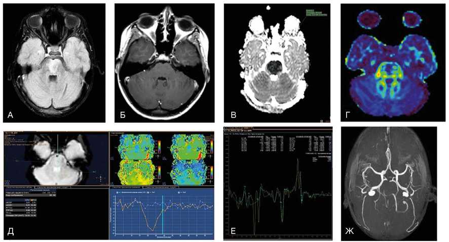
Примечание. При магнитно-резонансной томографии (МРТ) в варолиевом мосту визуализируется зона патологического гиперинтенсивного МР-сигнала на Flair (А) без признаков нарушения гематоэнцефалического барьера при внутривенном контрастировании в зоне поражения (Б). На диффузионных последовательностях (В) визуализируется цитотоксический отек с признаками ограничения диффузии и значениями измеряемого коэффициента диффузии 0,4x10 -3 мм2/с. При выполнении DTI (Г) данная зона демонстрирует снижение значений фракционной анизотропии до 0,25. При контрастной Т2*-перфузии отмечается отсутствие кровотока в зоне поражения (Д), что дополнительно подтверждено на бесконтрастной МР-ангиографии (Ж), где отсутствует визуализация тока крови по базилярной артерии. При МР-спектроскопии (Е) на фоне шума выраженные пики метаболитов в зоне поражения практически отсутствуют (данные Детского научно-клинического центра инфекционных болезней ФМБА, Санкт-Петербург).
вызван генетическими мутациями ядерной или митохондриальной ДНК [13–16]. Метаболическая уязвимость является важной составляющей поражения ствола мозга при этих заболеваниях [17]. Ткани и клетки с высокими метаболическими потребностями более восприимчивы к такого рода повреждению вследствие своей неспособности уменьшить потребление кислорода [18]. Клинически данные заболевания проявляются мультисистемным поражением, постановка диагноза затруднена вследствие широкой генетической и фенотипической гетерогенности.
Синдром Лея может возникать вследствие мутации митохондриальной ДНК, но большая часть случаев обусловлена мутациями ядерной ДНК, поражающей пируват-дегидрогиназный комплекс или любой из комплексов дыхательной цепи [19]. При МРТ ствола мозга регистрируются симметричные, иногда билатеральные очаги [20]. Наиболее часто поражаются черная субстанция, в особенности ее ретикулярная часть, покрышка моста и среднего мозга и нижние бугры четверохолмия. Также часто поражаются нижние ядра олив, но у детей, которые умирают на первом году жизни, такие изменения не описаны [20]. Билатеральная гипертрофическая дегенерация ядер олив была описана в 40% случаев у пациентов с заболеваниями обмена веществ, что заставляет предположить, что нижние ядра олив поражаются при синдроме Лея как из-за первичной метаболической уязвимости, так и от вторичной транссинаптической нейрональной дегенерации [21]. При синдроме Лея поражение ствола может также сопровождаться билатеральными и симметричными очагами в базальных ядрах и диффузной супратенториальной лейкоэнцефалопатией [22]. При синдроме Лея описывается также поражение мозжечка с формированием отека с петехиальным компонентом, что позволяет предполагать наличие микроангиопатии [20]. Гибель клеток Пуркинье и мозжечковая атрофия при синдроме Лея возникает, как предполагается, вследствие эксайтотоксичности. Эти изменения более характерны для других митохондриальных энцефалопатий, таких как миоклоническая эпилепсия с рваными красными волокнами, MERRF, MELAS, синдром Кернса–Сейра [23]. При этих синдромах помощь в дифференциальной диагностике оказывает информация о возрасте дебюта заболевания, клинической картине и особенностях супратенториального поражения. Однако дифференциальная диагностика может быть затруднена, поскольку поражения ствола были описаны при всех этих митохондриальных заболеваниях: напри- мер, при MERRF могут поражаться серое вещество вокруг сильвиевого водопровода и верхние ножки мозжечка, при синдроме Кернса–Сейра описаны билатеральные очаги в покрышке среднего мозга, а при MELAS — инфарктоподобные очаги в различных зонах ствола [24–26]. Кроме того, описывается переход синдрома Лея в MELAS [27].
Описано мультисистемное митохондриальное заболевание, специфически поражающее проводящие пути белого вещества, которое называется лейкоэнцефалопатией с поражением ствола головного мозга, спинного мозга и повышенным накоплением лактата (LBSL) [28]. Данное состояние ассоциировано с различными генетическими отклонениями, в частности с мутациями гена DARS2 [29]. При этом синдроме описано билатеральное симметричное поражение белого вещества медиальной петли, тригеминальной петли, пирамидных трактов, задних канатиков спинного мозга, верхних и нижних ножек мозжечка, белого вещества мозжечка; при МР-спектроскопии выявляется увеличение уровня лактата [28, 30].
Среди метаболических синдромов, поражающих ствол, центральный понтинный миелинолиз является МРТ-особенностью синдрома осмотической демиелинизации, метаболического синдрома, развивающегося у пациентов с тяжелыми электролитными нарушениями или быстрой коррекцией гипонатриемии [31, 32]. При этом состоянии в центральной части моста регистрируются Т2-гиперинтенсивные очаги с изменениями сигнала, связанными с поражением миелиновой оболочки, но не аксонов. В некоторых случаях появляется характерный «очаг в форме трезубца», для которого специфично отсутствие поражения кортикоспинальных трактов и вентролатеральной части моста. Такой очаг может распространяться до среднего мозга и средних ножек мозжечка [33]. При экстрапонтинном миелинолизе наблюдаются билатеральные симметричные Т2-ги-перинтенсивные очаги в области базальных ядер [22]. Это состояние является преходящим, и при восстановлении водно-электролитного баланса очаги, как правило, полностью исчезают [34].
У 20–30% младенцев с эпилепсией, получавших вигабатрин, описаны преходящие билатеральные симметричные Т2-гиперинтенсивные очаги в области покрышки, бледного шара и таламуса [35, 36]. Причины развития данного поражения неясны, однако известно, что наиболее выраженные МРТ-из-менения регистрируются через 3–6 мес от начала лечения [35].
клиническая; 20
Том 121 № 0 1
СТВОЛОВОЙ ЭНЦЕФАЛИТ У ДЕТЕЙ
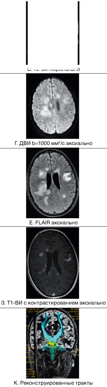
Термины «ромбоэнцефалит» и «стволовой энцефалит» используются для обозначения одного и того же поражения, вовлекающего субтенториальные структуры — ствол мозга и мозжечок [22]. Это состояние воспалительной природы может быть инфекционным, аутоиммунным или пара-неопластическим. Инфекционный ромбоэнцефалит может наблюдаться при любом вирусном или бак- териальном энцефалите; в 50% случаев регистрируется вовлечение супратенториальных структур [37]. Наиболее часто стволовой энцефалит вызывается энтеровирусами 71-го типа, листериями, вирусом простого герпеса [38, 39]. МРТ-находки при этой патологии неспецифичны. Очаги обычно множественные, асимметричные, Т2-гиперинтен-сивные и не всегда накапливающие контрастное вещество в режиме Т1 (рис. 2).
Рис. 2. Пациент М., 9 лет. МРТ: вирусный энцефалит, острый период течения заболевания. Этиология — герпес II типа (IgM с крови)
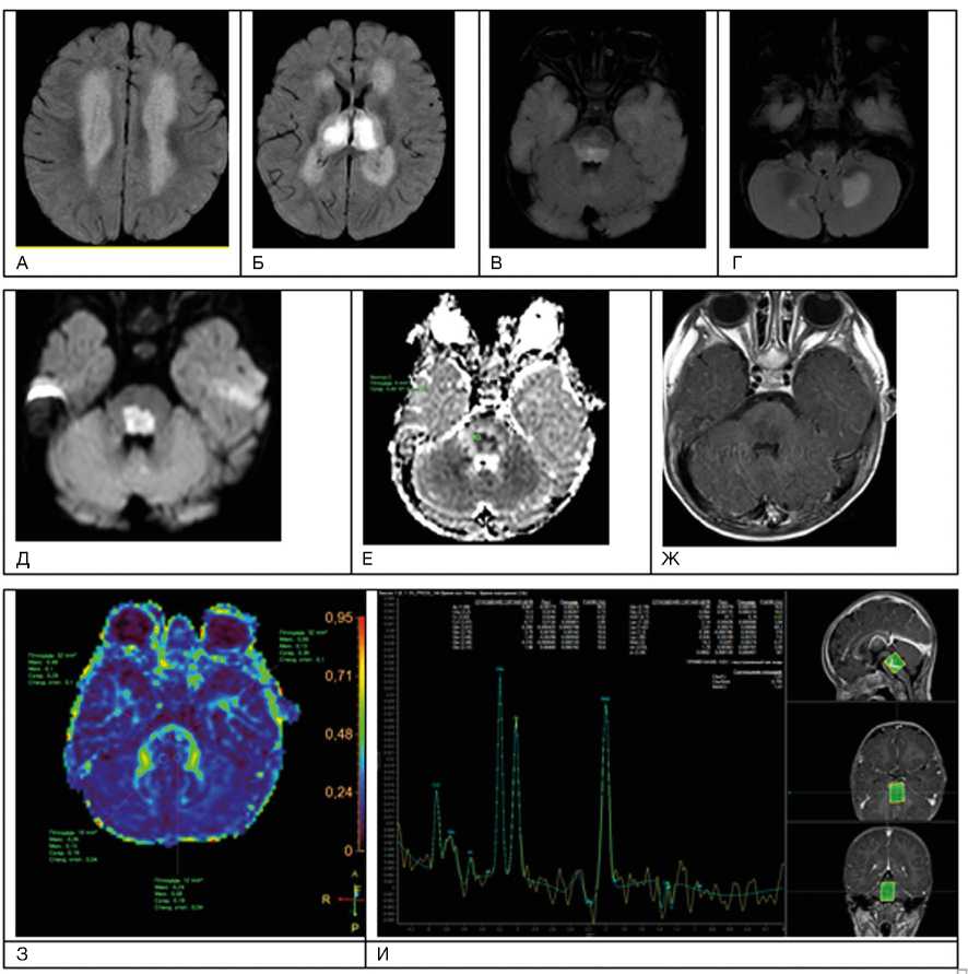
Примечание. А–И — последовательность Т2-flair. Имеется поражение зрительных бугров, глубокого перивентрику-лярного и субкортикального белого вещества лобных и теменных долей, задних отделов моста, глубоких отделов гемисфер мозжечка; Д, Е — с повышением сигнала за счет цитотоксического отека в стволе головного мозга при факторе взвешенности В1000 на диффузионно-взвешенных изображениях и признаками ограничения диффузии (измеряемый коэффициент диффузии снижен до 0,4 x 10 -3 мм2/с);Ж — при внутривенном контрастировании без нарушения гематоэнцефалического барьера в зоне поражения; З — при выполнении DTI МР-трактографии фракционная анизотропия в стволе снижена до 0,19; И — пики NAA и Cho практически одинаковые по высоте, т.е. косвенный признак снижения нейрональной плотности в зоне поражения, пик лактата (маркер анаэробного гликолиза) неубедителен на фоне шума (данные Детского научно-клинического центра инфекционных болезней ФМБА, Санкт-Петербург).
При некоторых вирусных энцефалитах (энцефалит Святого Луиса, японский энцефалит) регистрируется селективная уязвимость черной субстанции [40, 41]. Формирование абсцесса с кольцевым усилением сигнала после введения контрастного вещества наблюдается при поражении всех уровней ствола мозга, при инфекции, обусловленной Listeria monocytogenes [42]. При туберкулезе регистрируется милиарный паттерн [43]. Для обеих этих форм стволового энцефалита характерно формирование абсцессов, которые обычно расположены в мосту и выглядят как очаги с легкой Т2-гиперинтенсивно-стью сигнала вследствие формирования вазогенного отека. Центральная часть этих очагов может выглядеть изо- или гиперинтенсивной в Т2-режиме [44]. В случае формирования туберкуломы сигнал от центральной части очага — Т2-гипоинтенсивный [45]. Из-за гематогенного распространения распределение абсцессов обычно напоминает артериальную эмболию. Поражение ствола при энцефалите может быть ассоциированным с супратенториальными нарушениями, в некоторых случаях со специфическими симметричными билатеральными очагами в области базальных ядер.
Существует «только стволовой энцефалит», известный также как энцефалит Бикерстаффа, который, как предполагается, имеет воспалительную неинфекционную природу и в большинстве случаев аутоиммунную этиологию [46]. После того как была продемонстрирована патогенетическая роль антиганглиозидных антител, пациенты, у которых клинически наблюдался широкий спектр симптомов — от офтальмоплегии и атаксии до нарушения сознания и арефлексии, и которые ранее описывались как случаи симптома Миллера–Фишера, синдрома Гийена–Барре или энцефалита Бикерстаффа, стали расцениваться как больные синдромом анти-GQ1B антител [47–49]. В случае этих синдромов иммунная кросс-реакция, видимо, запускается предшествующим инфицированием Campylobacter jejuni и Mycoplasma pneumoniae [49, 50]. Энцефалит Бикерстаффа может имитировать глиому: для дифференциальной диагностики применяются исследование профиля метаболитов при МР-спектроскопии, повторная МРТ после терапии глюкокортикостероидами и анализ ликвора на ан-ти-GQ1B-антитела [48].
После обычных вирусных инфекций, таких как грипп А и В, парагрипп II, герпес-вирус VI типа, Коксаки вирус или энтеровирус, у иммунокомпетентных детей может развиться быстропрогрес- сирующая энцефалопатия, известная как острая некротизирующая энцефалопатия [51]. Она начинается в течение первых 4 сут от развития симптомов вирусной инфекции с летаргического состояния, которое затем в 50% случаев сменяется комой и судорогами. Среди пациентов, перенесших первый эпизод, у 50% развивается хотя бы одно обострение. Как семейные, так и спорадические формы развиваются у пациентов с миссенс-мутацией гена RAN-binding 2 [52]. Острая некротизирующая энцефалопатия считается другой нозологической формой, чем острый диссеминированный энцефаломиелит.
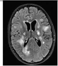
Острый диссеминированный энцефаломиелит в большинстве случаев — монофазное и мультифокальное воспалительное заболевание [38, 53, 54]. Стволовые очаги по данным МРТ невозможно дифференцировать от таковых при рассеянном склерозе, однако у таких пациентов чаще выявляются повреждения на уровне среднего мозга, кроме того, они более симметричны, билатеральны, чем при рассеянном склерозе [38, 55]. Билатеральное симметричное вовлечение мозжечка, базальных ядер, таламуса и относительная сохранность мозолистого тела подтверждают диагноз острого диссеминированного энцефаломиелита (рис. 3).
Кроме того, при повторных МРТ в периоде ре-конвалесценции очаги при остром диссеминированном энцефаломиелите уменьшаются в количестве, размерах, а также наблюдается ослабление интенсивности в режиме Т2. В 50% случаев очаги полностью исчезают [56]. При рассеянном склерозе часто происходит вовлечение ствола мозга. Очаги чаще расположены на дне 4-го желудочка, в периферических частях ствола, в особенности в мосту [57]. Субтенториальное расположение очагов считается специфичным для рассеянного склероза (пересмотренные критерии Мак-Дональда, 2017) [58]. Рассеянный склероз у детей наблюдается редко [59], тем не менее в дебюте заболевания вовлечение ствола и мозжечка, особенно у мальчиков, встречается чаще, чем у пациентов с дебютом в молодом возрасте [60, 61]. Это послужило основой для гипотезы, что при рассеянном склерозе преимущественно в иммунный процесс вовлечен более зрелый миелин [62]. Кроме того, необходимо отметить, что ствол мозга вовлекается в патологический процесс также при заболеваниях нейрооп-тикомиелитного спектра, что связано с образованием аутоантител к аквапорину-4 и в некоторых случаях в отсутствии этих антител [63].
клиническая; 20
тактика ™
в рассеянный склероз
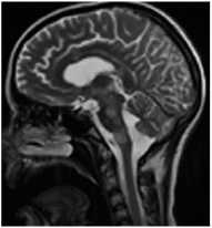
А. Т2-ВИ сагиттально
В. Т2-ВИ аксиально
Д. FLAIR аксиально
Ж. Т1-ВИ с контрастированием аксиально
И. Карта фракционной анизотропии
Рис. 3. Пациентка Р., 15 лет. МРТ: острый диссеминированный энцефаломиелит с трансформацией
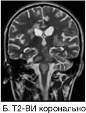
Примечание. Множественные перивентрикулярные, субкортикальные крупные очаги с нечеткими и неровными контурами, накапливающие контрастное вещество фрагментарно, преимущественно по краям (нарушение гематоэнцефалического барьера), с незначительным повышением МР-сигнала на ДВИ 1000 с/мм2 (отечные изменения). Внутренняя структура очагов неоднородная. Визуализируется поражение ствола мозга, спинного мозга и мозжечка. Ограничение фракционной анизотропии на уровне очагов (до 0,2) и снижение плотности реконструируемых трактов на этом уровне (данные Детского научно-клинического центра инфекционных болезней ФМБА России, Санкт-Петербург).
НЕЙРОДЕГЕНЕРАТИВНЫЕЗАБОЛЕВАНИЯ У ДЕТЕЙ
Дефицит кофактора молибдена является редким аутосомно-рецессивным нейродегенератив-ным заболеванием. Возможно, данная патология является не столько редкой, сколько трудно диагностируемой. При данном поражении развивается энцефалопатия с ранним дебютом или с поздним дебютом в атипичных случаях в виде общей задержки развития. Структурная МРТ и диффузионно-взвешенная МР-картина напоминает таковую при диффузном гипоксически-ишемическом поражении [64]. Тем не менее, в случае если наблюдается преимущественное вовлечение ножек мозга, бледного шара и субталамической области, это может заставить предположить описываемое состояние. Ранняя диагностика дефицита кофактора молибдена является критической для начала ранней поддерживающей и заместительной терапии, что позволяет предотвратить атрофию мозга [65].
Гипертрофическая дегенерация ядер оливы считается специфической формой транссинаптической гипертрофической дегенерации и развивается вследствие неспецифического повреждения денто-рубро-оливарного пути [66]. Хотя это состояние у детей считается редким, сообщается о его превалировании у пациентов с метаболическими заболеваниями, а также после операции по поводу опухолей задней черепной ямки [67, 68]. На МРТ выявляются Т2-гиперинтенсивные очаги, часто с билатеральным и симметричным увеличением оли-варных ядер. Они начинают появляться в течение месяца после острого события и в течение 3–4 лет имеют тенденцию к разрешению [69].
ОПУХОЛИ СТВОЛА МОЗГА У ДЕТЕЙ
МРТ-признаками глиомы ствола мозга являются Т2-гиперинтенсивные очаги с масс-эффектом, окружающие цистерны, 4-й желудочек, сильвиев водопровод и/или мозжечок. Чаще всего у детей развиваются отличающиеся по анатомии и клиническому течению 3 группы глиом ствола — это диффузная внутренняя понтинная глиома, экзофитиче-ская среднемозговая глиома и тектальная глиома [70]. Первый вариант имеет наихудший прогноз выживаемости (около 1 года) [71]. На МРТ отличительными особенностями являются перифокальный отек, отсутствие накопления контраста и масс-эф-фекта. Чаще всего возникает в возрасте 5–10 лет с развитием клинической триады — атаксии, поражения черепно-мозговых нервов и длинных трак- тов. В типичном случае поражение тотальное или субтотальное, чаще всего в мосту. Хотя отсутствие накопления контраста является характерной особенностью, в некоторых случаях наблюдается негомогенное частичное или периферическое накопление парамагнетика [72].
У детей младше 3 лет могут возникать солидные примитивные нейроэктодермальные опухоли. Они фокально, экзофитно расположены обычно в мосту, умеренно накапливают контраст и часто распространяются субарахноидально [73–75]. Опухоли ствола у детей также могут развиваться при неврофиброматозе 1-го типа [76, 77]. Чаще всего возникает астроцитома, которая растет менее агрессивно, чем у детей без неврофиброматоза [78]. У таких пациентов выявляются яркие Т2-гиперинтенсивные очаги, которые остаются стабильными и иногда исчезают у пациентов старше 12 лет [79]. Интракраниальное поражение ствола мозга может развиваться при гистиоцитозе клеток Лангерганса [80, 81]. Это редкое гранулематозное заболевание системы моноцит-макрофаг. Типичные клинические проявления при этом — литические очаги краниофациального скелета, вовлечение гипоталами-чески-питуитарной области, несахарный диабет. Интракраниально выявляются симметричные Т2-гиперинтенсивные очаги нейродегенерации и реже регистрируются массивные туморозные очаги [82].
ЗАКЛЮЧЕНИЕ
Поражение ствола мозга, изолированное или связанное с супратенториальными изменениями, возникает при широком спектре патологических состояний. Небольшой размер ствола, менее четкое различие между серым и белым веществом и определенное ограничение мультипараметри-ческой МРТ в случае исследования стволовых структур часто приводят к затруднениям в дифференциальной диагностике. Иногда большое диагностическое значение приобретают топография очага и интенсивность сигнала от него. Для целей дифференциальной диагностики основополагающее значение, тем не менее, играют клинические, эпидемиологические и лабораторные исследования.
ИСТОЧНИК ФИНАНСИРОВАНИЯ
Поисково-аналитическая работа проведена на личные средства авторского коллектива.
клиническая; 20
Том 121 № 0 1
Список литературы Магнитно-резонансная томография как инструмент дифференциальной диагностики при поражении ствола головного мозга у детей
- Ишков С.В., Левошко Л.И. Новые данные о проекционной анатомии отделов ствола головного мозга на основе компьютерно-томографических исследований // Морфология. - 2017. - Т.152. - №5. - С. 25-28.
- Bastianello S, Bozzao A, Paolillo A, et al. Fast spin-echo and fast fluid-attenuated inversion-recovery versus conventional spin-echo sequences for MR quantification of multiple sclerosis lesions. AJNR Am J Neuroradiol. 1997;18(4):699-704.
- Li C, Yan JL, Torheim T, et al. Low perfusion compartments in glioblastoma quantified by advanced magnetic resonance imaging and correlated with patient survival. Radiother Oncol. 2019;134:17-24. DOI: 10.1016/j.radonc.2019.01.008
- Kawanaka Y, Ando K, Ishikura R, et al. Delayed appearance of transient hyperintensity foci on T1-weighted magnetic resonance imaging in acute disseminated encephalomyelitis. Jpn J Radiol. 2019;37(4):277-282. DOI: 10.1007/s11604-018-00808-w
- Cai M, Zhang XF, Qiao HH, et al. Susceptibility-weighted imaging of the venous networks around the brain stem. Neuroradiology. 2015;57(2):163-169. DOI: 10.1007/s00234-014-1450-z
- Beller E, Keeser D, Wehn A, et al. T1-MPRAGE and T2-FLAIR segmentation of cortical and subcortical brain regions-an MRI evaluation study. Neuroradiology. 2019;61(2):129-136.
- DOI: 10.1007/s00234-018-2121-2
- Quattrocchi CC, Errante Y, Rossi Espagnet MC, et al. Magnetic resonance imaging differential diagnosis of brainstem lesions in children. World J Radiol. 2016;8(1):1-20.
- DOI: 10.4329/wjr.v8.i1.1
- Войтенков В.Б., Карташев А.В. Ретикулярная формация головного мозга в норме и патологии. - СПб.: Реноме, 2013. - 115 с.
- Lagman-Bartolome AM, Pontigon AM, Moharir M, et al. Basilar artery strokes in children: good outcomes with conservative medical treatment. Dev Med Child Neurol. 2013;55(5):434-439.
- DOI: 10.1111/dmcn.12092
- Toi H, Uno M, Harada M. et al. Diagnosis of acute brain-stem infarcts using diffusion-weighed MRI. Neuroradiology. 2003;45(6):352-356.
- DOI: 10.1007/s00234-002-0897-5
- Uziel G, Ghezzi D, Zeviani M. Infantile mitochondrial encephalopathy. Semin Fetal Neonatal Med. 2011;16(4):205-215.
- DOI: 10.1016/j.siny.2011.04.003
- Wong LJ. Mitochondrial syndromes with leukoencephalopathies. Semin Neurol. 2012;32(1):55-61.
- DOI: 10.1055/s-0032-1306387
- Nishino I, Spinazzola A, Hirano M. Thymidine phosphorylase gene mutations in MNGIE, a human mitochondrial disorder. Science. 1999;283(5402):689-692.
- DOI: 10.1126/science.283.5402.689
- Tang S, Wang J, Lee NC, et al. Mitochondrial DNA polymerase gamma mutations: an ever expanding molecular and clinical spectrum. J Med Genet. 2011;48(10):669-681.
- DOI: 10.1136/jmedgenet-2011-100222
- Scheper GC, van der Klok T, van Andel RJ, et al. Mitochondrial aspartylt-RNA synthetase deficiency causes leukoencephalopathy with brain stem and spinal cord involvement and lactate elevation. Nat Genet. 2007;39(4):534-539.
- DOI: 10.1038/ng2013
- Uluc K, Baskan O, Yildirim KA, et al. Leukoencephalopathy with brain stem and spinal cord involvement and high lactate: a genetically proven case with distinct MRI findings. J Neurol Sci. 2008;273(1-2):118-122.
- DOI: 10.1016/j.jns.2008.06.002
- Cavanagh JB. Selective vulnerability in acute energy deprivation syndromes. Neuropathol Appl Neurobiol. 1993;19(6):461-470.
- DOI: 10.1111/j.1365-2990.1993.tb00474.x
- Nagai T, Goto Y, Matsuoka T, et al. Leigh encephalopathy: histologic and biochemical analyses of muscle biopsies. Pediatr Neurol. 1992;8(5):328-332.
- DOI: 10.1016/0887-8994(92)90084-c
- Chen L, Cui Y, Jiang D, et al. Management of Leigh syndrome: Current status and new insights. Clin Genet. 2018;93(6):1131-1140.
- DOI: 10.1111/cge.13139
- Veiga MG, Marecos C, Duarte ST, et al. Leigh syndrome with atypical cerebellar lesions. eNeurological Sci. 2019;16:100-107.
- DOI: 10.1016/j.ensci.2019.100197
- Bindu PS, Taly AB, Sonam K, et al. Bilateral hypertrophic olivary nucleus degeneration on magnetic resonance imaging in children with Leigh and Leigh-like syndrome. Br J Radiol. 2014;87:2013047.
- DOI: 10.1259/bjr.20130478
- Quattrocchi CC, Longo D, Delfino LN, et al. MR differential diagnosis of acute deep grey matter pathology in paediatric patients. Pediatr Radiol. 2013;43(6):743-761.
- DOI: 10.1007/s00247-012-2491-2
- Sparaco M, Bonilla E, Di Mauro S, Powers J.M. Neuropathology of mitochondrial encephalomyopathies due to mitochondrial DNA defects. J Neuropathol Exp Neurol. 1993;52(1):1-10.
- DOI: 10.1097/00005072-199301000-00001
- Ito S, Shirai W, Asahina M, Hattori T. Clinical and brain MR imaging features focusing on the brain stem and cerebellum in patients with myoclonic epilepsy with ragged-red fibers due to mitochondrial A8344G mutation. AJNR Am J Neuroradiol. 2008;29(2):392-395
- DOI: 10.3174/ajnr.A0865
- Valanne L, Ketonen L, Majander A, et al. Neuroradiologic findings in children with mitochondrial disorders. AJNR Am J Neuroradiol. 1998;19(2):369-377.
- Castillo M, Kwock L, Green C. MELAS syndrome: imaging and proton MR spectroscopic findings. AJNR Am J Neuroradiol. 1995;16(2):233-239.
- Kori A, Hori I, Tanaka T, et al. Transition from Leigh syndrome to MELAS syndrome in a patient with heteroplasmic MT-ND3 m.10158T>C. Brain Dev. 2019;41(9):803-807.
- DOI: 10.1016/j.braindev.2019.05.006
- Schicks J, Schöls L, van der Knaap MS, Synofzik M. Teaching NeuroImages: MRI guides genetics: leukoencephalopathy with brainstem and spinal cord involvement (LBSL). Neurology. 2013;80(16):e176-e177.
- DOI: 10.1212/WNL.0b013e31828cf846
- Yelam A, Nagarajan E, Chuquilin M, Govindarajan R. Leucoencephalopathy with brain stem and spinal cord involvement and lactate elevation: a novel mutation in the DARS2 gene. BMJ Case Rep. 2019;12(1):32-35.
- DOI: 10.1136/bcr-2018-227755
- Lan MY, Chang YY, Yeh TH, et al. Leukoencephalopathy with brainstem and spinal cord involvement and lactate elevation (LBSL) with a novel DARS2 mutation and isolated progressive spastic paraparesis. J Neurol Sci. 2017;372:229-231.
- DOI: 10.1016/j.jns.2016.11.058
- Chinoy A, Wright NB, Bone M, Padidela R. Severe hypokalaemia in diabetic ketoacidosis: a contributor to central pontinemyelinolysis? Endocrinol Diabetes Metab Case Rep. 2019;2019(1):30-35.
- DOI: 10.1530/EDM-19-0034
- Bansal LR, Zinkus T. Osmotic demyelination syndrome in children. Pediatr Neurol. 2019;97:12-17.
- DOI: 10.1016/j.pediatrneurol.2019.03.018
- Alleman AM. Osmotic demyelination syndrome: central pontine myelinolysis and extrapontine myelinolysis. Semin Ultrasound CT MR. 2014;35(2):153-159.
- DOI: 10.1053/j.sult.2013.09.009
- Fuller K, Guerrero C, Kyin M, et al. The role of the interdisciplinary team in subacute rehabilitation for central pontine myelinolysis. Disabil Rehabil. 2019;1:1-7.
- DOI: 10.1080/09638288.2019.1579261
- Milh M, Villeneuve N, Chapon F, et al. Transient brain magnetic resonance imaging hyperintensity in basal ganglia and brain stem of epileptic infants treated with vigabatrin. J Child Neurol. 2009;24(3):305-315.
- DOI: 10.1177/0883073808324219
- Dracopoulos A, Widjaja E, Raybaud C, et al. Vigabatrin-associated reversible MRI signal changes in patients with infantile spasms. Epilepsia. 2010;51(7):1297-1304.
- DOI: 10.1111/j.1528-1167.2010.02564.x
- Skripchenko NV, Ivanova GP, Skripchenko EY, Murina EA. Panencephalitis in children in modern conditions: clinical, etiological and MRI-aspects. Zh Nevrol PsikhiatrIm S S Korsakova. 2019;119(6):20-31.
- DOI: 10.17116/jnevro201911906120
- Skripchenko EY, Ivanova GP, Karev VE, Skripchenko NV. [Difficulties of differential diagnosis of organic injury of the nervous system in children. (In Russ).]. Zh Nevrol PsikhiatrIm S S Korsakova. 2018;118(5):25-30.
- DOI: 10.17116/jnevro20181185225
- Jubelt B, Mihai C, Li TM, Veerapaneni P. Rhombencephalitis / brainstem encephalitis. Curr Neurol Neurosci Rep. 2011;11(6):543-552.
- DOI: 10.1007/s11910-011-0228-5
- Wasay M, Diaz-Arrastia R, Suss RA, et al. St Louis encephalitis: a review of 11 cases in a 1995 Dallas, Tex, epidemic. Arch Neurol. 2000;57(1):114-118.
- DOI: 10.1001/archneur.57.1.114
- Kalita J, Misra UK. The substantianigra is also involved in Japanese encephalitis. AJNR Am J Neuroradiol. 2000;21(10):1978-1980.
- Reynaud L, Graf M, Gentile I, et al. A rare case of brainstem encephalitis by Listeria monocytogenes with isolated mesencephalic localization. Case report and review. Diagn Microbiol Infect Dis. 2007;58(1):121-123.
- DOI: 10.1016/j.diagmicrobio.2006.11.001
- Nogueira Delfino L, Fariello G, Lancella L, et al. Central nervous system tuberculosis in non-HIV-positive children: a singlecenter, 6 year experience. Radiol Med. 2012;117(4):669-678.
- DOI: 10.1007/s11547-011-0743-0
- Ramalho J, Castillo M. Case of the season: brainstem abscess. Semin Roentgenol. 2008;43(3):168-170.
- DOI: 10.1053/j.ro.2008.03.001
- Akhaddar A, Mahi M, Harket A, et al. Brainstem tuberculoma in a postpartum patient. J Neuroradiol. 2007;34(5):345-346.
- DOI: 10.1016/j.neurad.2007.09.001
- Tan IL, Mowry EM, Steele SU, et al. Brainstem encephalitis: etiologies, treatment, and predictors of outcome. J Neurol. 2013;260(9):2312-2319.
- DOI: 10.1007/s00415-013-6986-z
- Odaka M, Yuki N, Hirata K. Anti-GQ1b IgG antibody syndrome: clinical and immunological range. J Neurol Neurosurg Psychiatry. 2001;70(1):50-55.
- DOI: 10.1136/jnnp.70.1.50
- Shahrizaila N, Yuki N. Bickerstaff brainstem encephalitis and Fisher syndrome: anti-GQ1b antibody syndrome. J Neurol Neurosurg Psychiatry. 2013;84(5):576-583.
- DOI: 10.1136/jnnp-2012-302824
- Ito M, Kuwabara S, Odaka M, et al. Bickerstaff's brainstem encephalitis and Fisher syndrome form a continuous spectrum: clinical analysis of 581 cases. J Neurol. 2008;255(5):674-682.
- DOI: 10.1007/s00415-008-0775-0
- Steer AC, Starr M, Kornberg AJ. Bickerstaff brainstem encephalitis associated with Mycoplasma pneumoniae infection. J Child Neurol. 2006;21(6):533-534.
- DOI: 10.1177/08830738060210061401
- Wang GF, Li W, Li K. Acute encephalopathy and encephalitis caused by influenza virus infection. Curr Opin Neurol. 2010;23(3):305-311.
- DOI: 10.1097/wco.0b013e328338f6c9
- Gika AD, Rich P, Gupta S, et al. Recurrent acute necrotizing encephalopathy following influenza A in a genetically predisposed family. Dev Med Child Neurol. 2010;52(1):99-102.
- DOI: 10.1111/j.1469-8749.2009.03405.x
- Caldemeyer KS, Smith RR, Harris TM, Edwards MK. MRI in acute disseminated encephalomyelitis. Neuroradiology. 1994;36(3):216-220.
- DOI: 10.1007/bf00588134
- Rossi A. Imaging of acute disseminated encephalomyelitis. Neuroimaging Clin N Am. 2008;18(1):149-161.
- DOI: 10.1016/j.nic.2007.12.007
- Lu Z, Zhang B, Qiu W, et al. Comparative brain stem lesions on MRI of acute disseminated encephalomyelitis, neuromyelitis optica, and multiple sclerosis. PLoS One. 2011;6(8):e22766.
- DOI: 10.1371/journal.pone.0022766
- Atzori M, Battistella PA, Perini P, et al. Clinical and diagnostic aspects of multiple sclerosis and acute monophasic encephalomyelitis in pediatric patients: a single centre prospective study. Mult Scler. 2009;15(3):363-370.
- DOI: 10.1177/1352458508098562
- Yousry TA, Grossman RI, Filippi M. Assessment of posterior fossa damage in MS using MRI. J Neurol Sci. 2000;172(Suppl 1):S50-S53.
- DOI: 10.1016/s0022-510x(99)00279-8
- Polman CH, Reingold SC, Banwell B, et al. Diagnostic criteria for multiple sclerosis: 2010 revisions to the McDonald criteria. Ann Neurol. 2011;69(2):292-302.
- DOI: 10.1002/ana.22366
- Лобзин Ю.В., Скрипченко Н.В., Иванова Г.П., Команцев В.Н. Диссеминированный лейкоэнцефалит и рассеянный склероз: причинно-следственная взаимосвязь // Саратовский научно-медицинский журнал. - 2013. - Т.9. - №2. - С. 170-178.
- Chabas D, Strober J, Waubant E. Pediatric multiple sclerosis. Curr Neurol Neurosci Rep. 2008;8(5):434-441.
- DOI: 10.1007/s11910-008-0067-1
- Ghassemi R, Antel SB, Narayanan S, et al. Lesion distribution in children with clinically isolated syndromes. Ann Neurol. 2008;63(3):401-405.
- DOI: 10.1002/ana.21322
- Ghassemi R, Narayanan S, Banwell B, et al. Quantitative determination of regional lesion volume and distribution in children and adults with relapsing-remitting multiple sclerosis. PLoS One. 2014;9(2):e85741.
- DOI: 10.1371/journal.pone.0085741
- Wingerchuk DM, Banwell B, Bennett JL, et al. International consensus diagnostic criteria for neuromyelitisoptica spectrum disorders. Neurology. 2015;85(2):177-189.
- DOI: 10.1212/WNL.0000000000001729
- Екушева Е.В., Данилов А.Б. Наследственная спастическая параплегия (обзор) // Журнал неврологии и психиатрии им. С.С. Корсакова. - 2002. - Т.102. - №8. - С. 44-52.
- Vijayakumar K, Gunny R, Grunewald S, et al. Clinical neuroimaging features and outcome in molybdenum cofactor deficiency. Pediatr Neurol. 2011;45(4):246-252.
- DOI: 10.1016/j.pediatrneurol.2011.06.006
- Екушева Е.В. Сенсомоторная интеграция при поражении центральной нервной системы: клинические и патогенетические аспекты: Автореф. дис.... докт. мед. наук. - М., 2016. - 48 с. Доступно https://search.rsl.ru/ru/record/01006661768. Ссылка активна на 14.12.2019.
- Sonam K, Khan NA, Bindu PS, et al. Clinical and magnetic resonance imaging findings in patients with Leigh syndrome and SURF1 mutations. Brain Dev. 2014;36(9):807-812.
- DOI: 10.1016/j.braindev.2013.10.012
- Mirabelli-Badenier M, Morana G, Bruno C, et al. Inferior olivary nucleus involvement in pediatric neurodegenerative disorders: does it play a role in neuroimaging pattern-recognition approach? Neuropediatrics. 2015;46(2):104-109.
- DOI: 10.1055/s-0035-1544185
- Tartaglione T, Izzo G, Alexandre A, et al. MRI findings of olivary degeneration.after surgery for posterior fossa tumours in children: incidence, time course and correlation with tumour grading. Radiol Med. 2015;120(5):474-482.
- DOI: 10.1007/s11547-014-0477-x
- Grimm SA, Chamberlain MC. Brainstem glioma: a review. Curr Neurol Neurosci Rep. 2013;13(5):346.
- DOI: 10.1007/s11910-013-0346-3
- Garzón M, García-Fructuoso G, Guillén A, et al. Brain stem tumors in children and adolescents: single institutional experience. Childs Nerv Syst. 2013;29(8):1321-1331.
- DOI: 10.1007/s00381-013-2137-1
- Guillamo JS, Doz F, Delattre JY. Brain stem gliomas. Curr Opin Neurol. 2001;14(6):711-715.
- DOI: 10.1097/00019052-200112000-00006
- Nowak J, Seidel C, Pietsch T, et al. Ependymoblastoma of the brainstem: MRI findings and differential diagnosis. Pediatr Blood Cancer. 2014;61(6):1132-1134.
- DOI: 10.1002/pbc.24915
- Zagzag D, Miller DC, Knopp E, et al. Primitive neuroectodermal tumors of the brainstem: investigation of seven cases. Pediatrics. 2000;106(5):1045-1053.
- DOI: 10.1542/peds.106.5.1045
- Екушева Е.В., Данилов А.Б., Вейн А.М. Синдром гемипареза: клинико-патофизиологический анализ // Журнал неврологии и психиатрии им. С.С. Корсакова. - 2002. - Т.102. - №11. - С. 18-28.
- Bilaniuk LT, Molloy PT, Zimmerman RA, et al. Neurofibromatosis type 1: brain stem tumours. Neuroradiology. 1997;39(9):642-653.
- DOI: 10.1007/s002340050484
- Guillamo JS, Créange A, Kalifa C, et al. Prognostic factors of CNS tumours in Neurofibromatosis 1 (NF1): a retrospective study of 104 patients. Brain. 2003;126(Pt 1):152-160.
- DOI: 10.1093/brain/awg016
- Ullrich NJ, Raja AI, Irons MB, et al. Brainstem lesions in neurofibromatosis type 1. Neurosurgery. 2007;61(4):762-766; discussion 766-767.
- DOI: 10.1227/01.NEU.0000298904.63635.2D
- Hervey-Jumper SL, Singla N, Gebarski SS, et al. Diffuse pontine lesions in children with neurofibromatosis type 1: making a case for unidentified bright objects. Pediatr Neurosurg. 2013;49(1):55-59.
- DOI: 10.1159/000355417
- Grois N, Fahrner B, Arceci RJ, et al. Central nervous system disease in Langerhans cell histiocytosis. J Pediatr. 2010;156(6):873-881.
- DOI: 10.1016/j.jpeds.2010.03.001
- Savardekar A, Tripathi M, Bansal D, et al. Isolated tumorous Langerhans cell histiocytosis of the brainstem: a diagnostic and therapeutic challenge. J Neurosurg Pediatr. 2013;12(3):258-261.
- DOI: 10.3171/2013.6.PEDS13132
- Prosch H, Grois N, Wnorowski M, et al. Longterm MR imaging course of neurodegenerative Langerhans cell histiocytosis. AJNR Am J Neuroradiol. 2007;28(6):1022-1028.
- DOI: 10.3174/ajnr.A0509
