Photometric recorder of the carotid artery pulse wave with a system for stabilizing the clamping force

Author: Berdnikov A.V., Matrosov G.V.
Journal: Cardiometry @cardiometry
Section: Report
Article in issue: 21, 2022.
Free access
Preventive monitoring, which increases the possibility of diagnosing and curing a disease at an early stage, is interesting and relevant in medical diagnostics. Recently, both minimally invasive and non-invasive diagnostic techniques have become widespread. They include the areas of cardiographic, encephalographic and myographic analyzers designed to study the state of the heart, the brain and striated muscles. The study of the functioning of the smooth muscles of the gastrointestinal tract is carried out using gastro-enterography methods. To obtain an integral assessment of the effectiveness of the circulatory system, the plethysmography method can be successfully applied, which allows modeling vascular tone and blood flow using models of hydrodynamics and the theory of elasticity. The present article analyzes the range of static (or dynamic) informative hemo-parameters that allow diagnosing at the initial stage arterial hypertension, aortic insufficiency or stenosis, atherosclerosis, diabetes mellitus and some other diseases. The photometric reflex absorption in the infrared range, followed by an assessment of blood flow in the aortas and arteries with recording of the shape of pulse waves were used as methods in our research work. The result of the work is a method and a system of computer control of the rod current acting on biological tissues (using the Bluetooth protocol, LabView environment, virtual digital filters and biomedical data archives) that allows non-invasively recording of the parameters of the carotid artery pulse wave.
System, stabilization, pulse wave, carotid artery
Short address: https://sciup.org/148324182
IDR: 148324182
Text of the scientific article Photometric recorder of the carotid artery pulse wave with a system for stabilizing the clamping force
clamping force. Cardiometry; Issue 21; February 2022; p. 66-69; DOI: 10.18137/cardiometry.2022.21.6669; Available from:
Non-invasive and technologically realizable methods for diagnosing the cardiovascular system are of great interest to the scientific medical community due to the availability of this type of research to the public in general (Omelyanovskiy, Avksentieva, et-al 2021) that increases the number of patients receiving treatment at the initial stage; including that with the help of telemedicine (Naberezhnaya, Zakharov, 2021; Tots-kaya, Pokrovskaya, 2018).
The plethysmography method belonging to this group of studies in its practical application is based on the concept of the vascular tone and the blood flow as the physiological functions, to the study of which elements of the theories of elasticity and hydrodynamics are applicable. This method is capable to produce a full range of informative hemostatic and hemodynamic parameters, which makes it possible to use it in the initial diagnosis of diseases such as arterial hypertension, aortic insufficiency, atherosclerosis, aortic stenosis, diabetes mellitus, Bottalo’s duct non-closure, obliterating endarteritis and others (Usanov, Skripal, et-al 2009).
Theoretical foundations and reference literature analysis
An introduction of modern methods and technologies for assessing the quality of medical care (Zheleznyakova, Seryapina, et-al 2020; Nicolay, Purkayastha, 2012; Harvey, 1996) also requires some new specific diagnostic techniques.
When using single signs for diagnostic studies on the pulse waveform, the most informative are the following: the absence of a dicrotic notch and the difference in the volumetric pulse on the arms and legs (Okorokov, 2007). Recording of pulse fluctuations can be carried out using photometric reflex-absorption sensors (Whitenett, Stewart, et-al 2003; Posani, Tripathi, et-al 2006) operating in the infrared range, the signal of which is proportional to the blood filling of the arterial vessels, lying under the sensor, at every moment of time.
Variants of devices for recording a pulse wave which exist today are designed as a clamping device attached to a finger or a toe, and they operate with transmitted radiation. This approach to the design of a plethysmographic meter, operating on reflected radiation, is associated with a significant drawback, which consists in the inability to control the clamping force of biological tissue containing blood vessels; this involves some changes in the blood flow in the investigated area and, as a consequence, leads to an increase in the measurement error. In addition, to assess the performance of the heart and the root sections of the circulatory system (for example, (Actual Problems of Prevention, 2015), it is of particular interest to assess blood flow in large aortas and arteries, as in vessels adjacent to the heart. This assessment requires recording pulse waves on large arteries, which cannot be ensured with the use of a clothespin-like sensor.
Materials and methods
Based on the above, the vector of scientific and engineering synthesis becomes clear: it is aimed at obtaining an alternative, improved, method for collecting and analyzing the parameters of pulse waves (Simonenko, Tuchin, 2007; Shurygin, 2000). An intermediate stage of this synthesis is the concept of an analyzer of the parameters of the carotid pulse waveform with a system for stabilizing the clamping force (Novikov, Matrosov, Berdnikov, 2018).
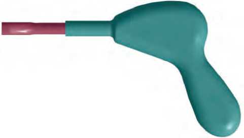
Figure 1. General view of the analyzer of the carotid artery pulse wave parameters with the clamping force stabilization system
The design of the device includes two main parts, namely, a housing and a measuring head. Sensitive elements are located inside the head: a strain-resistive transducer and a photometric sensor. In this case, the measuring head can move in the housing along its axis. This design solution provides for the kinematic system necessary for the implementation of the mech- anism for stabilizing the force of pressing the head against the tissues.
The mechanism for stabilizing the force of pressing the head to the tissues allows avoiding changes in the blood flow in the examined area of biological tissue and assumes that the rod of the electromagnet is rigidly connected to the measuring head, and the body of the electromagnet is rigidly connected to the body of the device. This mechanism is realized with the use of a solenoid electromagnet, a control logic circuit, a current setting system and a strain gauge circuit (Dai-chik, Prigorovsky, Khurshudov, 1989). Thus, by controlling the strength of the current in the winding of the electromagnet, it is possible to control the force with which the rod will act on the biological tissue.
External mechanical influences
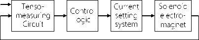
Figure 2. Structure of the clamping force stabilization mechanism
The control and correction of the pressure of the head of the device applied onto the tissue is necessary mainly to eliminate the negative effect of hand tremors in the operating personnel; since the tremor of the hands leads either to a loose contact of the pulse oximetry sensor with the tissue, or to local compression of the tissue and blood vessels therein, which will inevitably produce false data.
The device is powered autonomously from a battery installed in the handle of the device body. In the future, it is supposed that IoT sensors (Quandeng, 2013) can be used.
Results and discussion
The following sequence of research steps is assumed:
-
1. Smooth application of the sensitive surface of the device onto the area of the carotid artery location.
-
2. Gradual increase in the pressure applied to the handle in the longitudinal direction until a signal appears that the device is capable of delivering readings, that is, it enters its proper operating mode. In this case, the head rod will be in its central position and, therefore, has an equal stroke reserve for stabilization, both for clamping and removing the device.
-
3. Holding the device in its operating position, i.e. pressing onto the handle (if possible) with a force ap-
Issue 21. February 2022 | Cardiometry | 67
-
4. Check-up and analysis of data delivered by the device.
proximately equal to that which is applied at the moment the signal appears that the device enters its operating position.
The device, in addition to displaying brief hemodynamic data on the built-in display, allows streaming data to a PC via Bluetooth. The data obtained by the PC is then interpreted as a pulse wave graph using the capabilities provided by the NI LabView software package (Travis, Kring, 2008). In this case, it is possible to capture “raw data”, the filtering of which is carried out directly in the LabView environment by means of virtual digital filters (see Figure 3 herein). The pulse wave graph can be saved by a workstation or by a server in a medical office in order to archive biomedical data (Novikov, Berdnikov, Burmistrov, 2016).
A browser solution is also possible that significantly expands the scope of the device (Berdnikov, Ynusov, et-al, 2019), extending the accessibility, as well as computer simulation (Kaziev, 1990) in a graphic mode.
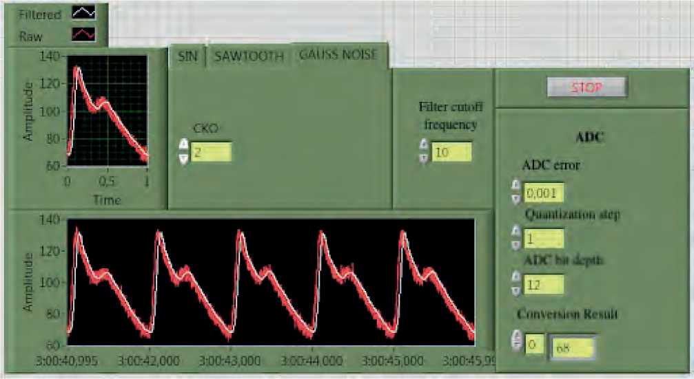
Figure 3. LabView plotter interface displaying graphs of noisy and filtered pulse waves (initial data generated in the simulator)
Conclusion
Thus, this offered device allows non-invasive recording followed by an analysis of the parameters of the carotid artery pulse waveform, while the accuracy of the analysis is increased due to the use of the specific system for stabilizing the force of pressing the measuring head to the biological tissue.
The proposed design solution makes it possible to implement the required kinematic system for stabilizing the force of pressing the head against the biological tissues.
Statement on ethical issues
Research involving people and/or animals is in full compliance with current national and international ethical standards.
68 | Cardiometry | Issue 21. February 2022
Conflict of interest
None declared.
Author contributions
The authors read the ICMJE criteria for authorship and approved the final manuscript.
References Photometric recorder of the carotid artery pulse wave with a system for stabilizing the clamping force
- Omelyanovskiy VV, et al. The main changes in the model of clinical and statistical groups in 2021. Medical technologies. Evaluation and Selection. 2021;2:9-21. https://doi.org/10.17116/medtech2021430219 [in Russian]
- Embankment IB, Zakharov DA. The role of the regional telemedicine center in the provision of medical care. Medical technologies. Evaluation and Selection. 2021;2:67-73. https://doi.org/10.17116/medtech20214302167 [in Russian]
- Totskaya EG, Pokrovskaya OB. Telemedicine as a mechanism for ensuring the availability of high-tech medical services and innovative technologies in health care. Emergency Doctor. 2018;6:71-7. [in Russian]
- Usanov DA, Skripal AV, et al. (2009). Methods and equipment for diagnosing the state of the cardiovascular system by the characteristics of the pulse wave. Saratov University Press, 96 p. [in Russian]
- Zheleznyakova IA, et al. Methodological approaches to the implementation of a quality control system for medical care in medical organizations. Medical technologies. Evaluation and selection. 2020;42(4):13-20. https://doi.org/10.17116/medtech20204204113 [in Russian]
- Nicolay CR, Purkayastha S, Greenhalgh A, et al. Systematic review of the application of quality improvement methodologies from the manufacturing industry to surgical healthcare. The British Journal of Surgery. 2012;99(3):324-35. https://doi.org/10.1002/bjs.7803
- Harvey G. Quality in health care: traditions, influences and future directions. International Journal for Quality in Health Care. 1996;8(4):341-50. https://doi.org/10.1093/intqhc/8.4.341
- Okorokov AN. (2007). Diagnostics of diseases of internal organs. Vol.7. Diagnosis of diseases of the heart and blood vessels. Moscow, 416 p. [in Russian]
- Whitenett G, Stewart G, Atherton K, Culshaw B, Johnstone W. Optical fibre instrumentation for environmental monitoring applications. J. Opt. A: Pure Appl. Opt. 2003;5:140-5.
- Posani KT, Tripathi V, Annamalai S, Weisse-Bernstein NR, Krishnaa S. Nanoscale quantum dot infrared sensors with photonic crystal cavity. Appl. Phys. Let, 2006;88:1–3.
- Topical issues of prevention (2015) of cardiovascular diseases and risk factor correction. Collection of materials for healthcare professionals. Tula Regional Center for Medical Prevention and Rehabilitation named after Ya.S. Stechkin. Tula, 55 p. [in Russian]
- Simonenko GV, Tuchin VV. (2007). Optical properties of biological tissues (teaching aid). p.48. [in Russian]
- Shurygin IA. (2000). Respiratory monitoring: pulse oximetry, capnography, oximetry. St. Petersburg: Nevsky Dialect. Moscow: BINOM, 301 p. [in Russian]
- Novikov MYu, Matrosov GV, Berdnikov AV. (2018). Intraoperative thoracic blood flow analyzer PI 2682831, published on March 22, 2018, Bulletin no. 9. [in Russian]
- Daichik ML, Prigorovsky NI, Khurshudov Kh. (1989). Methods and means of full-scale tensometry. Directory. Moscow: Mechanical engineering, 240 p. [in Russian]
- Quandeng G, (2013). Construction and Strategies in IoT Security System. Green Computing and Communications (GreenCom) IEEE International Conference on and IEEE Cyber, Physical and Social Computing, pp. 1129-1132. DOI:10.1109/Green-Com-iThings-CPSCom.2013.195
- Travis J, Kring J, (2008). LabVIEW for everyone. 3rd ed. Moscow: DMK Press. 800 p.
- Novikov MYu, Berdnikov AV, Burmistrov MV. Photometric information-measuring system for monitoring the degree of oxygen saturation of blood vessels of the bronchus during pneumonectomy. Successes of Modern Science. 2016;11(4):86-90. [in Russian]
- Berdnikov AV, Ynusov NA, Voroshin DI, Misiev DzKh. Optical deviсe for determining the viability of biological tissue. Amazoniia Investiga. 2019;8(24):193-8. [in Russian]
- Kaziev VM. Mathematical modeling and computational experiment. Informatics and education. 1990;5:24-8. [in Russian]
