
Potential of Diiodo-BODIPY Meso-Acid and Its Esters for Photodynamic Therapy of Microbial Biofilms
Journal: Journal of Stress Physiology and Biochemistry @jspb
Article in issue: 3 т.22, 2026.
Free access
Background. Nowadays, there is an increasing number of infectious diseases in humans and animals associated with formation of microbial biofilms, including fungal-bacterial consortia. Due to the diffusional barrier of the biofilm matrix, the biofilm-embedded cells are characterized by increased resistance to antimicrobials and the host immune system. In this study we assessed the ability of diiod derivatives of BODIPY luminophores to eradicate mono- and dual species communities of S. aureus and C. albicans. Results. The diiodo-BODIPY meso-butanoic acid, and its conjugate with isobornanethioethyl or pinanyl, were tested. The diiod derivative of BODIPY in the form of acid, when irradiated at 530 nm at concentrations of 2 μg/ml, resulted in complete cell death of planktonic staphylococci, and at 16 μg/ml completely eradicated its biofilm, but was non effective against Candida biofilms. Partial cell death was observed against S. aureus in mixed S. aureus and C. albicans biofilms at 8 μg/ml. The compound was able to penetrate biofilms as resolved by CLSM, but moderate antimicrobial activity was observed. Conclusion. The diiod-BODIPY acid of BODIPY luminophores are of interest for photodynamic therapy of staphylococcal biofilms. Their conjugation with terpenoids increases the rate of biofilm penetration, but the selection of a terpene radical is necessary to enhance antimicrobial activity.
Short address: https://sciup.org/143186162
IDS: 143186162
Text of the scientific article Potential of Diiodo-BODIPY Meso-Acid and Its Esters for Photodynamic Therapy of Microbial Biofilms
Despite the increasing updating of the arsenal of available antimicrobial’s, infections caused by resistant strains remain one of the most challenging problems in modern medicine. Among them, infections associated with formation of polymicrobial biofilms, communities of pathogens embedded into the matrix, are particularly difficult to treat. For example, dual species biofilms of Candida albicans and Staphylococcus aureus demonstrate resistance to antibiotics and antifungals up to 10-fold higher compared to their monospecies biofilms. Among reasons leading to this effect are the dense extracellular matrix, metabolic heterogeneity of the population, and complex cross-talk between species, which significantly limit drug penetration and efficacy (Nithyanand et al. , 2025; Van Dyck et al. , 2021).
Photodynamic therapy (PDT) is currently considered as one of the most promising alternative antimicrobial therapy. The method is based on the use of photoactive compounds that, when irradiated with light of a specific wavelength, generate reactive oxygen species (RO ), causing oxidative damage of microbial cells. This mechanism is independent of the target species and remains active under conditions close to real clinical scenarios (Tkaczyk et al. , 2025; Czucz Varga et al. , 2025). The excited photosensitizer, being high-energy, unstable, and short-lived, immediately begins to interact with surrounding molecules, primarily with oxygen present in tissues. This process leads to the generation of RO , which cause massive oxidative damage to cellular structures, consequently leading to the death of the target cells.
The main advantage of PDT is its spatial and temporal controllability: phototherapy is activated only in the irradiated areas, and only during irradiation, minimizing systemic side effects (Ghorbani et al. , 2018; Cui et al. , 2024). A second advantage of this approach is a multi-target mechanism of action, which significantly deminishes the development of resistance in pathogens (Cui et al. , 2024). Finally, PDT is capable of effectively overcoming the collective defense mechanisms of bacteria, including biofilms. Many photosensitizers are small in size, allowing them to penetrate the pores of the extracellular matrix of biofilms (Ghorbani et al. , 2018).
The PDT is able to facilitate the destruction of the biofilm matrix itself, which, in turn, facilitates the penetration of subsequent doses of the photosensitizer deeper into the community (Ghorbani et al. , 2018; Piskorz et al. , 2021).
Among the available photosensitizers, BODIPY derivatives are of particular interest. These compounds are characterized by high photostability, bright fluorescence, and, importantly for practical applications, can be easily structurally modified. The introduction of positively charged groups allows such molecules to effectively bind to the negatively charged surfaces of bacterial and fungal cells, ensuring their targeted accumulation at the affected area (Piskorz et al. , 2021). The introduction of a positive charge is critical for antimicrobial therapy: it enhances electrostatic interactions with negatively charged bacterial cell walls and fungal membranes, ensuring high local drug concentrations at the target site (Ordóñez-Hernández et al. , 2024; Piskorz et al. , 2021).
We previously synthesized meso -substituted BODIPY esters with monoterpenoids, which exhibited an optimal combination of spectral and biological properties (Guseva et al. , 2021; Guseva et al. , 2023). It was demonstrated that these luminophores are able to effectively penetrate to the pathogen cells and, depending on the terpene moiety, and specifically stain the organelles.
In this work we characterized the antimicrobial activity of photosensitizers based on diiod-BODIPY luminophores against various bacteria and yeasts C. albicans , including cells in biofilms.
MATERIALS AND METHODS
Diiod-BODIPY esters with (+)-pinanyl, isobornanethioethanol ( 1 , 2 , respectively) and diiod-BODIPY acid ( 3 ) (Figure 1) were obtained in accordance with the methods presented in the works (Guseva et al. , 2026; Eremeeva et al. , 2025).
The typical strain Staphylococcus aureus ATCC 29213 and the clinical isolates of Streptococcus sobrinus, Streptococcus pneumonia and Candida albicans 4940 were used in this study. Microorganisms were grown in LB medium (%: Tryptone – 1.0; yeast extract – 0.5; NaCl – 0.5; pH 7.5). LBA agar medium additionally contains 2% agar. A 24-h old biofilms were established in BM broth (%: Peptone – 0.7; glucose – 0.5; Mg O4×7H2O – 0.2; CaCl2 – 0.005) under static conditions at 37°C (Kayumov et al., 2015).
Antibacterial activity was determined by assessing the microbial growth in wells after treatment and CFUs counting in 96-well microtiter plates ( PL Lifescience). Luminophores were diluted in 96-well plates at concentrations ranging from 2 to 64 μg/mL. Each well was inoculated with 200 μL of bacterial culture (2-9 × 10⁷ CFU/mL) in LB broth and then kept for 120 min at room temperature with irradiation at 530 nm or in the dark as a negative control (Kromer et al. , 2023). Next, CFUs were counted (Baidamshina et al. , 2017). For that, a series of 10-fold dilutions of the liquid culture from each well were prepared in triplicate in 0.9% NaCl, and 3 μL of the suspension were transferred to LBA. CFU counts were performed on the last two drops containing 5-15 colonies and averaged. Alternatively, plates were incubated additional 90 min and the bacterial viability was assessed with Alamar Blue test. The concentration of compounds at which no viable cells could be detected with Alamar Blue test were considered as growth inhibiting concentration.
To determine the penetration of compounds into the cells and the biofilm, confocal laser scanning microscopy was performed using an Olympus IX83 inverted microscope equipped with a TEDYCON super-resolution platform. ingle and mixed biofilms were grown in 8-well slides (Ibidi, Germany) under static conditions for 24 hours in BM broth. The culture fluid was then removed, the test compounds were added at an amount equal to the bactericidal concentration, and the cultures were incubated for 120 minutes at room temperature with irradiation at 530 nm. Images were acquired using the TEDYCON smart control imaging system.
RESULTS AND DISCUSSION
Previously, we have demonstrated the ability of BODIPY derivatives to generate singlet oxygen when irradiated with visible light at a wavelength of 530 nm and, with an exposure time of 120 minutes, in turn leading to the death of microbial cells (Guseva et al., 2021). ince a significant number of infectious diseases are associated with the formation of biofilms, the matrix of which creates a diffusion barrier and thereby reduces the bioavailability of drugs for microorganisms within it, a number of photosensitizers based on diiod-BODIPY conjugated with terpene alcohol molecules (1, 2) and diiod-BODIPY acid (3) were synthesized (Figure 1).
For compounds 1 , 2 , and 3 , the availability to repress the microbial growth during 90 min after the treatment with irradiation at 530 nm for 120 min in a nutrient broth as well as and bactericidal and fungicidal activities were determined. The concentrations repressing the microbial growth for the compounds are shown in Table 1. As can be seen from the table, the maximum activity was observed for compound 3 , which at concentrations of 2 and 4 μg/ml inhibited the growth of S. aureus, S. pneumonia , and C. albicans . Compounds 1 and 2 exhibited moderate activity against S. aureus (MIC = 64 μg/ml) but were inactive against C. albicans and S. sobrinus at the concentrations studied. In the absence of irradiation, inhibition of bacterial growth was not observed, indicating the necessity of photo-dependent release of active oxygen under irradiation for the antimicrobial activity.
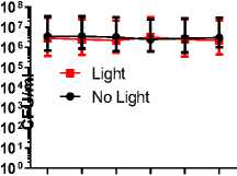
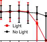
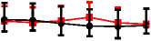
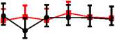
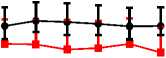
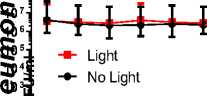
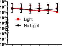
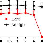
Then the bactericidal activity of these compounds was assessed by CFU counting in cultures irradiated for 120 min at 530 nm. As can be seen from the Figure 2, compound 3 at a concentration of 8 μg/mL provides complete cell death of S. pneumonia and C. albicans , while complete cell death of S. aureus was achieved at a concentration of 2 μg/mL. Thus, compound 3 appears to be of interest for photodynamic therapy of infections caused by S. aureus and C. albicans . Of note, in several works another diiod-BODIPY photosensitizers demonstrated bactericidal activity at 0.5-5 μM that corresponds to 1-10 μg/ml (Lincoln et al, 2017; Caruso et al. , 2012), reflecting that the efficiency of synthesized compounds is in the same range. By contrast, the fungicidal activity of photoditazine was observed at concentrations higher than 50 μg/ml (reviewed in emenov et al. , 2021) compared to 8 μg/mL for compound 3, suggesting its potential for the antifungal treatment.
ince many infectious diseases caused by S. aureus and C. albicans are often associated with the formation of biofilms, including fungal-bacterial ones, whose barrier properties can impede the penetration of compounds to the cells, we studied the antimicrobial activity of compound 3 against mono- and dual-species biofilms of S. aureus and C. albicans. ince compounds 1 and 2 demonstrated low efficiency against planktonic cells, they were not investigated as antibiofilm agents, despite the good biofilm-penetrating properties were reported for the BODIPY-terpene conjugates (Guseva et al., 2024). The biofilms were grown for 24 hours under static conditions, after which the medium was replaced, compound 3 was added at concentrations of 2-64 μg/mL, and irradiated with visible light (530 nm) for 2 hours. Control plates were incubated in the dark. Cell viability was determined by CFUs counting (Figure 3).
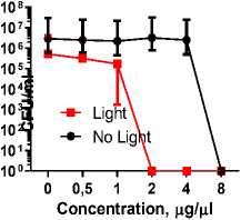
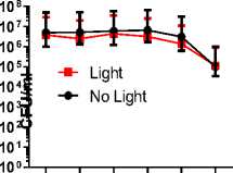
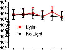
As can be seen in Figure 3, 3 readily penetrates S. aureus biofilm and leads to cell death at a concentration of 4 μg/mL. However, no bactericidal activity was observed against biofilms formed by C. albicans and a mixed culture.
Biofilm is a multicomponent system comprising both the cells themselves and an extracellular matrix consisting of various organic compounds, the barrier function of which can prevent compounds from penetrating the cells. Therefore, to understand whether the lack of antibacterial activity is due to the compound's inability to penetrate the biofilm matrix as well as to determine its intracellular localization, confocal laser scanning microscopy (CL M) was performed. Microscopy was made after 2 hours of irradiation with visible light (530 nm) of the 24-hour-old biofilm treated with the test compound. Compound 3 have the emission in the red channel, and therefore CFW (Calcofluor White) which stains the cell walls of staphylococci and yeast was additionally added and DiOC6 (3,3'-dihexyloxacarbocyanine iodide) which is reduced by membrane potential and thus is an indicator of cells viability. CFW was detected the blue channel, while DiOC6 was recorded in green channel (Figure 4).
The microscopy allows concluding that compound 3 has a selective tropism for cells in the biofilm. In C. albicans , it is slightly accumulated in some cells of the population, apparently, depending on the cell age or physiological state, as it was demonstrated for other BODIPY dyes (Guseva et al. , 2024), but the low intensity of the fluorescence indicates low efficiency of penetration into the cells. At the same time, the compound penetrates into S. aureus cells with greater efficiency, and apparently preferentially binds to the cell membrane (Guseva et al. , 2024).
Table 1. Concentrations of diiod derivatives of BODIPY repressing the microbial growth for 90 min under irradiation at 530 nm for 120 min (μg/mL).
|
Compound |
S. aureus |
C. albicans |
S. sobrinus |
S. pneumonia |
|
1 |
64 |
>64 |
>64 |
>64 |
|
2 |
64 |
>64 |
>64 |
>64 |
|
3 |
2 |
4 |
>64 |
4 |
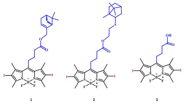
Figure 1. tructuresof BODIPYs 1–3.
Compound 1
Compound 2
Compound 3
(fl sj 3 (5
co
0 0,5 1 2 4 8
Concentration, µ g/ µ l
10 2 4
10 1 I
-■- Light
-•- No Light
0 0,5 1 2 4 8
Concentration, µ g/ µ l
108I
10 7 I
10 6 j
10 4 I
102 Э
10 1 j
(fl c <0 £
0 0,5 1 2 4 8
Concentration, µ g/ µ l
0 0,5 1 2 4 8
Concentration, µ g/ µ l
0 0,5 1 2 4 8
Concentration, µ g/ µ l
(/)1081 510 7 ,c10 6 c10 5 ■Q104 f 2 10 3 W)1025
+ Light
-•- No Light
CO10 0
107 i
10 6 I
10 4 I
10 1 I
-■- Light
-•- No Light
0 0,5 1 2 4 8
Concentration, µ g/ µ l
0 0,5 1 2 4 8
Concentration, µ g/ µ l
10 7 I
10 6 I
10 4 I
10 2 I
10 1 I
-■- Light
-•- No Light
10 8
100J---- 1-------- 1-------- 1-------- 1--------1--------r
0 0,5 1 2 4 8
Concentration, µ g/ µ l
0 0,5 1 2 4 8
Concentration, µ g/ µ l
"I---------1---------1---------1---------1---------г
0 0,5 1 2 4 8
Concentration, µ g/ µ l
108I
10 7 I
10 6 I
10 4 I
10 2 J
10 1 j
Concentration, µ g/ µ l
Figure 2. The amount of residual CFUs in cultures treated with diiod derivatives 1-3 of BODIPY with exposition to irradiation at 530 nm (light) or incubation in the dark (no light) for 120 min. Median values with range are shown (n=3).
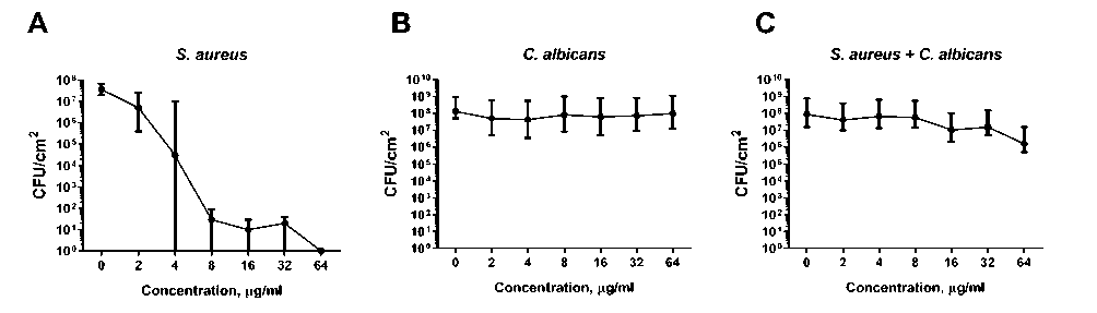
Figure 3. The amount of residual CFUs in S. aureus (A), C. albicans (B) mono- and mixed S. aureus - C. albicans (C) biofilms treated with compound 3 with exposition to irradiation at 530 nm for 120 min. Median values with range are shown (n=3).
C. albicans s. aureus S. aureus + C. albicans
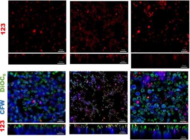
Figure 4. Confocal laser scanning microscopy of mono- and dual species biofilms of C. albicans and S. aureus after treatment with compound 3 followed by irradiation at at 530 nm. The scale bar is 10 µm.
CONCLUSIONS
Thus, compound 3 is of interest for the development of agents for photodynamic therapy of infections caused by S. aureus and C. albicans , as well as infections associated with the formation of S. aureus biofilms. Nevertheless, the improvement of the structure for better penetration into the yeasts as well as into the biofilm are required to increase the therapeutic efficiency.
CONFLICTS OF INTEREST
All authors declare that they have no conflicts of interest.
ACKNOWLEDGEMENTS
This research was funded by the Russian cience Foundation (grant no. 24-14-00194),