
Role of Ribosomal Protein S6 in Maintaining Translational Integrity in Plants under Osmotic Stress
Journal: Journal of Stress Physiology and Biochemistry @jspb
Article in issue: 3 т.22, 2026.
Free access
Regulation of protein synthesis is essential for cellular adaptation to environmental stress. In contrast to animals and yeast, plants lack several canonical mechanisms of rapid translational repression, suggesting the existence of alternative strategies for controlling ribosome activity. Increasing evidence indicates that discrete fragmentation of ribosomal RNA may represent one such mechanism in plant cells. In this study, we examined the role of ribosomal protein S6 (RPS6) in stress-associated 18S rRNA fragmentation in Arabidopsis thaliana. Wild-type (Col-0) and rps6a knockout protoplasts were subjected to osmotic stress (250 mM NaCl). Ribosomal RNA integrity was assessed by denaturing polyacrylamide gel electrophoresis followed by Northern blot hybridization using probes specific to the 5′ and 3′ termini of 18S rRNA. Osmotic stress induced significant accumulation of a 75-nt 5′-terminal fragment in wild-type cells. In the RPS6A knockout line, elevated basal levels of this fragment were observed even in the absence of stress. In contrast, the ~100-nt 3′-terminal fragment exhibited reduced abundance in the mutant background and showed distinct stress-dependent dynamics. These findings demonstrate that RPS6 contributes to maintaining 18S rRNA structural integrity and differentially modulates region-specific fragmentation under osmotic stress. The results support a model in which discrete 18S rRNA fragmentation represents a ribosome-centered layer of translational regulation integrated with stress-responsive signaling pathways in plants.
Short address: https://sciup.org/143186156
IDS: 143186156
Text of the scientific article Role of Ribosomal Protein S6 in Maintaining Translational Integrity in Plants under Osmotic Stress
Protein biosynthesis is one of the most energyconsuming processes in the cell. Therefore, under unfavorable conditions, most eukaryotic organisms employ mechanisms that enable rapid and reversible suppression of mRNA translation. In animals and yeast, these mechanisms have been extensively characterized and include phosphorylation of translation factors such as eEF2 and eIF2α, as well as regulation of translation initiation through eIF4E-binding proteins (4E-BPs). Together, these pathways efficiently reduce global mRNA translation while simultaneously allowing preferential synthesis of stress-responsive proteins (Irshad & Sharma, 2024; Baird & Wek, 2012; Hernández et al. , 2010; Browning & Bailey-Serres, 2015).
In plant cells, the situation differs fundamentally. Plants lack endogenous kinase activity toward eEF2 under both normal and stress conditions (Smailov et al. , 1993; Gallie et al. , 1997), and phosphorylation of eIF2α does not result in pronounced inhibition of mRNA translation (Zhigailov et al. , 2000; Shaikhin et al. , 1992). Moreover, orthologs of eIF4E-binding proteins have not been identified in plant genomes, and regulatory mechanisms well described in animals and yeast either operate only to a limited extent or are absent altogether. These observations indicate that plants rely on alternative strategies for regulating protein biosynthesis that differ from classical eukaryotic models (Browning & Bailey-Serres, 2015).
One such alternative mechanism of mRNA translation regulation may involve discrete fragmentation of ribosomal RNA, particularly 18S rRNA, a structural component of the small (40S) ribosomal subunit. We previously demonstrated that various stress conditions in plant cells induce highly specific, non-random cleavage of 18S rRNA, leading to the accumulation of defined 5′- and 3′-terminal fragments. Identified 5′ fragments were approximately 75 nt and 132–134 nt in length, while a 3′-terminal fragment of about 100 nt was also detected (Zhanybekova et al., 1996; Zhigajlov et al., 2022; Zhigailov et al., 2023). The reproducibility, specificity, and stress-induced accumulation of these fragments indicate that 18S rRNA cleavage represents a regulated process rather than nonspecific RNA degradation. Transcriptome-wide analyses further confirm that breaks in ribosomal RNAs occur discretely and affect defined regions of the molecule. Thus, 18S rRNA fragmentation in plants may serve a functional role in translational suppression under stress conditions (Chen et al., 2017; Henras et al., 2015).
A central component of translational regulation in eukaryotes is the Target of Rapamycin (TOR) signaling pathway. In animals, TOR coordinates cell growth, ribosome biogenesis, and protein synthesis, in part through activation of S6 kinase (S6K) and subsequent phosphorylation of ribosomal protein S6. This pathway is well characterized and is regarded as a key regulator balancing growth-related processes with stress responses (Dennis et al. , 1999; Thomas, 2000; Abraham, 2002).
Components of the TOR signaling pathway have also been identified in plants. In Arabidopsis thaliana , homologs of TOR, RAPTOR, and S6K have been characterized, and TOR activity has been shown to be essential for embryogenesis and normal plant growth (Menand et al. , 2002; Deprost et al. , 2007). RAPTOR1 interacts with TOR and S6K1, and S6K1 phosphorylates ribosomal protein S6. Moreover, plant S6K activity is sensitive to osmotic stress, supporting the involvement of the TOR–S6K pathway in stress-responsive regulation (Turck et al. , 2004; Robaglia et al. , 2012). A. thaliana contains two genes encoding functionally equivalent cytoplasmic ribosomal protein S6 isoforms, RPS6A and RPS6B . It has been shown that a non-phosphorylatable form of S6 largely retains the ability to support global translation, normal plant growth, and photosynthetic activity. This indicates that S6 phosphorylation is not strictly required for the basal function of the ribosome in plants (Creff et al. , 2010; Dasgupta, et al. 2024).
Thus, despite the presence of the TOR–S6K–S6 signaling pathway, its role in directly switching off translation in plants appears to be limited. This observation suggests that S6 should be considered not only as a target of signaling regulation but also as a structural component of the ribosome potentially involved in alternative mechanisms controlling translational activity.
Ribosomal protein S6 is located on the surface of the 40S subunit and interacts with functionally important domains of 18S rRNA (Creff et al. , 2010; Dasgupta, et al. 2024). Alterations in its state—whether through phosphorylation, loss of one isoform, or disruption of interactions with other ribosomal components—may affect the spatial organization of 18S rRNA and, consequently, its susceptibility to endonucleolytic cleavage. In this context, discrete 18S rRNA fragmentation can be viewed as a ribosome-centered mechanism of translational regulation, in which structural modification of the 40S subunit reduces translational activity without complete disassembly of the ribosomal machinery. We propose that RPS6 and the associated TOR–S6K pathway may act as modulators of this process, influencing the initiation or extent of 18S rRNA fragmentation under stress conditions.
To investigate stress-induced alterations in the plant translational apparatus, a protoplast-based system was employed. Protoplasts represent a convenient and reproducible model that allows analysis of cellular processes under controlled external stimuli. The absence of a cell wall increases sensitivity to stress and facilitates reagent accessibility, making protoplasts a versatile system for studying translational regulation and structural changes in ribosomes (Yoo et al. 2007; Sheen, 2001; Davey et al. 2005).
The aim of this study was to examine the role of ribosomal protein S6 in discrete 18S rRNA fragmentation in plants under stress conditions. To this end, we used wild-type and rps6a knockout lines of A. thaliana , combined with a protoplast system and analysis of 18S rRNA fragmentation by hybridization with DIG-labeled probes complementary to the 5′ and 3′ termini of the molecule.
MATERIALS AND METHODS
Plant material and growth conditions
Shoots of Arabidopsis thaliana wild-type Columbia-0 (Col-0) and the rps6a line (SALK_048825), kindly provided by Dr. L.A. Ryabova (University of Strasbourg, France), were used in this study. The A. thaliana rps6a line carries a knockout of the RPS6 gene encoding one of the ribosomal protein S6 isoforms. Genotypic characteristics of this line were described previously (Creff et al., 2010).
Protoplast isolation
Protoplasts were isolated from rosette leaves of 7– 14-day-old A. thaliana seedlings according to a standard protocol with minor modifications (Yoo et al. 2007). Leaf tissue was cut into strips approximately 0.5–1 mm wide and incubated in an enzymatic solution containing 1.5% (w/v) Cellulase R10 (Sigma-Aldrich), 0.4% (w/v)
Macerozyme R10 (Sigma-Aldrich), 0.4 M mannitol, 20 mM KCl, 20 mM MES (pH 5.7), and 10 mM CaCl₂.
Vacuum infiltration was performed at 0.1 bar for 1 min to facilitate enzyme penetration into leaf tissues. Samples were incubated overnight in the dark at 24–26 °C with gentle shaking. After enzymatic digestion, the suspension was filtered through a 70–75 μm nylon mesh and centrifuged at 1000 × g for 5 min.
The protoplast pellet was resuspended in chilled W5 washing buffer (154 mM NaCl, 125 mM CaCl₂, 5 mM KCl, 2 mM MES, pH 5.7). Protoplasts were incubated on ice for 30 min, centrifuged again, and resuspended in MMG buffer (0.5 M mannitol, 15 mM MgCl₂, 4 mM MES, pH 5.7). Protoplast concentration and viability were assessed microscopically using 1.0% Evans blue (Merck) staining and a hemocytometer (Beker et al. 1990).
Stress treatment of protoplasts
To evaluate the effect of osmotic stress, protoplasts were incubated in the presence of 250 mM NaCl. Control samples were incubated under identical conditions without the addition of the stress agent. Treatments were carried out at room temperature for the indicated time periods, after which protoplasts were immediately used for RNA isolation.
RNA isolation
Total RNA was extracted from protoplasts using TRI Reagent (Sigma-Aldrich) containing guanidine thiocyanate, according to the manufacturer’s instructions. The RNA pellet was washed with 75% ethanol, air-dried, and dissolved in RNase-free water. RNA concentration and purity were determined spectrophotometrically.
Denaturing RNA electrophoresis (PAGE)
RNA and rRNA fragmentation products were analyzed by electrophoresis under denaturing conditions in 10% polyacrylamide gels containing 8 M urea, prepared in 1× TBE buffer (89 mM Tris, 89 mM boric acid, 2 mM EDTA, pH 8.3).
Prior to loading, RNA samples were mixed with a denaturing RNA loading buffer containing formamide (Sigma-Aldrich), heated at 65 °C for 10 min, and rapidly cooled on ice.
Electrophoresis was performed at constant voltage until sufficient separation of RNA fragments was achieved. After electrophoresis, gels were stained in 0.5× TBE containing SYBR Gold (Thermo Fisher Scientific) to visualize the overall RNA profile. Imaging was performed using a Safe Image gel documentation system (Thermo Fisher Scientific) with a blue-red light source.
The same stained gel was subsequently used for RNA transfer onto a Hybond-N+ nylon membrane (Roche). Semi-dry transfer was carried out using a transblot apparatus (Sci-Plas) in 0.1× TBE buffer at 250 mA for 30 min. RNA was crosslinked to the membrane by UV irradiation using a UV crosslinker (UVP) for 2 min at 10 mJ/cm².
Probe synthesis and labeling
To detect 18S rRNA fragments, the following oligonucleotide probes were used: 5′18S (5′-
ACAAGCATATGACTACTGGCAGGATCAACCAGGTA) and 3′18S (5′-
CAATGATCCTTCCGCAGGTTCACCTACGGAAACCT) (Zhanybekova et al. , 1996), complementary to the 5′ and 3′ terminal regions of plant 18S rRNA, respectively.
Probes were labeled with digoxigenin (DIG) using the DIG Oligonucleotide 3′-End Labeling Kit (Roche) according to the manufacturer’s instructions. The resulting 5′18S-DIG and 3′18S-DIG probes were used for Northern blot analysis.
Northern blotting
Prior to hybridization, membranes were incubated at 55 °C for 60 min in hybridization buffer containing 50% formamide, 5× SSC, 0.1% N-lauroylsarcosine, 0.02% SDS, and 2% casein-based blocking reagent (Roche) to prevent nonspecific binding.
DIG-labeled probes were added to the same buffer, and hybridization was carried out overnight (12–16 h) at 55 °C with gentle agitation in a hybridization oven (HIR10M, Grant Boekel).
After hybridization, membranes were washed twice for 10 min in 2× SSC containing 0.1% SDS and twice for 15 min in 0.1× SSC containing 0.1% SDS.
Membranes were then incubated in blocking buffer (1× maleic acid buffer, pH 7.5, 2% blocking reagent) for 60 min at room temperature, followed by incubation with anti-DIG antibodies conjugated to alkaline phosphatase (Roche) diluted 1:5000 in the same buffer for 60 min.
After antibody incubation, membranes were washed twice for 15 min in 1× maleic acid buffer containing 0.3% Tween-20 and twice for 15 min in 1× maleic acid buffer.
For signal detection, membranes were incubated for 5 min in detection buffer (100 mM Tris-HCl, pH 9.5, 100 mM NaCl) and treated with a chemiluminescent substrate for alkaline phosphatase (CSPD, Roche). Membranes were exposed to X-ray film for 1 h, and films were developed using standard procedures.
Densitometric analysis and statistical evaluation
Densitometric analysis was performed using ImageJ software (version 1.42q). All measurements were carried out in three independent replicates. Statistical significance was assessed using Student’s t-test, with p < 0.05 considered significant compared to the control.
RESULTS AND DISCUSSION
To analyze the effect of stress factors on 18S rRNA integrity, a protoplast system of A. thaliana was used. Separation of RNA under denaturing conditions followed by hybridization with probes complementary to the 5′ and 3′ termini of 18S rRNA revealed the formation of discrete fragments that were absent or weakly represented in control samples.
Upon osmotic treatment (NaCl), accumulation of a 5′-terminal fragment approximately 75 nucleotides in length was observed (Fig. 1b). In wild-type protoplasts, this fragment was virtually undetectable under control conditions; however, its accumulation markedly increased after NaCl treatment. In the A. thaliana rps6a mutant line, an elevated level of the 5′-terminal 18S rRNA fragment was already detected in the absence of stress and further increased following osmotic treatment.
Analysis of 3′-terminal fragmentation of 18S rRNA is shown in Figure 2.
Quantitative analysis of Northern blot signals following densitometry (Table 1), normalized to 5.8S rRNA, demonstrated that in wild-type protoplasts the basal level of the 75-nt 5′-terminal 18S rRNA fragment was minimal. Osmotic stress (NaCl) induced a statistically significant increase in this fragment (p = 0.0199). In the rps6a mutant line, the level of the 75-nt fragment was significantly elevated even in the absence of stress compared to wild-type Col-0 protoplasts (p < 0.0001). Although NaCl treatment further increased accumulation of the 5′-terminal fragment in the mutant line, this increase did not reach statistical significance (p
= 0.1036). These findings indicate that ribosomal protein RPS6A contributes to limiting both basal and stress-induced discrete fragmentation of 18S rRNA.
Quantitative analysis of Northern blot signals corresponding to the 3′-terminal 18S rRNA fragment (~100 nt), normalized to 5.8S rRNA, revealed differences in the accumulation dynamics of this fragment compared with the 5′-terminal fragments. In wild-type protoplasts, the basal level of the 3′ fragment was high, whereas NaCl treatment led to a decrease in its relative intensity (p = 0.0044). In the rps6a mutant line, the level of the 100-nt 3′ fragment was significantly lower (p = 0.0042) than in the wild type. Most likely for this reason, NaCl treatment did not result in significant changes in the accumulation of the 100-nt 3′-terminal 18S rRNA fragment in the mutant background (p = 0.2559). These findings indicate that the accumulation of specific discrete 18S rRNA fragments in plant cells depends on the phosphorylation status of RPS6. Moreover, the stability of the 3′- and 5′-terminal regions of 18S rRNA appears to be regulated differently.
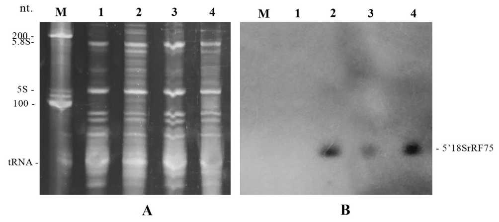
Figure 1. Distribution of 5′-terminal discrete fragments of 18S rRNA in Arabidopsis thaliana protoplasts A -SYBR-stained gel. B - Northern blot hybridized with the 5′18S-DIG probe. The arrow indicates the 5′-terminal discrete fragment of approximately 75 nt. Lane assignment: M — LowRange RNA ladder (Fermentas); 1 — RNA from wild-type (Col-0) protoplasts without stress treatment; 2 — RNA from Col-0 protoplasts treated with 250 mM NaCl; 3 — RNA from rps6a ( RPS6A knockout) protoplasts without stress treatment; 4 — RNA from r ps6a protoplasts treated with 250 mM NaCl.
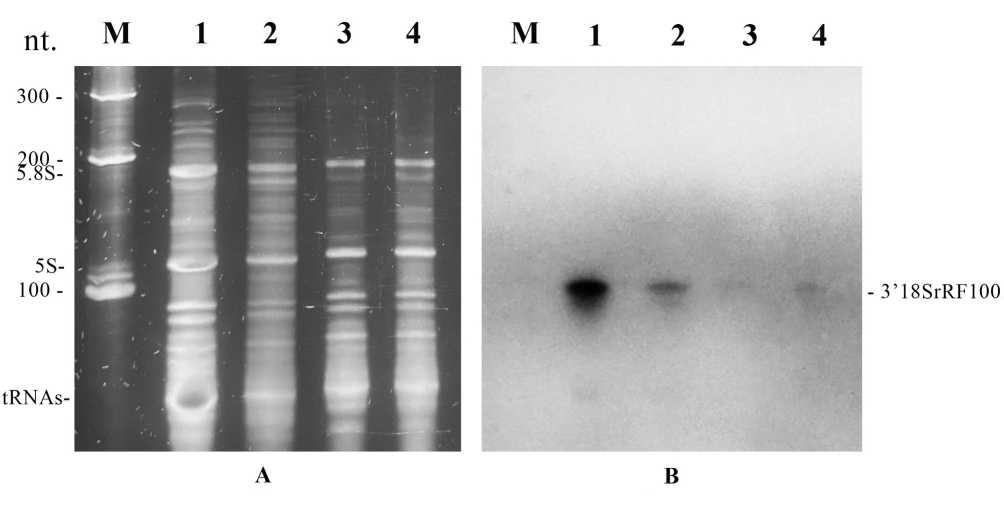
Figure 2. Distribution of 3′-terminal discrete fragments of 18S rRNA in Arabidopsis thaliana protoplast s. A -SYBR-stained gel. B - Northern blot hybridized with the 3′18S-DIG probe. The arrow indicates the 3′-terminal fragment of approximately 100 nt. Lane assignment: M — LowRange RNA ladder (Fermentas); 1 — RNA from wild-type (Col-0) protoplasts without stress treatment; 2 — RNA from Col-0 protoplasts treated with 250 mM NaCl; 3 — RNA from rps6a protoplasts without stress treatment; 4 — RNA from rps6a protoplasts treated with 250 mM NaCl.
Table1. Densitometric quantification of discrete 18S rRNA fragmentation (ImageJ 1.42q)
|
Parameter |
Type of protoplasts and incubation conditions |
|||
|
WT / no stress |
WT / 250 mМ NaCl |
s6a / no stress |
s6a / 250 mМ NaCl |
|
|
5′-terminal 18S rRNA fragmentation |
||||
|
S 5,8SrRNA |
9621,14 |
8529,03 |
9251,36 |
3956,24 |
|
S 5’18SrRF75 |
23,48 |
5559,63 |
1913,22 |
5398,92 |
|
S 5’18SrRF75 / S 5,8SrRNA |
0,002 ±0,002 |
0,685 ±0,170 |
0,207 ±0,001 |
1,612 ±0,852 |
|
3′-terminal 18S rRNA fragmentation |
||||
|
S 5,8SrRNA |
11838,12 |
3309,175 |
3696,03 |
2863,22 |
|
S 3’18SrRF100 |
19968,848 |
3186,347 |
265,29 |
367,82 |
|
S 3’18SrRF100/ S 5,8SrRNA |
1,68 ± 0,227 |
0,96 ± 0,017 |
0,07 ± 0,007 |
0,13 ± 0,032 |
Notes: WT — wild-type A. thaliana (Col-0) protoplasts; rps6a — protoplasts derived from A. thaliana plants carrying a knockout of the rps6a gene; S5.8SrRNA — densitometric intensity of the 5.8S rRNA reference band on the gel; S5′18SrRF75 — densitometric intensity of the 75-nt 5′-terminal 18S rRNA fragment band; S3′18SrRF100 — densitometric intensity of the 100-nt 3′-terminal 18S rRNA fragment band.
The data further demonstrate that discrete 18S rRNA fragmentation in plant cells represents a heterogeneous and multi-level process. Analysis of the 5′- and 3′-terminal fragments revealed fundamental differences in their regulation in response to stress. Whereas osmotic stress led to pronounced accumulation of the 75-nt 5′-terminal fragment, the dynamics of the ~100-nt 3′-terminal fragment were distinct. In wild-type protoplasts, the basal level of the 3′ fragment was relatively high under control conditions, while NaCl treatment resulted in a reduction of its relative abundance. This suggests that the formation and/or stability of different 18S rRNA fragments may be controlled by independent mechanisms that differ in their sensitivity to stress type and cellular physiological state.
Of particular interest is the influence of components of the TOR–S6K–S6 signaling pathway on the balance between distinct fragmentation events. In the rps6a mutant line, which lacks isoform A of the RPS6 protein, a marked reduction in discrete 3′-terminal 18S rRNA fragmentation was observed compared with wild-type A. thaliana (Col-0). This reduction may, in turn, affect overall mRNA translation levels in plant cells, since the integrity of the 3′ region of 18S rRNA is critical for translation initiation (Zhanybekova et al. , 1996). This consideration may help explain why the RPS6 phosphorylation pathway is conserved in plants.
Taken together, these results support the view that 18S rRNA fragmentation functions as a mechanism of mRNA translational regulation in plant cells under stress conditions.
CONCLUSIONS
The present study demonstrates that discrete 18S rRNA fragmentation in plant cells represents a regulated and multi-level process influenced both by environmental stress and by the structural status of ribosomal components. Using an Arabidopsis thaliana protoplast system, we showed that osmotic stress induces reproducible and region-specific changes in the accumulation of 18S rRNA fragments. The differential behavior of 5′- and 3′-terminal fragments indicates that fragmentation is not a nonspecific degradation event but rather a controlled process affecting defined structural domains of the small ribosomal subunit.
Our findings further indicate that ribosomal protein RPS6A plays a role in maintaining 18S rRNA integrity. Loss of RPS6A alters basal fragmentation patterns and modifies the stress response of specific rRNA regions. These results suggest that RPS6 contributes to the structural stabilization of the 40S subunit and modulates the susceptibility of 18S rRNA to cleavage under stress conditions. Importantly, the observed effects extend beyond classical views of RPS6 solely as a phosphorylation target within the TOR–S6K pathway, supporting its additional structural and regulatory functions within the ribosome.
The distinct regulation of 5′- and 3′-terminal fragments further implies that discrete 18S rRNA fragmentation may selectively influence ribosome functionality rather than simply reducing ribosome abundance. Given the functional importance of the 3′ region of 18S rRNA in translation initiation, structural alterations in this domain may affect translational efficiency and contribute to adaptive reprogramming of protein synthesis under stress.
Taken together, our data support a model in which ribosome-centered structural modulation constitutes an additional layer of translational regulation in plants. Discrete 18S rRNA fragmentation may therefore act as a mechanism linking environmental stress perception, signaling cascades, and functional remodeling of the translational apparatus. Future studies aimed at identifying the nucleases responsible for site-specific cleavage and the factors controlling fragment stability will be essential for elucidating how this mechanism integrates into broader stress-response networks.
ACKNOWLEDGMENTS
The authors thank Dr. L.A. Ryabova (Université de Strasbourg) for kindly providing A. thaliana seed lines used in this study.
This work was supported by the Committee of Science of the Ministry of Science and Higher Education of the Republic of Kazakhstan under grant AP26195475 “The fragmentation of 18S rRNA as a new molecular mechanism of plant stress response” and program BR27195585 “Creation of new domestic test systems and search for potential biomarkers for the diagnosis of socially significant” (2025–2027).
CONFLICTS OF INTEREST
All authors declare that they have no conflicts of interest.