Structural myocardial catastrophe under the influence of chronic neurogenic pain due to development of B16 melanoma in female mice

Author: Shikhlyarova A.I., Frantsiyants E.M., Kaplieva I.V., Zhukova G.V., Neskubina I.V., Nepomnyashchaya E.M., Trepitaki L.K., Kozel Y.Yu., Yengibaryan M.A., Vereskunova A.A., Babieva S.M., Legostaev V.M., Kharagezov D.A., Volkova V.L., Kit O.I.
Journal: Cardiometry @cardiometry
Section: Review
Article in issue: 22, 2022.
Free access
Topicality. Most of the inhabitants on the planet have directly experienced certain manifestations of pain, and in the case of chronic pain sensations, some significant changes may occur not only in the psycho-emotional sphere of a human individual, but also some functional alterations in the life maintenance systems. There are two global problems of medicine which are associated with oncology and cardiovascular pathology, and, taking into account the prevalence of pain symptoms of various etiologies, a pathological triangle is formed, each side of which affects the overall configuration thereof. There is a need to understand these pathological relationships in order to determine the real possibilities of breaking them and correcting such common complex life situations. Any attempt to create a concept and visually verify the real pathological changes in the heart, when modeling a combination of chronic neurogenic pain and a malignant process, is relevant that is the aim of this research work. We targeted the morphological picture of the heart in female mice with chronic neurogenic pain (CNP) and the growth of B16 melanoma and found a rather tough scenario of unfolded events. Materials and methods. We used female mice of the C57BL/6 line with a normal genotype. Chronic neurogenic pain (CNP) was produced by bilateral ligation of the sciatic nerve. Against that background, all animals were subcutaneously transplanted with melanoma B16/F10. After decapitation, the isolated heart preparations were carried out according to the stages of morphological preparation for staining sections with hematoxylin-eosin, followed by morphological examination of the structure with the Leica DM LS2 microscope furnished with an Olympus optical.C-5050 Zoom video camera and Morfotest software. Photographing was conducted with magnifications x10, x40, x100. Results. A pronounced pro-oncogenic effect of pain stimulation of the neurogenic nature was revealed, which consisted in an earlier manifestation of the tumor growth, large-scale metastasizing even to atypical target organs, and the formation of a pre-terminal state at an earlier time. At the same time, morphological correlates of prolonged damage to the heart at the level of the valves and the ventricular wall were determined, the key elements of which were ischemia, total longitudinal splitting of muscle fiber bundles, blood filling of large vessels, hemorrhages, deep cell dystrophy of cardiomyocytes, myolysis, macrofocal necrosis (myomalacia), an accumulation of necrotic masses, fibrosis and eosinophilic infiltration. Visualization of the nuclei of cardiomyocytes indirectly indicated the switch of the death program to the non-apoptotic pathway, i.e. necrosis as a result from the “tumor-caused” depletion of the energy reserves of cardiomyocytes. Conclusion. The simulated hypertrophied manifestations of myocardial catastrophe in the experiment expand the concept of a prolonged infarction and provide a basis for predicting and preventing a negative course of events in difficult patients with persistent pain syndromes and comorbid pathology against the background of the development of a malignant process.
Chronic neurogenic pain, b16 melanoma, heart, morphological changes in the myocardium, female mice
Short address: https://sciup.org/148324607
IDR: 148324607 | DOI: 10.18137/cardiometry.2022.22.2837
Text of the scientific article Structural myocardial catastrophe under the influence of chronic neurogenic pain due to development of B16 melanoma in female mice
Alla I. Shikhlyarova, Elena M. Frantsiyants, Irina V. Kaplieva, Gаlina V. Zhukova, Irina V. Neskubina, Evgenia M. Nepomnyashchaya, Lidia K. Trepitaki, Yulia Yu. Kozel, Marina A. Yengibaryan, Alexandra A. Vereskunova, Stella M. Babieva, Vladislav M. Legostaev, Dmitriy A. Kharagezov, Viktoria L. Volkova, Oleg I. Kit. Structural myocardial catastrophe under the influence of chronic neurogenic pain due to development of B16 melanoma in female mice. Cardiometry; Issue 22; May 2022; p. 28-37; DOI: 10.18137/car-diometry.2022.22.2837; Available from: http://www.cardiometry. net/issues/no22-may-2022/structural-myocardial-catastrophe
Cardiovascular diseases are the most common causes of disability and death in the world, and the number of patients of this category of diseases has been increasing. Heart failure (HF) is a progressive myocardial remodeling disorder characterized by the impaired cardiac function and circulatory congestion [1]. Acute myocardial infarction (MI) is a dramatic, confusing, stressful event. Despite the fact that various therapeutic and preventive measures are being implemented, there has not been a significant reduction in cardiovascular disease (CVD) achieved worldwide. The World Health Organization (WHO) predicts that cardiovascular disease deaths in the world will rise from 17.1 million in 2016 to 23.4 million in 2030 [2]. Studies have shown that the occurrence rate of this complication in Russia is 3-4 times higher than in the countries of the European Union and the USA [3]. Myocardial infarction leads to myocardial ischemia, blockage of microcirculation, apoptosis of cardiomyocytes and necrosis in the area of infarction that leads to vicarious fibrosis of the heart [1].
Chronic systemic inflammation is a recognized cardiovascular risk factor which is based on the immune activation as the main pathophysiological factor [4]. Chronic intestinal inflammation caused by Crohn’s disease and ulcerative colitis is associated with an increased risk of coronary, cerebrovascular, and peripheral arterial diseases [5]. Approximately one in every three individuals worldwide experiences a chronic and painful disease [6]. Osteoarthritis (OA) and chronic low back pain are the most common diseases that contribute significantly to the overall burden of the life years lived with disability [7]. In Japan, for example, chronic pain from osteoarthritis affects approximately 25.3 million people aged over 40, and the occurrence of chronic leukemia among people over 50 is estimated at about 24.8% [8]. Chronic neurogenic pain adversely affects people’s health by limiting their physical and functional capabilities; therefore, chronic pain control is of paramount importance. A meta-analysis showed that patients with chronic pain had a 24% higher risk of developing cardiovascular disease than those without the latter [9]. The male gender and comorbidities were found to increase the incidence of acute myocardial infarction [10].
Previously, we have shown that the reproduction of chronic neurogenic pain (CNP) produced by bilateral ligation of the sciatic nerves has a stimulating effect on the growth of experimental melanoma in females and promotes an early tumor appearance and metastasizing, as well as a reduction in the life span of animals [11]. In addition, it has been determined that chronic neurogenic pain associated with a malignant process aggravates the dysfunction of the mitochondria of the heart cells with destabilization of the respiratory chain mediated by free radical oxidation processes [12, 13]. Morphological studies of areas of damage to the heart muscle show that in the myocardium detected are hemorrhage, foci of necrosis, ruptures of some individual cells, focal infiltration of leukocytes, fibrinous necrosis of the walls of blood vessels, dilated heart cavities, and blood clots in the lumen [14]. Microscopy of smears of a suspension of cardiac mitochondria at 2-3 weeks of the melanoma growth against the background of CNP has revealed a process of cold glow of high brightness, i.e. chemiluminescence [15]. It is interesting that the detected abnormality of the contractile function of the heart in the dynamics of the standard growth of B16 / F10 melanoma, regarded as manifestations of tumor stress, or rather, the systemic effect made by the tumor on the body, has not been accompanied by signs of damage to the heart wall in macroscopic and morphological examination.
Knowledge of the mechanisms connected with the progression of myocardial infarction associated with chronic neurogenic pain is necessary for the development of new treatment technologies.
The aim of our research work is to study morphological changes in the heart of female mice with the development of a malignant tumor (B16/F10 melanoma model) under the conditions of chronic neurogenic pain.
Materials and methods
In this research work, female mice of the C57BL/6 line with a normal genotype with an initial individual weight of 20–25 g were used. The animals were supplied by the Andreevka Research Center for Biomedical Technologies at the Federal Medical and Biological Agency (Moscow Region). All mice were divided into experimental groups as follows: the mice with B16/ F10 melanoma as the comparison group; the mice with chronic neurogenic pain (CNP) + B16/F10 melanoma as the main test group.
The laboratory animals (mice) were kept under natural light conditions with free access to water and food. Work with animals was carried out in accordance with the rules of the “European Convention for the Protection of Vertebrate Animals used for Experimental and other Scientific Purposes” (Directive 86/609/EEC), as well as in accordance with the International Guiding Principles for Biomedical Research Involving Animals and Order No. 267 “Approval of the Rules of Laboratory Practice” dated June, 19, 2003 issued by the Ministry of Health of the Russian Federation.
Model of chronic neuropathic pain
All manipulations with the animals were carried out in a box. Tools, utensils and hands were disinfected in the conventional way, observing all asepsis conditions.
The model of chronic neurogenic pain (CNP) was reproduced by applying a ligature to the sciatic nerve on both sides under xylazolethyl anesthesia [16]. Anesthesia: xyl-zoletyl, 10 minutes before the main anesthesia; premedication: xylazine (Xila preparation) intramuscularly, at a dose of 0.05 ml/kg of body weight (according to the instructions), then after 10 minutes, Zoletil-50 was administered at a dose of 10 mg per 100 g of body weight. On post-operative day 14, mechanical allodynia and hyperalgesia were measured.
-
2 weeks after the reproduction of chronic neurogenic pain in the mice of the main group, the B16/F10 melanoma cells were transplanted into the C57BL/6 strain mice by standard subcutaneous injection of tumor suspension under the right shoulder blade in a volume of 0.5 ml of cell suspension in a 1:10 dilution with physiological saline solution. The animals of the comparison group were transplanted with the B16/ F10 melanoma cells in a similar way, but without reproduction of CNP. The tumor strain of murine melanoma B16/F10 was obtained from the Federal State Budgetary Institution “NN Blokhin National Medical Research Center of Oncology” at the Ministry of Health of the Russian Federation.
After decapitation, the isolated heart preparations were harvested according to the stages of morphological preparation for staining sections with hematoxy-lin-eosin, followed by the morphological examination of the structure with the Leica DM LS2 microscope furnished with an Olympus optical C-5050 Zoom video camera and Morfotest software. Photographing was conducted with magnifications x10, x40, x100.
Results
Our experiments made it possible to give some visual representations of the state of the heart in case of 30 | Cardiometry | Issue 22. May 2022
systematic neurogenic pain as a background condition for the development of oncological pathology.
Appearance of primary tumors
As shown in Table 1 given herein, the subcutaneous tumors appeared in the female mice of the main group with CNP as early as 1 week after inoculation, and after 2 weeks in the mice of the comparison group. That was a significant gap in the reduction of the duration of the latent period thereof, which might indicate the formation of conditions for a pro-oncogenic influence of the pain factor. The average volume of subcutaneously transplanted B16 melanoma in the female mice clearly confirmed that stimulating dynamics.
Direct indicators of average tumor volumes
The minimum size of the newly recorded subcutaneous tumors in the mice of the main group at week 1 was 2 times greater than that found in the comparison group at week 2. At the same time, the maximum tumor size at week 3 of the experiment in the mice from the comparison group (without pain) was 2.3 times greater than that recorded in the mice with pain (see Table 1 herein). However, in that case, the majority of mice with pain at week 3 were reported to be in a pre-terminal state, while we dealt with a completely different type of situation with the mice from the comparison group.
Additional signs of tumor growth stimulation
Despite the fact that in the mice with pain in the preterminal period, the volume of the tumor increased less intensively, some important features of pathogenesis modulated by pain were revealed such as the occurrence of a 2-focus growth already from week 1 in a considerable number of mice. In addition, in the mice from the main group, B16 melanoma demonstrated its more aggressive behavior, since it metastasized more often, sometimes to several organs, and affected atypical metastasis zones. Metastases in the mice of the main group were recorded as early as 1 week after the inoculation, and in the mice from the comparison group 4 weeks after thereof. Moreover, in the mice under the CNP conditions, the tumor metastasized not only to its typical sites, namely the liver, the spleen, and the lungs, but also to atypical regions like the heart and the uterus.
Table 1. Effect produced by chronic pain on the melanoma B16 growth in female mice
|
Week 1 |
Melanoma В16 |
Melanoma В16+pain |
||||||
|
Week 2 |
Week 3 |
Week 4 |
Week 1 |
Week 2 |
Week 3 |
|||
|
Number of mice |
22 |
22 |
22 |
16 |
28 |
22 |
10 |
|
|
о g о |
average size, cm3 |
no |
0.85±0.12 |
2.75±0.73 |
4.69±0.86 |
0.70±0.25 |
1.65±0.27 |
2.50±0.49 |
|
minimum size, cm3 |
– |
0.12 |
0.44 |
1.29 |
0.24 |
1.01 |
1.35 |
|
|
maximum size, cm3 |
– |
1.48 |
8.77 |
9.30 |
1.35 |
2.22 |
3.75 |
|
|
number of mice with tumor ulceration |
– |
no |
2 |
2 |
no |
6 (3–5 mm) |
6 (2–9 mm) |
|
|
number of mice with 2 tumor nodes |
– |
– |
– |
– |
8 |
8 |
2 |
|
|
metastases |
n/a |
n/a |
n/a |
avail. |
avail. |
avail. |
avail. |
|
|
metastasis zones |
– |
– |
– |
spleen (2) |
heart (2) |
heart (4), lungs (4) |
heart (6), lungs (4), liver (2), uterus (2) |
|
|
number of mice in pre-termi-nal state |
– |
– |
4 |
2 |
2 |
8 |
6 |
|
Morphological changes in the myocardium
From the presented data on the strong stimulating effect produced by pain on the development of melanoma B16, it was clear that some events associated with the early appearance of the B16 tumor in the female mice unfolded as early as at week 1, and the pre-termi-nal state in them was already identified. This indicated that even before the B16 melanoma transplantation, chronic pain had already sufficiently weakened or depleted the reserve capabilities of the functional systems in the organism. One of the main morphological manifestations of the response by the heart to the combined stressful effects of pathogenic factors, namely CNP plus the tumor progression, was a significant pathological change in the aortic and mitral valve systems and, as a result, hypertrophy of the myocardial ventricular wall recorded already 1 week after the B16 melanoma transplantation (see Figure 1 herein).
As a result from the prolonged stressogenic effect made by pain and the added loading on the heart due to the tumor manifestation in its early period, a disorder of the structural organization of the aortic and mitral valve systems was observed. There was a thickening of the valve leaflets, their edge fusioning with the formation of commissures, and, as a result, stenosis of the mitral foramen developed with progression of fibroplastic endocarditis. Hyperplastic changes in the valves which occured along the edge of the valves due to continuous traumatization by blood flow probably contributed to hindering blood flow in the pulmonary circulation and producing a heavier load on the left atrium and the right ventricle. The left atrium, undertaking an intensive operation, noticeably expanded, and its wall hypertrophied. We believe that the pathogenesis of the acquired valvular defects, first of all, is closely related to the etiology of CNP, and the growth of an aggressive tumor completes the disorganization of the heart structures.
On the heart preparations at low magnification of the microscope, at some places clearly visible was the structure of the epicardium, which, being a visceral sheet of the pericardium, had the structure of a thin serous membrane lined with mesothelium. In some areas, a detachment of the epicardium with the formation of pockets or loose ends was found. The endocardium included a layer of endothelium facing the cavity of the heart, a subendothelial layer with structures similar to Purkinje fibers with nuclei, sarcoplasm and bundles of myofibrils. The next layer was formed by the myocardium, where some pathological structural changes were clearly presented (see Figure 2 herein)
As noted above, narrowing of the ascending aorta, which was observed in the mice with chronic pain and the tumor growth, resulted in myocardial hypertrophy. In that case, longitudinal splitting of muscle fiber bundles, increased fibrosis, and filling of the gaps with detritus and blood were observed in quite significant areas.
Based on the video sequence of the images (see Figure 3 herein), we can detect not only the process of disorganization of the connective tissue, but even more pronounced pathological processes in the pericardium. It is known that pericarditis is an inflammation of the outer shell of the heart that is not considered as an independent disease. Its etiology may be associated with necrobiotic processes in the myocardi-
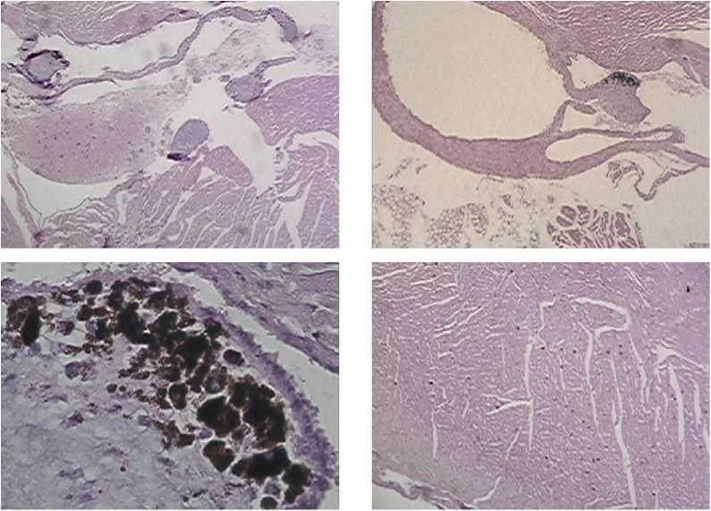
Figure 1. Fragments of the mouse heart at week 1 of B16 melanoma growth under the CNP conditions: significant thickening of the aortic and mitral valve cusps, fibrosis and metachromasia against the background of stenosis of the aortic opening for blood flowing with hypertrophy of the ventricle walls. Stained with hematoxylin-eosin, magnification x5, x10, x100.
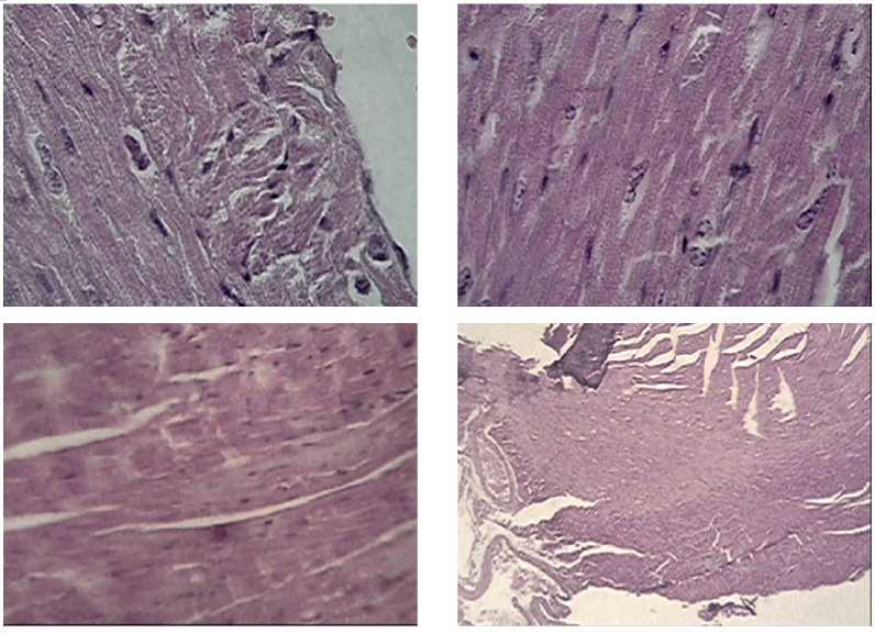
Figure 2. Fragments of the mouse heart at week 1 of B16 melanoma growth under the CNP conditions: thinned epicardial layer with endothelium and subendothelial elastic layer bordering the myocardium. Multiple small extensive foci of necrobiotic lesions of cardiomyocytes. Disappearance of transverse striation pattern. Stained with hematoxylin-eosin, magnification x5, x100.
um (infarction). However, a substantial role in the development of pathological changes in the pericardium is played by factors of a traumatic stressogenic nature (traumatic pericarditis), the spread of a cancerous tumor (cancerous pericarditis).
32 | Cardiometry | Issue 22. May 2022
In the light of the above studies, the presence of the root cause of pericarditis in the form of a not relievable pain stimulation in the process of the constant compression of the sciatic nerves and the territorial proximity of the metastatic melanoma nodes, most often in
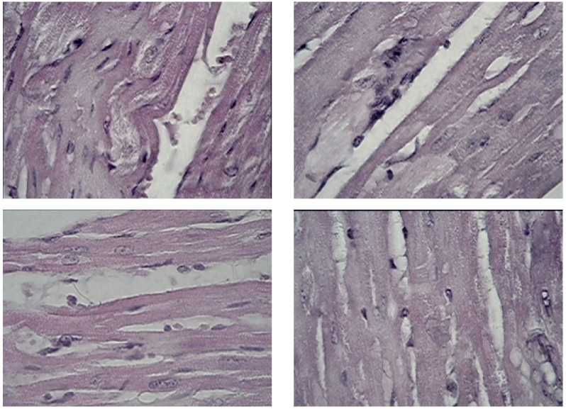
Figure 3. Fragments of the mouse heart at week 2 of B16 melanoma growth under the CNP conditions: development of loose fibrous connective tissue with foci of myxomatosis and metachromasia, longitudinal splitting of muscle fiber bundles, increased fibrosis, filling of gaps with detritus and blood. Stained with hematoxylin-eosin, magnification x 40, x100.
the lungs and even in the pericardium, in our opinion, can fully explain the pathogenesis of pericarditis. The infarcted area of the myocardium, located subpericar-dially, has led to the development of fibrinous pericarditis. It may be assumed that, along with the transition to the pericardium of inflammatory and oncological factors from the neighboring organs (lungs) by the hematogenous or lymphogenous pathway, the dominance of the neurogenic pathway of myocardial trophism is also formed due to failures of its own conducting systems of autoregulation, as well as stressogenic impulses of the nerve trunk of the sympathetic system and the vagus nerve of the parasympathetic system of the organism. It follows that in the mechanism of the occurrence of structural and functional disorders of the heart, the reactivity of the organism, the virulence of a pathogenic factor and the state of the intramural nervous system assumes unarguable significance.
Microscopy of histological sections of the myocardium indicated some pronounced dystrophic changes in cardiomyocytes (see Figure 4 herein). In the visual fields, there were signs of cloudy swelling and fatty degeneration up to myolysis and necrosis. As noted, the processes might be of focal and diffuse nature, often localized in the subendocardial layers of the myocardium. Microscopically, in addition to the changes in muscle fibers, plethora, edema of the myocardial stro- ma, and pronounced cellular dystrophy were detected. The impression was given that the heart muscle was flabby, the cavities were dramatically expanded, and the muscle was faint as evidenced by weak staining of the cells. Along with edema, accumulations of leukocytes, eosinophils, lymphocytes, plasma cells, and histiocytes were found. Among the features of cell reactions, we could highlight eosinophilic infiltration, which was found to be like allergic eosinophilic myocarditis. Thus, we might assert the damage to the myocardium is of non-coronary origin.
Particular attention was paid to the condition of the nuclei of cardiomyocytes. In ischemic heart disease, division of the nuclei of the muscle cells of the heart by mitosis or amitosis can be extremely rarely observed, and mitoses in this case are of pathological type and culminate in the generation of mutiple micronuclei that might be expected in cardiomyocytes. However, in the gallery of images presented to our thoughtful reader (see Figure 5 and 6), in our opinion, it is practically impossible to identify the areas of mitotically dividing cells. On the contrary, for a period of time when chronic neurogenic pain had already prepared the negative scenario for some events in the heart, including the mechanisms of pathological pathways for replenishing the cell population, the subsequent melanoma transplantation altered finally not only the life
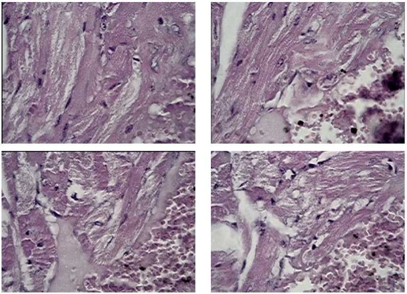
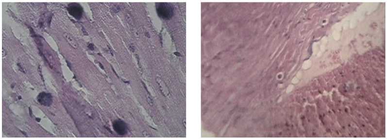
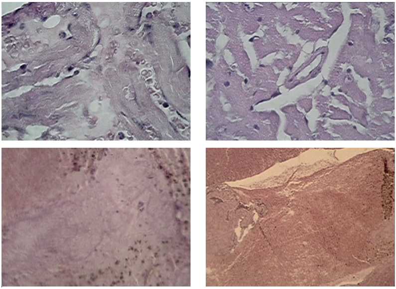
Figure 4. Fragments of the mouse heart at week 3 of B16 melanoma growth under the CNP conditions: hemorrhages, necrobiosis, basophilia, fibrosis in the media of branches of the coronary arteries of the heart, significant fields of necrotically altered tissue. Stained with hematoxylin-eosin, magnification x5,x10,x40,x100.
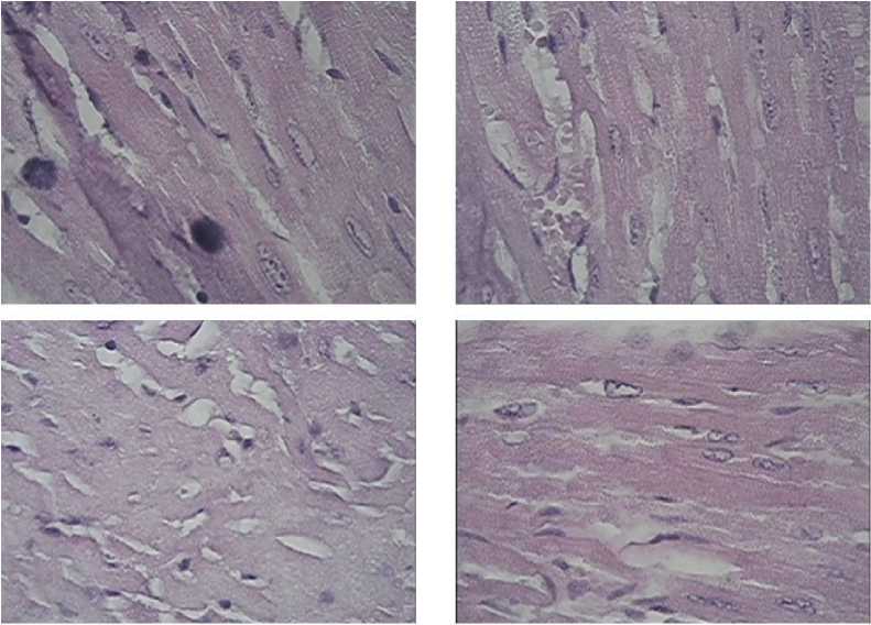
Figure 5. Fragments of the mouse heart at week 3 of B16 melanoma growth under the CNP conditions: eosinophilic infiltration, hemorrhages, fibrosis, erasure of the cellular structure of cardiomyocytes, and nuclear degeneration. Stained with hematoxylin-eo-sin, magnification x 100.
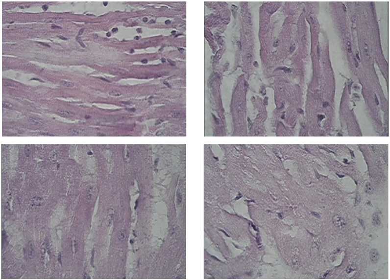
Figure 6. Fragments of the mouse heart at week 3 of B16 melanoma growth under the CNP conditions: stratification and degeneration of the structure of myocardial fibers with the prevalence of necrotic processes. Stained with hematoxylin-eosin, magnification x 100.
program, but also the cell death program. Apparently, we may believe that with a prolonged, rather than an acute form of infarction, cells commit to their death by following no longer the path of an organized, energy-dependent, process of apoptosis with characteristic cell shrinkage, nuclear fragmentation and formation of apoptotic bodies. Forced by the influx of the negative influence made by the tumor, the programmed mode of apoptosis switches to the non-apoptotic pathway of cell death, i.e. necrosis. One of the explanations for this phenomenon may be the stoppage of the apoptotic program of death of the surviving heart tissue as a result from the “tumor-caused” depletion of the energy reserves of cardiomyocytes (ATP), which occurs before the morphological features become fully evident [17].
On the studied preparations, a pathological pattern of death of cardiomyocytes was observed, accompanied by clarification of nuclei with large chromatin grains appearing as a dispersed or linear-type structure along the nuclear membrane. Attention was drawn to the presence of vasculization and the loss of clear contours of disconnected fibers, a spilled space of necrotically altered tissue against the background of activation of fibrinolysis in the intercellular space.
We failed to detect manifestations of a regenerative reaction in the heart with extensive areas of myocardial infarction in the female mice with the progression of B16 melanoma under the conditions of chronic neurogenic pain. There was also no filling of necrosis areas with full-fledged muscle tissue. The revealed changes in the heart were irreversible.
Conclusions
On the basis of the obtained materials of our morphological studies, we have tried to formulate our concept and visually verify the real pathological changes in the heart in the female mice when modeling common life situations integrating chronic neurogenic pain and a malignant process. Despite the different etiology, the pathogenesis of chronic pain includes multiple multifocal manifestations, especially when it refers to highly organized structures of the organism: the nervous and cardiovascular systems with a high risk of coronary and cerebrovascular diseases. It is believed that an increase in the incidence of acute myocardial infarction is associated to a greater extent with the male gender aspect, but the question of changes in the reactivity of the cardiovascular system in females with CNP and the tumor growth remains open. We have targeted the morphological picture of the heart in female mice with B16 melanoma transplanted under the skin and found a rather tough scenario of the events, which, in general, can be outlined by several generalizing conclusions as given below:
-
1. Continuous pain stimulation of a neurogenic nature has a significant pro-oncogenic effect, including an earlier manifestation of the tumor growth and large-scale metastasizing even to atypical target organs.
-
2. Chronic neurogenic pain triggers the action of the mechanisms of destructive changes in the myocar- 36 | Cardiometry | Issue 22. May 2022
-
3. Exhausting pain stimulation in combination with an aggressive tumor growth can considerably accelerate the rate of reduction in the organism resistance, suppression of the life maintenance systems, provoking the development of severe heart failure and the formation of a pre-terminal state at an earlier time.
-
4. One of the leading vectors of the pathological influence made by chronic neurogenic pain is aimed at weakening the mechanisms responsible for the cell population replenishment, and the subsequent melanoma transplantation changes not only the life program, but also the cell death program. Obviously, with a prolonged, rather than an acute form of infarction, the cells commit to their death following no longer the path of an organized energy-dependent process of apoptosis, but by switching to a non-apoptotic path of the cell death, i.e. necrosis as a result from the “tumor-caused” depletion of energy reserves of cardiomyocytes.
dium before tumor cell transplantation, therefore, as early as 1 week after the melanoma inoculation and the formation of one or two tumor nodes, harsh heart damage has been observed. The valve system suffers that leads to ventricular wall hyperplasia, ischemia, longitudinal splitting of muscle fiber bundles, hemorrhages, cellular dystrophy, myolysis, multifocal necrosis, fibrosis, eosinophilic infiltration. Thus, not an acute, but a prolonged myocardial infarction is realized.
The morphological study of the changes found in the heart in the female mice using the pathological models of chronic neurogenic pain and the malignant tumor growth has made it possible to reproduce and trace the catastrophe of the highly organized cellular system responsible for the implementation of the hemodynamic processes. The simulated hypertrophied manifestations of the catastrophe in the experiment give grounds to predict and prevent the negative course of events in difficult patients with persistent pain syndromes and comorbid pathology against the background of the development of a malignant process.
Statement on ethical issues
Research involving people and/or animals is in full compliance with current national and international ethical standards.
Conflict of interest
None declared.
Author contributions
The authors read the ICMJE criteria for authorship and approved the final manuscript.
References Structural myocardial catastrophe under the influence of chronic neurogenic pain due to development of B16 melanoma in female mice
- Gabriel-Costa D. The pathophysiology of myocardial infarction-induced heart failure. Pathophysiology. 2018; 25: 277–284.
- Huang J, Wang Z, Hu Z, Jiang W, Li B. Association between blood vitamin D and myocardial infarction: A meta-analysis including observational studies. Clinica Chimica Acta. 2017; 471: 270–5.
- Agarkov N, et al. The information content and predictive value of cardiac markers in myocardial infarction in the elderly. Adv. Gerontol 2020; 33(1): 82–6.
- Hofmann R, Bäck M. Gastro-Cardiology: A Novel Perspective for the Gastrocardiac Syndrome. Frontiers in cardiovascular medicine. 2021; 8: 764478. https://doi.org/10.3389/fcvm.2021.764478
- Wu H, et al. Inflammatory bowel disease and cardiovascular diseases: a concise review. Eur Heart J Open. 2021; 10: 1093/ehjopen/oeab029
- Briggs AM, Woolf AD, Dreinhöfer K, et al. Reducing the global burden of musculoskeletal conditions. Bull World Health Organ. 2018; 96(5): 366. doi: 10.2471/BLT.17.204891. International Association for the Study of Pain. Musculoskeletal pain fact sheets. https://www.iasp-pain.org/Advocacy/Content.aspx-?ItemNumber=1101. Assessed 01 Dec 2020.
- WHO. Musculoskeletal conditions. https://www.who.int/news-room/fact-sheets/detail/musculoskeletal-conditions. Assessed 02 Nov 2020.
- Iizuka Y, et al. Prevalence of chronic nonspecific low back pain and its associated factors among middle-aged and elderly people: an analysis based on data from a musculoskeletal examination in Japan. Asian Spine J. 2017; 11(6): 989. doi: 10.4184/asj.2017.11.6.989.
- Wang H, et al. Osteoarthritis and the risk of cardiovascular disease: a meta-analysis of observational studies. Sci Rep. 2016;6:39672. doi: 10.1038/srep39672.
- Kikuchi S, et al. Database Analysis on the Relationships Between Nonsteroidal Anti-inflammatory Drug Treatment Variables and Incidence of Acute Myocardial Infarction in Japanese Patients with Osteoarthritis and Chronic Low Back Pain. Advances in therapy. 2021; 38(3): 1601–1613. https://doi.org/10.1007/s12325-021-01629-6.
- Kit OI, et al. Effect of chronic neuropathic pain on the course of the malignant process of B16/f10 melanoma in male mice. News of Higher Educational Institutions. North Caucasian region. Series: Natural Sciences. 2019; 1 (201):106-1. [in Russian]
- Frantsiyants ЕМ et al. The functional state of cardiomyocyte mitochondria in a malignant process against the background of comorbid pathology in the experiment. South Russian Journal of Cancer. 2021; 2(3): 13-22. https://doi.org/10.37748/2686-9039-2021-2-3-2. [in Russian]
- Kit ОI, et al. The processes of self-organization of mitochondria during the growth of experimental tumors under conditions of chronic neurogenic pain. News of higher educational institutions. North Caucasian region. Series: Natural Sciences. 2019; 2 (202): 97-105. [in Russian]
- Shikhlyarova A.I., et al. Relationship between comorbid pathology and tumor progression. Morphological portrayal of internal organs in modeling the growth of Guerin’s carcinoma under diabetic conditions. Cardiometry. 2022; 21:18-26.
- Frantsiyants E.M., et al. Content of apoptosis factors and self-organization processes in the mitochondria of heart cells in female mice C57BL/6 under growth of melanoma b16 / f10 linked with comorbid pathology. Cardiometry. 2021;18:121-130.
- Kotieva IM, et al. An increase in the degree of malignancy of melanoma against the background of chronic pain in female mice. Malignant tumors. 2017; 7(3S1): 123-4. [in Russian]
- Saprunova VB, Bakeeva LE, Yaguzhinsky LS. Ultrastructure of cardiomyo cyte mitochondria under anoxic conditions on a model of surviving heart tissue. Mitochondria in pathology. Pushchino. 2001. 71-4. [in Russian]
