The advent of elastic stable intramedullary nailing for the treatment of long bone fractures in skeletally immature patients

Author: Andreacchio Antonio, Alberghina Flavia, Canavese Federico
Journal: Гений ортопедии @geniy-ortopedii
Section: Оригинальные статьи
Article in issue: 4 т.27, 2021.
Free access
Introduction Management of pediatric long bones fractures is a complex and rapidly evolving field. Traditionally, casting and conservative techniques played a key-role in the management of fractures in skeletally immature patients. However, the surgical approach has evolved steadily over the past four decades or so and increasing evidence has been published supporting the advantages of fixation techniques over conservative methods. The purpose of this narrative review is to outline how innovations in orthopedic surgery have changed the rationale of treating long bones fractures in children and adolescents with focus on surgical techniques, particularly elastic stable intramedullary nailing (ESIN). Material and methods We aimed to describe the main trends in pediatric long bones fractures management and to identify its specificities and difficulties as well as the best standard of care. Results The introduction of ESIN has profoundly influenced the management of pediatric upper and lower extremity fractures. Overall, in comparison to conservative techniques, advantages of ESIN include minimally invasiveness, short hospital stay, primary bone union, early mobilization and progressive weight bearing, and good outcome with low complication rate. Moreover, the flexible nail can be used as a closed reduction tool itself. Conclusions Irrespective of the technique performed, the key-concepts remain 1) the proper understanding of the injury to treat; 2) the identification the main characteristics of the patient; 3) the pros and cons of each technique; and 4) the potential complications.
Long bone fracture, surgery, operative treatment, children, adolescents
Short address: https://sciup.org/142230225
IDR: 142230225 | UDC: 616.71-001.5-089.227.84-053.2 | DOI: 10.18019/1028-4427-2021-27-4-406-412
Применение эластичных стабильных интрамедуллярных стержней для лечения переломов длинных костей у пациентов с незрелым скелетом
Введение. Лечение переломов длинных костей у детей - сложная и быстро развивающаяся область травматологии. Традиционно гипсование и консервативные методы играли ключевую роль в лечении переломов у пациентов с незрелым скелетом. Тем не менее, хирургический подход неуклонно развивался в течение последних четырех десятилетий, и было опубликовано все больше данных, подтверждающих преимущества методов фиксации над консервативными методами. Цель данного описательного обзора - показать, как инновации в ортопедической хирургии изменили обоснование лечения переломов длинных костей у детей и подростков с акцентом на хирургические методы, в частности, на эластичные стабильные интрамедуллярные стержни (ESIN). Материалы и методы. Авторы стремились описать основные тенденции в лечении переломов длинных костей у детей и выявить их особенности и трудности, а также выбрать лучший стандарт лечения. Результаты. Внедрение ESIN значительно повлияло на тенденции в лечении переломов верхних и нижних конечностей у детей. В целом, по сравнению с консервативными методами, преимущества ESIN заключаются в минимальной инвазивности, сокращении сроков пребывания в стационаре, обеспечении первичного сращения костей, возможности ранней мобилизации пациента и постепенного увеличения нагрузки на конечность. Также указанный метод обусловливает достижение хорошего результата с незначительным показателем осложнений. Более того, гибкий гвоздь может использоваться как закрытый редукционный инструмент. Выводы. Независимо от выполняемой техники ключевыми позициями остаются: 1) адекватная оценка травмы, которую необходимо лечить; 2) выявление основных характеристик пациента; 3) плюсы и минусы каждой техники; 4) возможные осложнения.
Text of the scientific article The advent of elastic stable intramedullary nailing for the treatment of long bone fractures in skeletally immature patients
Management of pediatric long bones fractures is a complex and rapidly evolving field. It has been estimated that about 50 % and 30 % of boys and girls, respectively, sustain a fracture during their childhood; the distal radius is the most frequently injured bone, followed by the humerus [1, 2]. In recent years, more and more children have become involved in sport and recreational activities, and have been using electric motion devices, exposing themselves to increased risk of fracture [3]. Preventable injuries occurring during sport and recreational activities have been reported to be the cause of up to 39 % of all pediatric fractures [1, 2].
Traditionally, casting and conservative techniques play a considerable role in the management of pediatric fractures [4, 5]. However, for displaced fractures, especially in older patients with limited remodeling potential, surgical treatment may offer better clinical, functional, and radiographic outcome [6–9].
The approach to treat long bones fractures in children and adolescents has evolved steadily over the past four decades or so, and increasing evidence supports the advantages of fixation techniques over conservative modalities. The key-factors influencing the choice of treatment include: i) fracture location and pattern; ii) age and iii) weight of the patient. Additional factors to take into account are the mechanism of injury, presence of associated injuries, soft tissue involvement, expected patient and family compliance, and surgeon’s preference.
In particular, the development and the diffusion of the elastic stable intramedullary nailing (ESIN) technique has
significantly impacted the management of upper and lower extremity long bone fractures in children and adolescents.
The purpose of this narrative review is to outline how innovations in pediatric orthopedic surgery have modified the rationale of treating long bones fractures in skeletally immature patients with focus on surgical techniques, particularly elastic stable intramedullary nailing (ESIN).
METHODS
The review includes clinical studies and reviews concerning pediatric long bones fracture treatment written in English over the last ten years. The search was performed using PubMed, Google Scholar, Scopus, Medline and Cochrane Library databases from January 2010 up to January 2021. A combination of search terms including ‘fracture’, ‘surgical treatment’, ‘children’, ‘elastic nailing’, ‘ESIN’ was utilized. Exclusion criteria consist of studies that were case-reports, studies including adult patients.
The references of all selected articles were also reviewed to find potential articles that were missed.
Any remaining non-eligible articles were excluded, and duplicate articles were removed.
The following data were extracted from the included papers: author(s), year of publication, title, journal, sample size in the hospital setting, length of follow-up, characteristics of the study population (age, sex, fracture site), surgical treatment characteristics (approach, mean of fixation), definition of functional outcomes and related score or questionnaire used. Studies published in the last 5 years are listed in Table 1 and 2.
Cited lower limb clinical studies published over the past 5 years
Table 1
|
Title |
Year |
Patients (n) |
Fracture |
Type of Fixation |
Complications |
|
Extra-articular proximal femur fractures in children and adolescents treated by elastic stable intramedullary nailing [35] |
2019 |
24 |
Proximal femur |
ESIN |
20 % malalignment (all < 10 degrees) |
|
Intraoperative Issues and Clinical and Radiographic Outcomes of Femur Fractures Treated With Flexible Nails: A Comparison of Cases Utilizing Skeletal Traction and a Traction Table to Cases Using Manual Traction Only [25] |
2020 |
69 |
Femoral shaft |
ESIN |
LLD < 2 cm; 1 non-union; 1 valgus deformity) |
|
Complications of elastic stable intramedullary nailing of femoral shaft fractures in children weighing fifty kilograms (one hundred and ten pounds) and more [30] |
2016 |
20 |
Femoral shaft |
ESIN |
45 % (2 delayed union; 2 malunion; 2 nail tip bursitis; 1 nail migration; 1 non-union; 1 septic pseudoarthrosis) |
|
Displaced distal femur metaphyseal fractures: clinical and radiographic outcome in children aged 6-16 years treated by elastic stable intramedullary nailing [36] |
2020 |
24 |
Distal femur |
ESIN |
20% (3 non-union; 2 secondary displacement) |
|
Displaced tibia shaft fractures in children treated by elastic stable intramedullary nailing: results and complications in children weighing 50 kg (110 lb) or more [31] |
2016 |
26 |
Tibial shaft |
ESIN |
15 % (2 nail tip bursitis; 1 nail migration; 1 compartment syndrome) |
|
Elastic stable intramedullary nailing for severely displaced distal tibial fractures in children [37] |
2016 |
21 |
Tibial shaft |
ESIN |
23 % (2 LLD; 2 delayed union; 1 restriction of the dorsal hallux extension) |
ESIN – elastic stable intramedullary nailing; LLD – Leg length discrepancy
Cited upper limb clinical studies published over the last 5 years
Table 2
|
Title |
Year |
Patients (n) |
Fracture |
Type of Fixation |
Complications |
|
Outcome of Conservative Versus Surgical Treatment of Humeral Shaft Fracture in Children and Adolescents: Comparison Between Nonoperative Treatment (Desault's Bandage), External Fixation and Elastic Stable Intramedullary Nailing [6] |
2017 |
26 |
Humeral shaft |
Ex-Fix ESIN |
34 % (5 secondary displacement; 2 refracture; 1 infection; 1 ulnar nerve deficit) |
|
Evaluation of upper extremity function of displaced diaphyseal humeral fractures in children treated by elastic stable intramedullary nailing: preliminary results [7] |
2016 |
16 |
Humeral shaft |
ESIN |
18 % (1 displacement secondary to a fall; 1 refracture; 1 osteomyelitis) |
|
Displaced humeral shaft fractures in children and adolescents: results and adverse effects in patients treated by elastic stable intramedullary nailing [8] |
2016 |
38 |
Humeral shaft |
ESIN |
10 % (1 secondary displacement; 2 refracture; 1 osteomyelitis) |
|
Functional outcome of displaced intercondylar fractures of the humerus in children and adolescents [9] |
2017 |
18 |
Distal humerus (T-condylar) |
ESIN |
50 % (4 loss of movement; 2 radial humeral OA; 1 valgus deformity; 2 varus deformity) |
|
Long-term results of elastic-stable intramedullary nailing (ESIN) of diaphyseal forearm fractures in children [43] |
2019 |
90 |
Diaphyseal forearm |
ESIN |
not mentioned |
|
Risk factors for refracture of the forearm in children treated with elastic stable intramedullary nailing [44] |
2019 |
267 |
Diaphyseal forearm |
ESIN |
4 % (11 refracture) |
|
Functional outcome of displaced radial head fractures in children treated by elastic stable intramedullary nailing [45] |
2017 |
24 |
Radial head |
ESIN |
8 % (1 pseudoarthrosis; 1 nail displacement) |
|
Functional and radiological outcome in patients with acute Monteggia fracture treated surgically: a comparison between closed reduction and external fixation versus closed reduction and elastic stable intramedullary nailing [47] |
2020 |
26 |
Monteggia fracture |
Ex-Fix ESIN |
26 % (1 pin-tract infection; 2 HO; 3 dysesthesia; 6 residual pain and limited motion) |
ESIN – elastic stable intramedullary nailing; OA – osteoarthritis; HO – heterotopic ossifications
The era of ESIN
More than 30 years after its introduction, the ESIN technique, also known as FIN (Flexible Intramedullary Nailing) method, Métaizeau technique or Nancy technique, has become a widespread method of treating paediatric long bone fractures [10, 11]. Initially, the technique was used for the treatment of diaphyseal fractures of upper and lower extremity only, and aimed i) to achieve a good fracture alignment, ii) to avoid damage to the physis or the periosteum, iii) to decrease the time of cast immobilization and iv) to facilitate weight bearing.
The ESIN technique is based on the so called three-point contact: the two nails with a curvature in the opposite direction and three contact points on the bone (two at the metaphysis and one at the fracture site) produce equal forces acting with an opposite moment (Fig. 1). The construct stabilizes the fracture by neutralizing shear forces and allows only compression forces to act at the level of the fracture site thus promoting callus formation and bone healing [12]. Moreover, the flexible nail itself can be used as a closed reduction tool [13].
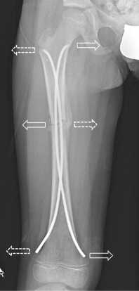
Fig. 1. ESIN three-point fixation: dotted arrows indicate the forces of the side nail; continuous arrows indicate the forces of the medial nail. Each nail has three points of contact: entry point, curvature apex (fracture site), anchor point
Nail size (NS) is selected as a function of the medullary canal diameter (MCD) measured at the narrowest point of the diaphysis. Lascombes et al. recommend selecting a NS/ MCD ratio of 40 % in lower extremity fractures and have advised against ESIN in children with a MCD >10 mm [11]. In the humerus, the appropriate nail size is determined by the equivalence: nail diameter = 33 % of the MDC [14]. As the upper limbs are not weight-bearing, smaller diameter nails can provide sufficient stability as well as an easier crossing of the fracture site during insertion. In the forearm, only one nail is required for each bone and the NS should be between 40 % and 50 % of the MDC.
Both stainless steel and titanium elastic nails are available on the market. Compared to a titanium nail of the same diameter, a nail made of steel has a lower elasticity while its elastic return force is twice as high. In addition, it has greater curvature stiffness: the rigidity of a steel nail corresponds to that of a titanium nail with 0.5 mm higher diameter. Consequently, due to its mechanical properties, steel is considered a more suitable choice for adolescents while titanium appears to be more indicated in younger children [15].
Compared to ESIN, conservative treatment requires prolonged periods of immobilization, is frequently unable to achieve and maintain an adequate reduction, especially in heavier patients (obese or adolescents) or in case of severely unstable fractures. Secondary problems, though not less important, are а limited ability to check the soft tissues and the risk of pressure sores. Furthermore, cases of peroneal paralysis, compartmental syndrome, excessive shortening and non-union have been reported in pediatric femur shaft fractures treated conservatively [16].
Lower extremity
Femur and tibia diaphyseal fractures
Shemshaki et al. randomized 46 children aged 6 to 12 years of age to receive skeletal traction followed by hip spica cast or TEN for simple femoral-shaft fracture; the authors reported better results in terms of hospital stay, time to start walking with support or independently, returning to school, and parent’s satisfaction in the TEN group [17]. Their findings agreed with the other studies on the same topic [12, 18, 19].
Lewis at al. [20] recently analyzed the financial impact of closed femur fractures in 3 to 6-year-olds treated with immediate spica casting versus intramedullary fixation. In contrast to previous literature, they observed longer hospital stays, significantly greater hospital charges, longer follow-up and more clinic visits in children treated with intramedullary nailing compared with spica casting. However, children aging 3 to 6 years can be successfully treated conservatively and the choice to perform ESIN in patients younger than 6 years may be considered by several authors as an overtreatment [11, 21, 22]; irrespective of age, patients less than 35 kg of weight can be treated conservatively.
Similarly, displaced fractures of the tibia diaphysis in children are frequently treated with ESIN. As for the femur, the advantages include minimally invasive surgery with a short hospital stay, primary bone union, early mobilization and weight bearing, and good outcome with low complication rates [23–25].
Overall, looking at the literature of the past two decades or so, ESIN has become the gold standard of treatment of femoral and tibial midshaft fractures in children between 6 and 12 to 15 years of age; however, there are concerns about their use in patients weighing more than 50 kg (110 pounds).
The prevalence of childhood obesity is increasing in most of industrialized countries and becoming a serious global public health problem [26]. As confirmed by recent studies, about 20 % of European school-age children are overweight and 5 % are obese; in North America, even higher figures as 30 % and 15 % have been reported, respectively [27, 28].
Unsurprisingly, higher rates of complications have been reported in overweight children treated with ESIN for a lower limb fracture, being unstable fracture pattern, higher age and higher weight identified as risk factors associated with poor outcomes [29–32]. For example, Moroz et al. found that patients weighing more than 49.3 kg were some five times more likely than lighter patients (less than 49.3 kg) to have poor outcomes following ESIN for femur shaft fractures [33]. Similarly, Canavese et al., analyzing the outcome of children weighing 50 kg (110 pounds) or more treated with ESIN for displaced femur shaft fracture, concluded that complication rate was higher in heavier children (67 % in children weighing 55 kg or more, 35 % in children weighing 50 to 54 kg and 12.8 % in children weighing less than 50 kg) [30]. In contrast to data concerning displaced femoral shaft fractures, there is no evidence to confirm that overweight presents a risk factor for higher complication rate and poor outcome for displaced tibial shaft fractures [22, 31].
As suggested by Marengo et al., the correlation between outcome and weight may depend on the bone’s anatomy [31]. More to the point, assuming that femur and tibia are rigid bodies, the elastic deformations involved would be negligible. Both bones are constrained at both top (by the hip for the femur and by the knee for the tibia) and bottom (by the knee for the femur and by the ankle for the tibia). The upshot is that the main force acting on both bones is therefore weight. Through the femur, the weight vector counts two components: one along the anatomical axis of the femur and the other perpendicular to it, whereas for the tibia, the weight vector runs right through the anatomical axis. Moreover, the femur bone is solicited by lateral forces acting that could help improving the procurvatum, whereas the tibia does not have to bear lateral forces as the weight vector only acts in one direction, the axis of the bone [22–31].
Proximal femur fractures
Initially used in the treatment of midshaft fractures, over the decades, ESIN indications expanded to include also metaphyseal fractures of both upper and lower extremity (Fig. 2). Moreover, following this trend, slight variations of the standard nail design have been introduced, without changing the technique of insertion.
ESIN can be successfully used in the treatment of pediatric extra-articular proximal femur fractures or subtrochanteric fractures as well [34–35]. However, to contrast multiple and vigorous displacement forces acting on both proximal and distal fracture fragments the elastic nails should be advanced more proximally, the lateral nail to the femoral neck just below the proximal physis and the medial nail to the greater trochanter with the tip fixed to the cortical bone. This configuration improves fracture stability and prevents displacement, in particular malrotation [34–35].
Distal femur and tibia metaphyseal fractures
The use of ESIN in metaphyseal fractures may be challenging due to a relatively short length of the distal fragment and the proximity of the growth plate [36] (Fig. 3). For instance, the use of ESIN in the distal tibia is technically demanding and not free of complications [37] but the method has shown good results and it is a valid and effective alternative to casting and other fixation techniques [38].
Similarly, in many centers, ESIN has gradually replaced conservative treatment and pinning in various displaced pediatric upper limb fractures.
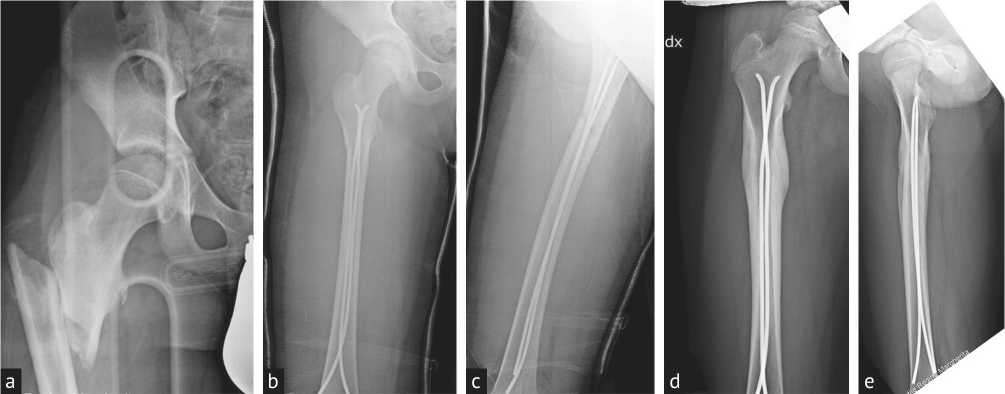
Fig. 2. Proximal femur shaft fracture in a 10-year-old boy (a) treated with ESIN. Post-operative radiographs (b-c) and radiographs 6 months after surgery (d–e)
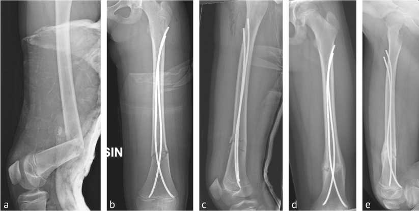
Fig. 3. Distal femur shaft fracture in a 10-year-old girl (a) treated with ESIN. Post-operative radiographs (b–c) and radiographs 4 months after surgery (d–e)
Upper extremity
Proximal and diaphyseal humerus fractures
To reduce the time of immobilization and the risk of severe complications, the use of ESIN has been strongly supported in recent years for proximal humerus and diaphyseal humerus fractures [6-8] (Fig. 4). Although surgical indications and type of surgery are not clearly defined and still debated [6, 39], ESIN fixation provides stable fixation, good rotational control and early mobilization. A meta-analysis from Palhavn et al. (14 articles; all Level-IV) compared non-operative and surgical treatment [40]. Despite both techniques can achieve similar functional outcome, the authors observed that older patients, and patients with more severely displaced fractures, tended to have poorer functional outcome, regardless of the type of treatment, and concluded that surgery should be preferred in children older than 13 years of age with severely displaced proximal humerus fractures.
Canavese et al. [6] compared clinical and radiographic outcomes of a series of 36 patients with displaced humeral shaft fractures who underwent Desault’s bandage, external fixation, or ESIN. The authors observed that surgical treatment can achieve better radiologic outcomes than conservative treatment. However, conservative treatment was as efficacious as surgical treatment apart from the higher posttreatment pain and the length of time for immobilization [6].
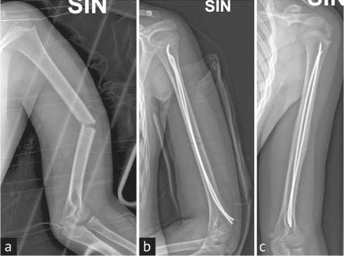
Fig. 4. Midshaft humeral fracture in an 11-year-old boy (a) treated with ESIN. Post-operative radiograph (b) and radiograph 3 months after surgery (c)
Distal metaphyseal fractures of the humerus
The management of distal humerus metaphyseal fractures may be problematic due to the relatively short length of the distal fragment, and the resultant instability. Surgical treatment of such injuries traditionally consisted of closed or open reduction and internal fixation with Kirschner wires [41]. Nevertheless, the technique of fixation has not been standardized and different pin configurations, including cross-pins, lateral entry pins, and medial pins have been described, as well as high rate of loss of fixation, especially in patients with transverse fractures [42]. Marengo et al. [41] reported their experience in treating 14 patients (mean age 9.7 years) using anterograde ESIN technique observing no loss of fixation or refracture during follow-up (28.1 months) although one patient developed cubitus varus.
Forearm fracture
The vast majority of pediatric diaphyseal forearm fractures can be successfully treated with closed reduction and cast immobilization. However, when non-operative management fails, or when it is not applicable (open fractures), ESIN may provide stable reduction and subsequent union with short operating time, excellent cosmesis, minimal soft tissue dissection, ease of hardware removal, and early motion [43]. Reported complications following intramedullary fixation of upper extremity fractures include hardware migration, infection, loss of reduction, nerve injury, decreased ROM, synostosis, muscle entrapment, and delayed union [44].
Radial head and neck fracture
Radial head and neck fractures is a peculiar pediatric fracture pattern in which closed reduction and ESIN stabilization is particularly effective [45]. In contrast to percutaneous pinning in which closed reduction is achieved directly through the fracture site (with potential damage to the extensor tendon and posterior interosseous nerve), the ESIN technique allows reduction and fixation through the medullary canal from the distal radius. A long 1.4 to 1.8 mm nail with the proximal 1-cm extremity bent at 30° is inserted and passed from distal to proximal into the radial neck. The nail is then rotated and advanced so the tip can be passed into the displaced radial head and held in subchondral bone. The nail is then rotated again until radial head regains its anatomical position [46]. The risk of the technique includes extensor pollicis longus tendon and superficial radial nerve damage.
Monteggia fracture
In Monteggia fractures, fixation is usually needed in case of failed reduction or instability of the radial head. Restoration of ulnar length, alignment, and reduction of the radiohumeral joint can be achieved either with ulna K-wiring, ESIN, plate or external fixator [47]. However, the insertion of the elastic nail curved toward the opposite side of the radial head helps to maintain the radiohumeral reduction and stabilize the ulna fracture.
Complications
Overall, complications of ESIN technique include skin irritation due to the nail prominence, nail migration, infection, delayed healing, angular malalignment or malrotation and compartmental syndrome [14, 30, 48]. Pandya et al. identified three factors associated with the development of compartmental syndrome after tibial flexible nailing: weight > 50 kg, comminuted/complex fracture patterns, and presentation with preoperative neurologic deficits in the absence of compartmental swelling [49]. Nevertheless, reduction defects, consolidation problems and malunions are most likely secondary to an incorrect nail/medullary canal diameter ratio selection [14].
The above-mentioned limitations of the ESIN technique outline the importance of considering other fixation techniques, mostly in adolescents and in overweight patients with length-unstable fracture patterns; alternative options include external fixation, submuscular plating, rigid intramedullary fixation [50–52].
CONCLUSION
In summary, over the past four decades or so, the approach to pediatric long bones fractures has changed, and progressively evolved towards surgical management. The main causes are the increasing prevalence of skeletal injuries, the demand for shorter time of hospital stay and recovery, and the greater offer of implants specifically designed for skeletally immature patients.
All these factors are leading to a more focused and hopefully improved level of care. However, irrespective of the technique performed, the key-concepts remain unchanged: 1) proper understanding of the injury to treat; 2) identification of the main characteristics of the patient; 3) pros and cons of each technique; and 4) potential complications.
References The advent of elastic stable intramedullary nailing for the treatment of long bone fractures in skeletally immature patients
- Limb Lengthening Forum [сайт]. URL: https://www.limblengtheningforum.com/ (дата обращения: 29.07.2020).
- Коррекция ног : [сайт]. URL: https://legscorrection.ru/forum/index.php (дата обращения: 29.07.2020).
- Хирургия нижних конечностей : [сайт]. URL: http://varus-valgus.net/ (дата обращения: 29.07.2020).
- Федеральный закон «Об основах охраны здоровья граждан в Российской Федерации»: текст с изменениями и дополнениями на 2020 год. М. : Эксмо, 2020. 96 с. (Законы и кодексы).
- Эстетическая и реконструктивная хирургия нижних конечностей / под ред. А.А. Артемьева. М. : ГЭОТАР-Медиа, 2008. 248 с.
- Маркер Н.А. Выбор тактики и результата ортопедической коррекции голени при косметической деформации : автореф. дис. ... канд. мед. наук. Новосибирск, 2009. 35 с. URL: http://medical-diss.com/docreader/288249/a?#?page=1 (дата обращения: 29.07.2020).
- Кулеш П.Н., Соломин Л.Н. Коррекция формы ног по эстетическим показаниям (обзор литературы) // Гений ортопедии. 2013. № 2. C. 117-123.
- Comparison of PACS and Bone Ninja mobile application for assessment of lower extremity limb length discrepancy and alignment / A.T. Whitaker, M.G. Gesheff, J.J. Jauregui, J.E. Herzenberg // J. Child. Orthop. 2016. Vol. 10, No 5. P. 439-443. DOI: 10.1007/s11832-016-0761-5
- Paley D. Principles of deformity correction. Herzenberg J.E., editor. Springer Science & Business Media. 2002. 806 p.
- Основы чрескостного остеосинтеза аппаратом Г. А. Илизарова / под ред. Л.Н. Соломина. СПб. : МОРСАР АВ, 2005. 544 с.
- Solomin L.N., The Basic Principles of External Skeletal Fixation Using the Ilizarov and Other Devices. 2nd Edition / Solomin L.N., R.R. Vreden Russian Research Institute of Traumatology and Orthopaedics (St. Petersburg, Russia), editors. Milan: Springer-Verlag. 2012. 1593 p. DOI: 10.1007/978-88-470-2619-3_18
- The Art of Limb Alignment / S.C. Standart, J.E. Herzenberg, J.D. Conway, N.A. Siddiqui, P.K. McClure. 9th ed. Baltimore : Rubin Institute for Advanced Orthopedics, Sinai Hospital of Baltimore. 2020. 247 p.
- Correction of tibia vara with six-axis deformity analysis and the Taylor Spatial Frame / D.S. Feldman, S.S. Madan, K.J. Koval, H.J. van Bosse, J. Bazzi, W.B. Lehman // J. Pediatr. Orthop. 2003. Vol. 23, No 3. P. 387-391.
- Программа моделирования Legscorrection. URL: https://legscorrection.ru/prog.php (дата обращения: 29.07.2020).
- Каплунов О.А., Каплунов А.Г., Шевцов В.И. Косметическая коррекция формы и длины ног. М. : ГЭОТАР-Медиа, 2010. 160 с. (Приложение на компакт-диске - программа «Коррекция формы ног»).
- Coventry M.B. Osteotomy of the upper portion of the tibia for degenerative arthritis of the knee. A preliminary report // J. Bone Joint Surg. Am. 1965. Vol. 47. P. 984-990.
- Proximal tibial osteotomy. A new fixation device / A. Miniaci, F.T. Ballmer, P.M. Ballmer, R.P. Jakob // Clin. Orthop. Relat. Res. 1989. No 246. P. 250-259.
- Lobenhoffer P., Agneskirchner J.D. Improvements in surgical technique of valgus high tibial osteotomy // Knee Surg. Sports Traumatol. Arthrosc. 2003. Vol. 11, No 3. P. 132-138. DOI: 10.1007/s00167-002-0334-7.
- Segev E. Updates on preoperative planning, limb deformity analysis and surgical correction for the growing children: EPOS Meeting Marseille 2015 proceedings // J. Child. Orthop. 2016. Vol. 10, No 6. P. 493-498. DOI: 10.1007/s11832-016-0795-8
- Hambardzumyan V., Herzenberg J.E. Bone Ninja app as a body image simulation tool for shared decision-making // J. Limb Lengthen. Reconstr. 2019. Vol. 5, No 2. P. 105-110. URL: https://www.jlimblengthrecon.org/text.asp?2019/5/2/105/274579
- Шевцов В.И., Немков В.А., Скляр Л.В. Аппарат Илизарова. Биомеханика. Курган : Периодика, 1995. 165 с.
- Etude des résultats à moyen terme d'une série de 33 allongements des membres inférieurs selon la technique de H. Wagner / J. Caton, P. Dumont, J. Bérard, C.R. Michel // Rev. Chir. Orthop. 1985. Vol. 71. P. 44-48.
- Paley D. Problems, obstacles, and complications of limb lengthening by the Ilizarov technique // Clin. Orthop. Relat. Res. 1990. No 250. P. 81-104.
- Donnan L.T., Saleh M., Rigby A.S. Acute correction of lower limb deformity and simultaneous lengthening with a monolateral fixator // J. Bone Joint Surg. Br. 2003. Vol. 85, No 2. P. 254-260. DOI: 10.1302/0301-620x.85b2.12645
- Лечение детей с деформациями длинных трубчатых костей нижних конечностей методом чрескостного остеосинтеза с использованием аппарата Орто-СУВ: анализ 213 случаев / В.А. Виленский, А.А. Поздеев, Т.Ф. Зубаиров, Е.А. Захарьян, А.П. Поздеев // Ортопедия, травматология и восстановительная хирургия детского возраста. 2016. Т. 4, № 4. C. 21-32. DOI: 10.17816/PTORS4421-32
- Do small changes in rotation affect measurements of lower extremity limb alignment? / A.A. Jamali, J.P. Meehan, N.M. Moroski, M.J. Anderson, R. Lamba, C. Parise // J. Orthop. Surg. Res. 2017. Vol. 12, No 1. P. 77. DOI: 10.1186/s13018-017-0571-6
- The effect of circular external fixation on limb alignment / S. Sabharwal, S. Badarudeen, E. McClemens, E. Choung // J. Pediatr. Orthop. 2008. Vol. 28, No 3. P. 314-319. DOI: 10.1097/BP0.0b013e3181653ba2
- Sokal R.R., Rolf F. J. Biometry: The Principles and Practices of Statistics in Biological Research. 3rd ed. New York : W.H. Freeman & Co. 1994. 880 p.
- Preoperative planning of medial opening wedge high tibial osteotomy using 3D computer-aided design weight-bearing simulated guidance: Technique and preliminary result / B. Chernchujit, S. Tharakulphan, R. Prasetia, N. Chantarapanich, C. Jirawison, K. Sitthiseripratip // J. Orthop. Surg. (Hong Kong). 2019. Vol. 27, No 1. P. 2309499019831455. DOI: 10.1177/2309499019831455
- Климов О.В., Новиков К.И., Аранович А.М. Моделирование формы нижних конечностей у пациентов с варусной деформацией голеней // Гений ортопедии. 2008. № 2. C. 50-53.
- Косметические аспекты оперативной коррекции диспластических варусных деформаций нижних конечностей / А.С. Баринов, А.А. Воробьев, С.С. Зайцев, П.С. Царьков // Клиническая медицина. 2010. № 4. С. 57-60.
- Correction of proximal tibia varus with external fixation / K. Ashfaq, A.T. Fragomen, J.T. Nguyen, S.R. Rozbruch // J. Knee Surg. 2012. Vol. 25, No 5. P. 375-384. DOI: 10.1055/s-0031-1299659
- Gradual correction of idiopathic genu varum deformity using the Ilizarov technique / Y.E. Park, S.H. Song, H.N. Kwon, M.A. Refai, K.W. Park, H.R. Song // Knee Surg. Sports Traumatol. Arthrosc. 2013. Vol. 21, No 7. P. 1523-1529. DOI: 10.1007/s00167-012-2074-7
- Современное состояние эстетической ортопедии нижних конечностей: проблемы и перспективы / А.А. Артемьев, Н.В. Загородний, А.Н. Ивашкин, З.М. Бытдаев, М.Д. Абакиров // Клиническая практика. 2015. № 1. C. 4-9.
- Biomechanical and functional improvements gained by proximal tibia osteotomy correction of genu varum in patients with knee pain / R.J. Da Cunha, A.P. Kraszewski, H.J. Hillstrom, A.T. Fragomen, S.R. Rozbruch // HSS J. 2020. Vol. 16, No 1. P. 30-38. DOI: 10.1007/s11420-019-09670-6.
- Saragaglia D., Rubens-Duval B., Chaussard C. Computer-assisted combined femoral and tibial osteotomy for severe genu varum: early results in 16 patients. // Rev. Chir. Orthop. Reparatrice Appar. Mot. 2007. Vol. 93, No 4. P. 351-356. DOI: 10.1016/s0035-1040(07)90276-7
- Simultaneous bilateral correction of genu varum with Smart Frame / B. Ozkul, Y. ^amurcu, S. Sokucu, U. Yavuz, Y.E. Akman, B. Demir B. // J. Orthop. Surg. (Hong Kong). 2017. Vol. 25, No 2. P. 2309499017713915. DOI: 10.1177/2309499017713915
- Coventry M.B., Ilstrup D.M., Wallrichs S.L. Proximal tibial osteotomy. A critical long-term study of eighty-seven cases // J. Bone Joint Surg. Am. 1993. Vol. 75, No 2. P. 196-201. DOI: 10.2106/00004623-199302000-00006
- Соломин Л.Н., Кулеш П.Н. Анализ показателей референтных линий и углов при изменении формы ног с использованием чрескостного остеосинтеза (предварительное сообщение) // Травматология и ортопедия России. 2011. № 2. С. 62-69.
