
The Role of the Aromatic Hydrocarbon Receptor in the Pathogenesis of Atopic Dermatitis: Molecular and Genetic Aspects
Journal: Journal of Stress Physiology and Biochemistry @jspb
Article in issue: 3 т.22, 2026.
Free access
The aryl hydrocarbon receptor (AhR) is an evolutionary transcription factor that acts as the most important sensor of various exogenous and endogenous molecules. Recent data indicate that AhR is involved in several physiological processes, such as cell physiology, host defense, proliferation and differentiation of immune cells and detoxification. Moreover, AhR has been reported to be involved in the development and maintenance of several pathological conditions. In recent years, an increasing number of studies have been accumulating emphasizing the regulatory role of AhR in skin physiology. However, there is evidence of both beneficial and harmful effects of AHR signaling. Currently, most of the evidence concerns inflammatory skin diseases, in particular atopic dermatitis, psoriasis, acne and purulent hydradenitis. This review examines the role of AhR in the pathogenesis of atopic dermatitis, the mechanisms of skin homeostasis and the therapeutic significance of its pharmacological modulation in this skin inflammatory disease.
Short address: https://sciup.org/143186165
IDS: 143186165
Text of the scientific article The Role of the Aromatic Hydrocarbon Receptor in the Pathogenesis of Atopic Dermatitis: Molecular and Genetic Aspects
Atopic dermatitis (AD) is a common and heterogeneous eczematous skin disorder characterized by Th2-skewed cutaneous inflammation, barrier dysfunction, and chronic pruritus (Fritsche et al. , 2007; Furue et al. , 2018a, 2018b, 2019a). Frequent relapses accompanied by intense itching impair patients' quality of life and reduce treatment satisfaction. The lifetime prevalence of AD reaches 20% in the general population (Furue et al. , 2018b). Skin barrier dysfunction is associated with reduced production of terminal differentiation molecules, such as filaggrin (Fritsche et al. , 2007; Furue et al. , 2018b). Compromised skin barrier integrity also facilitates increased microbial colonization, notably by Staphylococcus aureus, which further exacerbates Th2-skewed skin inflammation (Fritsche et al. , 2007; Van den Bogaard et al. , 2013). Furthermore, certain autoimmune diseases are comorbid with AD (Van den Bogaard et al. , 2013).
ecently, the concept of a ‘pure’ endotype of AD, devoid of any prior or concurrent respiratory comorbidities, has been distinguished from its "mixed" counterpart, which is associated with sensitization to aeroallergens or food allergens. This implies that elevated serum IgE levels may be associated with the disease but are not a mandatory parameter for diagnosis.
The skin constitutes the outermost surface of the body and is vulnerable to numerous external chemicals and internal substances. To maintain homeostasis, skin cells-including keratinocytes, sebocytes, fibroblasts, dendritic cells, and other immune cells-express several chemical sensors, such as the aryl hydrocarbon receptor (Ah ), pregnane X receptor, constitutive androstane receptor, and peroxisome proliferator-activated receptors (Fritsche et al. , 2007; Furue et al. , 2018a, 2018b, 2019). Among these chemical receptors, AH has garnered particular attention due to its crucial roles in photoaging, epidermal differentiation, and immunomodulation (Kennedy et al. , 2013; Van den Bogaard et al. , 2013; Furue et al. , 2018b, 2019a, 2019b).
Ah , also known as the dioxin receptor, binds polyaromatic hydrocarbons and environmental dioxins with high affinity, inducing oxidative stress through the generation of a wide array of reactive oxygen species ( OS) (Kennedy et al. , 2013; Van den Bogaard et al. , 2013; Furue et al. , 2019b). Furthermore, Ah is a promiscuous receptor activated by numerous exogenous and endogenous ligands, including photoinduced chromophores, phytochemicals, and microbial bioproducts (Geng et al. , 2006; Furue et al. , 2017; Kiyomatsu-Oda et al. , 2018; Mitamura et al. , 2018; Takemura et al. , 2018). Many Ah ligands exhibit antioxidant activity by activating the antioxidant transcription factor nuclear factor erythroid 2-related factor 2 (N F2) (Mitamura et al. , 2018; Seo et al. , 2019). Involvement of aryl hydrocarbon receptors in the pathogenesis of atopic dermatitis
AD is a multifactorial disease involving genetic predisposition, environmental factors, and immune dysregulation. At the core of AD pathogenesis lies chronic type 2 inflammation, also referred to as T2 inflammation. Nearly half of all patients with moderate-to-severe AD carry at least one filaggrin mutation (Geng et al. , 2006; Kennedy et al. , 2013; Furue et al. , 2019b).
Currently, two hypotheses explain the pathophysiological mechanisms underlying AD: the "outside-in" (skin barrier dysfunction) and "inside-out" (immune dysregulation) models (Kiyomatsu-Oda et al. , 2018). According to the former, the "outside-in" model, AD is initiated by impairment of the epidermal skin barrier, which leads to the penetration of antigens (including allergens), activation of immune system cells, and subsequent development of T2 inflammation. Conversely, the "inside-out" model views AD as a disease stemming from immune dysregulation, where activation of Th2 lymphocytes, type 2 innate lymphoid cells (ILC2s), and other immune cells, along with their production of T2 cytokines, including IL-4 and IL-13, results in skin damage and further perpetuates T2 inflammation.
T2 cytokines, characteristic of this inflammatory type, activate signaling and effector pathways that mediate the pathological skin changes observed in AD (Mitamura et al., 2018). IL-4 and IL-13 play particularly pivotal roles in AD pathogenesis, not only mediating immune dysregulation by directing naive T-helper cell differentiation towards Th2 cells and promoting immunoglobulin class switching to IgE in B cells, but also stimulating epidermal thickening, fibrosis, and reduced production of antimicrobial peptides, barrier proteins (e.g., filaggrin), and skin lipids, including ceramides, thereby underpinning the characteristic skin lesions of AD (Mitamura et al., 2018; Takemura et al., 2018).
Bacterial and fungal skin infections also play a significant role in sustaining chronic skin inflammation in AD, contributing to the persistence of inflammation and further compromising skin barrier function.
ecently, the hygiene hypothesis has received considerable attention in understanding the mechanisms underlying AD development. It is established that the aryl hydrocarbon receptor (Ah ), a ligand-dependent transcription factor, mediates the biochemical and toxic effects of xenobiotics, environmental stressors, endogenous ligands, microbial products, and physiological compounds, such as tryptophan derivatives (Van den Bogaard et al., 2013; Furue et al., 2018a). ecent evidence indicates that Ah participates in numerous physiological processes, including xenobiotic metabolism, cell cycle regulation, proliferation, development, and immune responses, playing a key role in signaling networks (Kennedy et al., 2013; Van den Bogaard et al., 2013; Furue et al., 2019b). Moreover, Ah involvement has been described in the pathogenesis of various diseases (Kennedy et al., 2013). Ah signaling appears to play a crucial role in maintaining skin homeostasis by regulating the metabolism of environmental toxins, oxidative stress, photo-induced responses, keratinocyte differentiation, epidermal barrier function, melanogenesis, and the cutaneous immune network (Geng et al., 2006; Kiyomatsu-Oda et al., 2018; Mitamura et al., 2018). Several studies have demonstrated that the positive or negative biological consequences of Ah activation in the skin are highly dependent on the presence or absence of a pathological state, the specific ligand triggering Ah activation or inhibition, and other contributing factors (Furue et al., 2017; Takemura et al., 2018). In healthy skin, Ah is constitutively active, and canonical and non-canonically mediated signaling processes are tightly balanced (Seo et al., 2019).
In skin exposed to a xenobiotic Ah ligand, canonical Ah signaling can become dominant, leading to a range of adverse effects, such as increased expression of reactive intermediates, aging, or skin cancer development. Conversely, in chronically inflamed skin diseases, such as AD and psoriasis, high levels of non-canonical Ah partner molecules are expressed (Seo et al. , 2019).
Genetic aspects of the aryl hydrocarbon receptor
The aryl hydrocarbon receptor (Ah )/Ah nuclear translocator (A NT) system constitutes a sensitive sensor for low-molecular-weight xenobiotic chemicals of exogenous and endogenous origin, including dioxins, phytochemicals, microbial bioproducts, and tryptophan photoproducts. Ah and A NT are abundantly expressed in the skin. Upon activation, the Ah /A NT axis enhances skin barrier functions and accelerates terminal epidermal differentiation by increasing filaggrin expression. Furthermore, Ah activation induces oxidative stress. However, some Ah ligands simultaneously activate the nuclear factor erythroid 2-related factor 2 (N F2) transcription factor, a master regulator of antioxidant enzymes that counteracts oxidative stress. The immunoregulatory system governing T-helper 17/22 (Th17/22) and regulatory T (Treg) cells is also modulated by the Ah system. Notably, Ah agonists, such as tapinarof, are currently employed as therapeutic agents for psoriasis and AD.
The aryl hydrocarbon receptor (Ah ) was initially named for its function in xenobiotic metabolism, particularly of compounds containing aromatic hydrocarbons (Furue et al., 2018a). In 1976, a study by Poland et al. demonstrated that 2,3,7,8-tetrachlorodibenzo-p-dioxin (TCDD) bound with high affinity to a cellular molecule in mouse liver cells, a key step leading to the definition of hepatic absorption of the compound (Furue et al., 2019a). Subsequent studies revealed that Ah is evolutionarily conserved in both its domain structures and functions, being expressed in various tissues and playing diverse roles in homeostasis (Furue et al., 2018b). Following ligand binding, Ah translocates from the cytoplasm to the nucleus, where it associates with the Ah nuclear translocator (A NT); this complex subsequently binds to the xenobiotic-responsive element (X E) (Fritsche et al., 2017). The Ah /A NT complex functions as a transcription factor responsible for the expression of genes belonging to the cytochrome P450 family, notably CYP1A1, CYP1A2, and CYP1B1 (Van den Bogaard et al., 2013). Early research on Ah primarily focused on toxicological aspects resulting from its activation by dioxins, which are environmental toxins (Kennedy et al., 2013; Furue et al., 2019b). However, extensive research over the past two decades has identified numerous endogenous Ah ligands and elucidated numerous physiological functions of the receptor (Geng et al., 2006). Notably, genetic studies in mice have shown that Ahr deficiency causes cardiac hypertrophy and epidermal hyperplasia, among other abnormalities, underscoring that Ah is important for normal development in vivo (Kiyomatsu-Oda et al., 2018; Mitamura et al., 2018) .
Structure of AhR
Ah belongs to the basic helix-loop-helix (bHLH)/PAS protein family. Its primary structure can be divided into three distinct domains: the N-terminal bHLH domain, the Per-A NT-Sim (PAS) domains (A and B), and the C-terminal transactivation domain (TAD) (Mitamura et al., 2018; Takemura et al., 2018). Although the three-dimensional structure of Ah remains unavailable, the crystal structure of the Ah –A NT– X E complex has been determined (Seo et al., 2019). Based on analysis of the complex structure and studies of homologous proteins of the bHLH-PAS family, it is suggested that the stability of heterodimerization between Ah and A NT, as well as interdomain interactions of Ah , are primarily governed by the bHLH and PAS domains (Arima et al., 2018). The Ah activation axis involves ligand association, nuclear translocation, and binding to the canonical xenobiotic response element (X E) of target genes (Igarashi et al., 2019). The PAS-B domain, a conserved ligand-binding pocket, functions to perceive xenobiotic signals (Jung et al., 2018). Mutagenesis studies demonstrate that murine residues Ala375, His285, and Gln377 play key roles in ligand binding (Komura et al., 2018). In contrast to PAS-B, the PAS-A domain primarily controls the specificity and stability of heterodimerization with A NT. Biochemical and deletion analyses indicate that an α-helical structure at the N-terminus forms an essential dimer interface, along with hydrophobic contacts with residues in the opposing PAS-A domain, to maintain stability between Ah and A NT (Takeuchi et al., 2018). The bHLH domain specifically recognizes and interacts with the consensus X E sequence (TTGCGTG) via two N-terminal α-helices and a flexible linker (Williams et al., 2008; Seo et al., 2019). Human Ah residues His39, Ser36, and Arg40 within these helices play important roles in forming phosphate and/or hydrogen bonds with corresponding nucleotide bases on the target DNA strand.
Studies on AH gene polymorphisms have revealed that polymorphisms AH rs10249788 and rs2066853 are found in patients with AD, psoriasis, and healthy control individuals, but no significant differences in genotype or allele frequencies were detected among the three groups (Li et al., 2019). However, AH rs2066853 (AG + AA) or rs10249788 (CT + TT) genotypes constitute risk factors for developing a severe dry skin phenotype, and combined genotypes rs10249788 (CT + TT) and rs2066853 (AG + AA) lead to a higher risk of severe skin dryness in Chinese AD patients (Li et al., 2019). rs10249788 is located in the promoter region of AH , where nuclear factor 1C (NF1C) binds and suppresses AH transcription and protein expression (Li et al., 2013). Notably, NF1C preferentially associates with the C allele compared to the T allele at rs10249788. Thus, subjects with the rs10249788 (CC) allele express less Ah than those with the rs10249788 (TT) allele (Li et al., 2013). Indeed, AH m NA levels for the TT genotype are 1.7-fold higher than for the CC genotype (Liu et al., 2015). No significant differences in Ah production were observed between CC and CT genotypes (Liu et al., 2015). Concomitant with elevated Ah levels, cells with the TT genotype express significantly higher levels of CYP1A1, IL-24, and IL-1β (Liu et al., 2015). Interestingly, IL-24 suppresses filaggrin expression via STAT3 activation (Kim et al., 2014).
Immunohistochemical studies and real-time PC analyses for Ah have been reported in AD (Kim et al. , 2014; Hong et al. , 2016). Hong et al. demonstrated increased expression of both Ah and A NT without induction of CYP1A1 in lesional skin of AD patients compared to healthy skin (Hong et al. , 2016). Alternatively, Kim et al. showed elevated expression of A NT and CYP1A1, but not Ah , in AD lesional skin (Kim et al. , 2014). Since a Th2-skewed environment effectively reduces filaggrin and other barrier-related molecules, the upregulation of Ah /A NT may be compensatory to mitigate Th2-mediated filaggrin reduction. A recent study demonstrated the possibility that a Th2-skewed environment reduces the production of endogenous Ah ligands, such as indole-3-aldehyde, by the commensal skin microbiota (Takeuchi et al. , 2018; Yu et al. , 2019). Collectively, these results suggest that most Ah molecules likely lack physiological ligands in the Th2-prone environment of AD. Consequently, rapidly metabolized Ah ligands, such as FICZ and indole-3-aldehyde, appropriately activate the Ah /A NT/FLG axis and may be beneficial in AD treatment (Yu et al. , 2019). However, robust and prolonged activation of the Ah /A NT/FLG axis by slowly metabolized dioxins and environmental pollutants may exacerbate barrier dysfunction and worsen the course of AD (Jung et al. , 2018).
Opportunities for AhR-Targeted therapy in atopic dermatitis
Although the pathogenetic significance of Ah and its gene polymorphisms in AD remains unclear, recent clinical trials employing the topical Ah ligand Tapinarof have reported its efficacy in AD ( ichardson et al. , 1988; Bissonnette et al. , 2012; Smith et al. , 2017). Tapinarof (5-[(E)-2-phenylethenyl]-2-[propan-2-yl]benzene-1,3-diol, WBI-1001, GSK2894512, or bentivimod) is a naturally occurring (but currently fully synthetic) hydroxylated stilbene produced by bacterial symbionts of entomopathogenic nematodes ( ichardson et al. , 1988; Bissonnette et al. , 2012; Zang et al. , 2016; Smith et al. , 2017).
Tapinarof is a high-affinity Ah ligand with antioxidant activity via N F2 activation and OS-scavenging structure (Smith et al. , 2017) (Fig. 1). Tapinarof has attracted increased attention because its topical application is effective for AD patients in clinical trials (Bissonnette et al. , 2012; Iwamoto et al. , 2019; Peppers et al. , 2019). Tapinarof activates the AH /CYP1A1 axis and increases filaggrin and involucrin expression (Smith et al. , 2017). Even in AD patients with barrier disruption, systemic absorption of topical tapinarof is limited and likely decreases during the treatment course, paralleling treatment success in restoring barrier dysfunction (Peppers et al. , 2019). Overall, topical tapinarof is well-tolerated, but frequent adverse effects include headaches and folliculitis (Peppers et al. , 2019).
Role of AhR in skin functioning
Ah influences skin physiology through its ability to mediate the UVB stress response and anti-apoptotic signaling in response to UV (Zang et al., 2016; Bissonnette et al., 2018). In keratinocytes, Ah is activated in response to UVB, leading to upregulation of CYP1A1 and CYP1B1 expression and activation of epidermal growth factor receptor (EGF ) signaling (Zang et al., 2016; Bissonnette et al., 2018). The mechanism involves endogenous photoproducts generated upon UVB exposure acting as Ah agonists (Miake et al., 2019). For instance, certain metabolites of tryptophan, an essential amino acid that acts as the most potent natural chromophore absorbing ultraviolet radiation, represent a group of ligands for Ah involved in skin cancer induction and progression (Koch et al., 2017). Furthermore, the endogenous Ah ligand FICZ has been found to be produced in human keratinocytes following UVB exposure (Edamitsu et al., 2019; Miake et al., 2019). However, the effect of Ah in the presence of UVB appears to be dual. On one hand, Ah seems to contribute to the UV stress response system that drives adaptive changes (Morita, 2018). On the other hand, Ah is involved in the induction of regulatory T cells (Tregs) and the maintenance of their suppressive activity (Ortiz-Salvador, Pérez-Ferriols, 2017). The first activity appears to depend on Ah activation in dendritic cells, while the second effect occurs in the Tregs themselves (Ortiz-Salvador, Pérez-Ferriols, 2017). Thus, Ah can be added to the list of molecular targets that UV exploits to exert immunosuppressive effects. Another factor by which Ah contributes to skin homeostasis is activation by ligands from the skin microflora. FICZ has been detected in skin scales from patients suffering from inflammatory skin diseases associated with yeasts of the genus Malassezia, a commensal skin microorganism that can become pathogenic. Other high-affinity Ah agonists identified in these patient-derived extracts include indirubin, indolo[3,2-b]carbazole (ICZ), tryptanthrin, malassezin, and pityriacitrin (Ortiz-Salvador, Pérez-Ferriols, 2017). Notably, Ah :A NT signaling has been reported to play a key role in regulating skin barrier structure and function (Zang et al., 2016; Koch et al., 2017; Smith et al., 2017; Peppers et al., 2019). In particular, activation of the axis by environmental ligands such as dioxins accelerates terminal epidermal differentiation by increasing the production of aberrant skin barrierforming proteins in vivo and in vitro (Zang et al., 2016; Bissonnette et al., 2018). Conversely, both Ah - and A NT-deficient mice exhibited severe impairments in keratinization and skin barrier function (Geng et al., 2006; Kennedy et al., 2013). The mechanisms by which Ah signaling enhances skin barrier function are not fully understood. However, it has been demonstrated that AH :A NT initiates the expression of the transcription factor OVO-like 1 (OVOL1), which subsequently enhances the expression of filaggrin (FLG), hornerin (HN N), and loricrin (LO ), proteins specific to fully differentiated keratinocytes (KCs) and corneocytes (Miake et al., 2019; Koch et al., 2017). Finally, in skin cells, Ah appears to modulate the expression of genes such as metalloproteinases, which are necessary for cell motility during development and skin renewal (Edamitsu et al., 2019).
Role of AhR in mediating the effects of phototherapy for atopic dermatitis
In a murine dermatitis model, topical application of FICZ activated Ah and significantly reduced dermatitis severity and histological signs of inflammation, with decreased IL-22 gene expression in chronic dermatitis induced by mite antigen (Kiyomatsu-Oda et al., 2018). Furthermore, topical FICZ restored dermatitis-induced downregulation of filaggrin (Kiyomatsu-Oda et al., 2018). CCL17 and CCL22 are crucial chemokines for Th2 cell recruitment (Kiyomatsu-Oda et al., 2018). IL-4/IL-13 stimulates dendritic cells to produce CCL17 and CCL22 via STAT6 activation and promotes Th2 cell recruitment in lesional skin in AD (Kiyomatsu-Oda et al., 2018). Glyteer (soybean tar glycerin) inhibits IL-4/IL-13-mediated STAT6 activation and subsequent CCL17 and CCL22 production in dendritic (Kiyomatsu-Oda et al., 2018). Moreover, the pruritogenic Th2 cytokine IL-31 synergistically enhances IL-4/IL-13-mediated CCL17 and CCL22 production in dendritic cells, as IL-4/IL-13 increase the expression of the IL-31 receptor A (IL31 A) (Mitamura et al., 2018). Glyteer again attenuates IL-4/IL-13-mediated upregulation of IL31 A and subsequent CCL17 and CCL22 production by inhibiting STAT6 activation [35]. Coal tar is known to inhibit STAT6 activation via the N F2 antioxidant pathway (Van den Bogaard et al., 2013). Ligation of Ah by FICZ also reduces the expression of the type 1 IgE Fc receptor in Langerhans cells (Koch et al., 2017).
Although antioxidant Ah ligands are therapeutic in dermatitis, excessive Ah activation through genetic manipulation in transgenic mice or treatment with dioxin induces pruritic dermatitis, most likely due to abnormally accelerated keratinization, epidermal acanthosis, nerve fiber elongation, and production of the pruritogenic molecule artemin (Edamitsu et al. , 2019; Morita, 2018; Furue et al. , 2019b). Consequently, excessive Ah activation is detrimental to the skin. In parallel, ovalbumin-induced delayed-type hypersensitivity is enhanced by topical application of benzopyrene, with increased expression of IL-5, IL-13, and IL-17 in lymph node cells (Hong et al. , 2016).
Since FICZ is an endogenous photoproduct of UVB (Fritsche E. et al. , 2007), the barrier effects of FICZ may explain, at least in part, why UVB phototherapy is effective for treating AD and psoriasis (Morita, 2018; Ortiz-Salvador et al. , 2017). In mammals, UVB triggers a multifaceted signaling pathway collectively known as the UVB response, which encompasses both acute (e.g., DNA repair, sunburn, immunosuppression, and tanning)
and chronic effects (e.g., photoaging and photocarcinogenesis). At the cellular level, UVB-stimulated signal transduction regulates replication arrest and DNA repair, gene expression, and, when damage is irreparable, apoptotic cell death, which is induced to protect the host from accumulation of potentially mutagenic keratinocytes (Fritsche et al. , 2007; Van den Bogaard et al. , 2013).
Ultraviolet radiation is transduced via absorption of UV photons by chromophores. Genomic DNA and photosensitive molecules that generate reactive oxygen species are considered primary cellular chromophores for UVB, but various other molecules, including aromatic amino acids, can absorb UVB and serve as signaling effectors of the cell damage response. Although pathways linking DNA damage to the biological effects of UVB have been scrutinized, the proximal cytoplasmic effectors of UVB that are responsible for activating key signal transducers such as mitogen-activated protein kinases (MAPKs) have been difficult to identify. Fritsche et al. provide compelling evidence that the leading molecular event driving the UVB signaling pathway resides in the cytosol and consists of ligand-dependent activation of the aryl hydrocarbon receptor (Ah ) (Fritsche et al. , 2007).
Importantly, ex vivo studies with wild-type or Ah -null fibroblasts showed that serum from mice whose skin was exposed to a 15-minute UVB dose, but not control serum, contained agonist activity for 30 minutes after UV irradiation, inducing Ah -dependent gene expression. Moreover, 15-minute UV exposure of skin induced sitespecific Ah DNA binding and target gene regulation in vivo within 3–6 hours after irradiation in blood and peripheral tissues, including the gut. These results indicate that cutaneous exposure of mice to a single minimal erythemal dose of UVB induces rapid Ah signaling in many peripheral organs, providing compelling evidence that moderate sun exposure may exert endocrine control of immunity via Ah .
To test the effects of single moderate UV doses on Ah signaling in the in vitro and in vivo studies described below, an irradiation protocol was used that generated 1.2 kJ/m² or 2.5 kJ/m² after 15- or 30-minute exposure, equivalent to approximately 1–2 minimal erythemal doses in mice. It has also been shown that the irradiation level used causes no or only moderate increases in circulating vitamin D metabolite levels in vivo and thus represents a low physiological dose. In vitro experiments were conducted to examine the kinetics of UV-induced Ah nuclear translocation using a protocol that achieved complete separation of nuclear lamin and cytoplasmic actin. A narrowband (311 nm) UV source was used for in vitro studies, as broadband UVB induces elevated levels of cell death in vitro even at limited exposure. Under these conditions, a single 10– 30 min exposure induced Ah nuclear translocation in well-differentiated SCC25 squamous carcinoma cells and THP-1 cells to a degree similar to that induced by Ah agonists. A similar degree of Ah nuclear translocation was also observed in HaCaT keratinocytes after a single 15-minute UVB exposure. In other control experiments, a single UV dose had no effect on the subcellular localization of the vitamin D receptor after 1 hour, although addition of the Ah activator kynurenine led to Ah accumulation in the nucleus over the same period, indicating no general effect on nucleocytoplasmic shuttling resulting from UV exposure. Light-activated Ah signaling has been observed in various cultured cells since the first report by Payne in 1976, and the functional significance of tryptophan as a UVB chromophore for generating an endogenous photoproduct that activates Ah , accounting for the effect of light on cells, has been repeatedly documented (Fritsche et al., 2007; Van den Bogaard et al., 2013; Kennedy et al., 2013; Geng et al., 2006). To further investigate the effect of UVB exposure on Ah activation, we analyzed the expression of the well-characterized Ah target gene CYP1A1 4 hours after a 15-minute UVB exposure, which revealed induction in SCC25 cells that was approximately 2-fold higher than that induced by FICZ over the same period. Induction of CYP1A1 expression was also observed in similar experiments with HaCaT keratinocytes and human THP-1 macrophages. Ah -specific induction of CYP1A1 expression was observed in SCC25 cells exposed for 5– 20 min to the same dose of broadband UV radiation. Furthermore, a single narrowband UV dose resulted in sustained CYP1A1 expression for at least 72 hours after exposure in SCC25 epithelial cells. The effects of treatment on Ah target gene regulation were further confirmed by western blot analysis of CYP1A1 protein, which revealed increased expression over a 24-hour period following a 15-minute UVB exposure.
The role of Ah in UVB-stimulated gene expression was confirmed by receptor depletion in SCC25 cells, which abrogated induced CYP1A1 and the Ah target gene IL1B expression. These studies were performed using either a single si NA targeting Ah or pooled si NAs recognizing fully independent sites in the transcript, with similar results. The efficacy of the single si NA was confirmed in previous studies (Memari et al., 2019), while the pooled si NAs were validated by western blotting with two different anti-Ah antibodies. A similar knockdown experiment in SCC25 cells abrogated induction of CCL1 and S100A9, previously identified as target genes (Memari et al., 2019). Finally, chromatin immunoprecipitation (ChIP) assays were used to test the effects of UV irradiation on Ah DNA binding at previously characterized X Es in the promoters of CYP1A1, IL10, and IL23A, all of which, along with the Ah target gene IL22, are induced by Ah agonists or by UVB. A 15-minute UV exposure enhanced Ah DNA binding to target promoter X Es in SCC25, HaCaT, and THP1 cells.
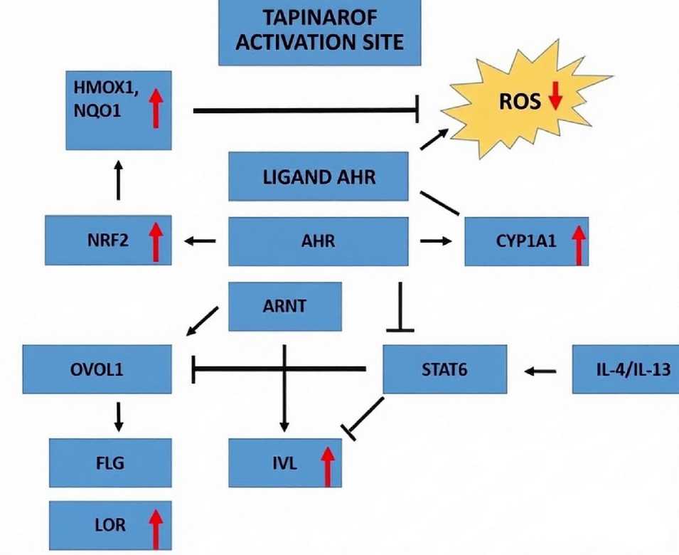
Figure 1. Aryl-carbohydrate receptor (AH metabolic effects and tapinarof action points (red words and arrows)
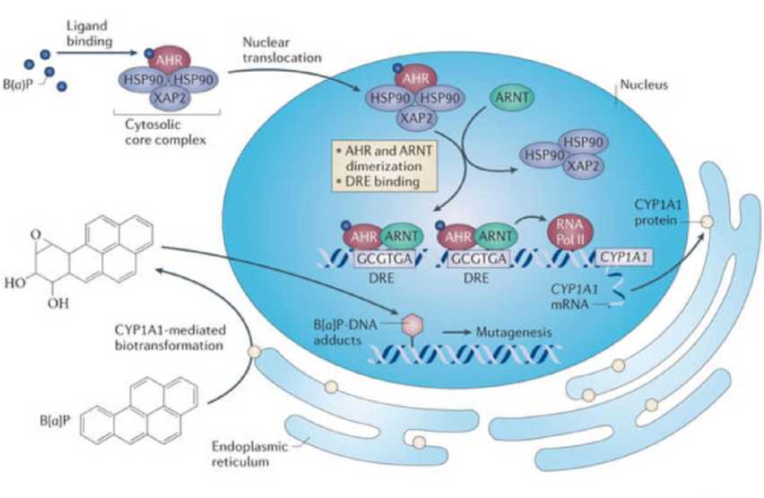
Figure 2. Functioning of the Ah -A NT system in mammalian cells
Collectively, the above in vitro experiments demonstrate that moderate UVB exposure induces Ah signaling within the cell.
UV irradiation of rodent and human skin induces Ah function locally and may induce CYP1A1 or other hepatic monooxygenases. To test the effect of a single moderate UVB dose on AH signaling in mice, a UV source (peak at 313 nm) was used that better reflects the composition of sunlight than narrowband UV. It was shown that a single 15-minute UV exposure (1.2 kJ/m²) in mice induced expression of the AH target genes Cyp1a1, Il22, and Il23a in the skin (Fig. 2), consistent with previous studies using a higher UV dose (Fritsche E. et al. , 2007). To extend these results and test whether AH ligands are rapidly released into the bloodstream following UVB exposure of mouse skin, serum was collected from control male or female animals or from UVB-exposed animals 30 min after a 15-min irradiation and incubated.
CONCLUSIONS
In recent years, a growing number of research has underscored the regulatory role of Ah in skin physiology. Furthermore, Ah in keratinocytes perceives environmental stimuli and contributes to the pathology of atopic dermatitis. It is hypothesized that the mechanisms of atopic dermatitis may operate via Ah activation. Consequently, assessing its role in the pathogenesis of dermatoses, including atopic dermatitis, is a logical endeavor. However, evidence exists for both physiological and pathological effects of Ah signaling. Therefore, an understanding of Ah function within the relevant metabolic cascades leading to disease is required. Currently, most data pertain to inflammatory skin disorders, particularly atopic dermatitis, psoriasis, acne, and hidradenitis suppurativa. Indeed, altered Ah function appears to be associated with both skin barrier disruption and the release of proinflammatory cytokines-two key factors in most chronic inflammatory conditions. These observations suggest that agents targeting Ah may be effective in treating this pathology. At present, the Ah agonist tapinarof has demonstrated efficacy in clinical trials for some of these diseases; however, broader clinical validation is warranted. Diverse experimental studies are also essential to fully establish the role of Ah in both physiological and pathological skin mechanisms, as well as to identify novel Ah - targeted molecules for the treatment of skin, inflammatory, and other diseases.
CONFLICTS OF INTEREST
All authors declare that they have no conflicts of interest.