
Myocarditis as a concomitant condition in patients with post-infarction myocardial fibrosis and acute decompensated heart failure
Journal: Siberian Journal of Clinical and Experimental Medicine @cardiotomsk
Section: Клинические исследования
Article in issue: 2 т.41, 2026.
Free access
Background. Acute decompensated heart failure (ADHF) in patients with post-infarction cardiosclerosis, even with previously performed optimal coronary revascularization, is typically considered a manifestation of ischemic heart disease progression. However, in clinical practice acute decompensation can also be influenced by non-ischemic mechanisms, including myocarditis. Such a combination of conditions appears particularly significant in patients with reduced left ventricular ejection fraction (LVEF) and a history of coronary revascularization in the absence of signs of ongoing myocardial ischemia. Aim: To assess the prevalence of myocarditis and myocardial viral antigen expression in patients hospitalized with ADHF, reduced LVEF, post-infarction myocardial fibrosis, and a history of coronary revascularization. Material and Methods. The study included 26 patients admitted with ADHF. Acute coronary syndrome and other conditions, such as anemia, pneumonia, sepsis, influenza, decompensation of diabetes mellitus, tumors, cachexia, or severe valvular heart stenosis, capable of independently causing HF decompensation were excluded. Median LVEF was 29.5% (IQR 22.0–32.0). All patients underwent transthoracic echocardiography, invasive coronary angiography, and right ventricular endomyocardial biopsy. Histological and immunohistochemical (IHC) analyses were performed to identify myocardial inflammation and viral antigen expression. Endomyocardial biopsy of the right ventricle was performed, with three myocardial samples obtained from each patient, followed by pathohistological and immunohistochemical examination. Results. Myocarditis was diagnosed in 18 patients (69%), including viral myocarditis in 13 (50%), viral-autoimmune myocarditis in 3 (12%), and autoimmune myocarditis in 1 patient (4%). Viral antigen expression in the myocardium was detected in 89% of cases. The frequency of viral antigen detection was as follows: enterovirus – 77%, human herpesvirus type 1 – 8%, type 2 – 8%, type 6 – 50%, Epstein-Barr virus – 23%, cytomegalovirus – 8%; parvovirus B19 and adenovirus were not detected. Viral antigen expression was observed in 94% of patients with myocarditis (except for one case of autoimmune myocarditis) and in 75% of patients without histological signs of myocardial inflammation. Conclusion. In patients with ADHF, reduced LVEF, post-infarction cardiosclerosis, and a history of coronary revascularization, myocarditis was detected in 69% of cases and represented a frequent comorbid condition. In the absence of myocardial ischemia, inflammation of the myocardium may contribute to the development of ADHF, which should be considered during diagnostic evaluation.
Short address: https://sciup.org/149151449
IDS: 149151449 | UDC: 616.12-008.46+616-005.8-06]:616.127-002 | DOI: 10.29001/2073-8552-2026-41-2-94-102
Миокардит как сочетанное заболевание у пациентов с постинфарктным кардиосклерозом при острой декомпенсации сердечной недостаточности
Введение. Острая декомпенсация сердечной недостаточности (ОДСН) у пациентов с постинфарктным кардиосклерозом даже при ранее выполненной оптимальной коронарной реваскуляризации, как правило, рассматривается как проявление прогрессирования ишемической болезни сердца. Вместе с тем в клинической практике острая декомпенсация может быть обусловлена и неишемическими механизмами, в частности миокардитом. Такое сочетание заболеваний представляется особенно значимым у пациентов со сниженной фракцией выброса левого желудочка и коронарной реваскуляризацией в анамнезе при отсутствии признаков текущей ишемии миокарда. Цель: оценить частоту выявления миокардита и экспрессии вирусных антигенов в миокарде у пациентов с ОДСН, сниженной фракцией выброса левого желудочка, постинфарктным кардиосклерозом и коронарной реваскуляризацией в анамнезе. Материал и методы. В исследование были включены 26 пациентов, госпитализированных с ОДСН. Критериями исключения являлись острый коронарный синдром и состояния, способные самостоятельно вызвать декомпенсацию сердечной недостаточности, включая анемию, пневмонию, сепсис, грипп, декомпенсацию сахарного диабета, онкологические заболевания, кахексию и выраженные пороки клапанов сердца. Медиана фракции выброса левого желудочка составила 29,5% (22,0; 32,0). Всем пациентам выполнена трансторакальная эхокардиография, инвазивная коронарогафия, эндомиокардиальная биопсия правого желудочка. Для выявления воспаления миокарда и экспрессии вирусного антигена проведено патогистологическое и иммуногистохимическое исследование. Результаты. Миокардит был диагностирован у 18 пациентов (69%), включая вирусный миокардит – у 13 (50%), вирусно-аутоиммунный – у 3 (12%) и аутоиммунный – у 1 пациента (4%). Экспрессия вирусных антигенов в миокарде выявлялась в 89% случаев. Частота обнаружения вирусных антигенов составила: энтеровирус – 77%, вирус герпеса человека 1-го типа – 8%, 2-го типа – 8%, 6-го типа – 50%, вирус Эпштейна – Барра – 23%, цитомегаловирус – 8%; парвовирус B19 и аденовирус выявлены не были. Экспрессия вирусных антигенов отмечалась у 94% пациентов с миокардитом (за исключением одного случая аутоиммунного миокардита) и у 75% пациентов без гистологических признаков воспаления миокарда. Заключение. У пациентов с ОДСН со сниженной фракцией выброса левого желудочка, постинфарктным кардиосклерозом и коронарной реваскуляризацией в анамнезе миокардит выявлялся в 69% случаев и представлял собой частое сочетанное заболевание. В отсутствие ишемии миокарда его воспаление может способствовать развитию ОДСН, что следует учитывать при проведении диагностики.
Text of the scientific article Myocarditis as a concomitant condition in patients with post-infarction myocardial fibrosis and acute decompensated heart failure
Введение . Острая декомпенсация сердечной недостаточности (ОДСН) у пациентов с постинфарктным кардиосклерозом даже при ранее выполненной оптимальной коронарной реваскуляризации, как правило, рассматривается как проявление прогрессирования ишемической болезни сердца. Вместе с тем в клинической практике острая декомпенсация может быть обусловлена и неишемическими механизмами, в частности миокардитом. Такое сочетание заболеваний представляется особенно значимым у пациентов со сниженной фракцией выброса левого желудочка и коронарной реваскуляризацией в анамнезе при отсутствии признаков текущей ишемии миокарда.
Цель : оценить частоту выявления миокардита и экспрессии вирусных антигенов в миокарде у пациентов с ОДСН, сниженной фракцией выброса левого желудочка, постинфарктным кардиосклерозом и коронарной реваскуляризацией в анамнезе.
Материал и методы . В исследование были включены 26 пациентов, госпитализированных с ОДСН. Критериями исключения являлись острый коронарный синдром и состояния, способные самостоятельно вызвать декомпенсацию сердечной недостаточности, включая анемию, пневмонию, сепсис, грипп, декомпенсацию сахарного диабета, онкологические заболевания, кахексию и выраженные пороки клапанов сердца. Медиана фракции выброса левого желудочка составила 29,5% (22,0; 32,0). Всем пациентам выполнена трансторакальная эхокардиография, инвазивная коронарогафия, эндомиокардиальная биопсия правого желудочка. Для выявления воспаления миокарда и экспрессии вирусного антигена проведено патогистологическое и иммуногистохимическое исследование.
Результаты . Миокардит был диагностирован у 18 пациентов (69%), включая вирусный миокардит – у 13 (50%), вирусно-аутоиммунный – у 3 (12%) и аутоиммунный – у 1 пациента (4%). Экспрессия вирусных антигенов в миокарде выявлялась в 89% случаев. Частота обнаружения вирусных антигенов составила: энтеровирус – 77%, вирус герпеса человека 1-го типа – 8%, 2-го типа – 8%, 6-го типа – 50%, вирус Эпштейна – Барра – 23%, цитомегаловирус – 8%; парвовирус B19 и аденовирус выявлены не были. Экспрессия вирусных антигенов отмечалась у 94% пациентов с миокардитом (за исключением одного случая аутоиммунного миокардита) и у 75% пациентов без гистологических признаков воспаления миокарда.
Заключение . У пациентов с ОДСН со сниженной фракцией выброса левого желудочка, постинфарктным кардиосклерозом и коронарной реваскуляризацией в анамнезе миокардит выявлялся в 69% случаев и представлял собой частое сочетанное заболевание. В отсутствие ишемии миокарда его воспаление может способствовать развитию ОДСН, что следует учитывать при проведении диагностики.
Background
Acute decompensated heart failure (ADHF) represents a clinical syndrome with sudden or worsening heart failure (HF) symptoms that typically demand hospital admission or emergency care. Despite advances in modern cardiology, ADHF remains associated with high mortality and substantial functional impairment, particularly in patients with advanced structural heart disease and prior myocardial infarction. Inhospital death rates average around 7.5% [1], and 1-year mortality surpasses 27.2% after discharge [2]. Coronary artery disease (CAD) drives much of ADHF. After myocardial infarction (MI), harmful left ventricular (LV) remodeling often triggers advancing systolic dysfunction and HF. Persistent ischemia then activates neurohormonal and inflammatory cascades [3-5], worsening cardiac impairment and reducing left ventricular ejection fraction (LVEF). Myocarditis involves inflammation of the heart muscle, with patterns that vary widely and often go undercounted in populations. The estimated incidence ranges from 10 to 20 per 100,000, though actual rates likely exceed that. Clinical presentation is highly variable and often nonspecific: patients might show none or face severe outcomes like pulmonary edema, cardiogenic shock, life-threatening arrhythmias, or sudden death. Diagnostic methods and mechanistic insights have advanced, yet prognosis remains poor, especially with LV systolic dysfunction, ADHF, or key arrhythmias [6]. Clinical suspicion of myocarditis is based on a combination of findings: recent viral infection, chest pain resembling infarction, palpitations, HF traits, ECG changes, ventricular rhythms, and unstable hemodynamics. Laboratory markers of cardiac damage and failure, along with typical echo or cardiac magnetic resonance imaging (MRI) patterns, strengthen the diagnosis. In advanced ischemic disease after MI, these signs often get blamed on CAD alone. This can hide overlapping myocarditis or delay its detection [7]. Research now aims to unravel myocarditis pathways and progression in those with weakened LV function, stressing long-term results [6]. Cardiotropic viruses play a central role, with enteroviruses, adenoviruses, herpes types, cytomegalovirus, and Epstein-Barr virus leading as causes of heart inflammation [8-10]. Large studies show cardiac MRI raises acute myocarditis detection from 5% to 13% in cases with angina-like pain and high troponin levels [7]. Endomyocardial biopsy stands as the benchmark; it provides direct views of inflammation and causes. Without clear signs, diagnosis proves tough, more so in structural heart disease. Given the high prevalence of CAD, its coexistence with myocarditis represents a clinically relevant but often overlooked scenario. Thus, CAD without clear ongoing ischemia should not serve as the only reason for ADHF and LV systolic dysfunction. Targeted studies remain limited, drawn mostly from case reports and small groups [11, 12].
The aim of this study was to evaluate the prevalence of myocarditis and the frequency of detection of antigens of cardiotropic viruses in heart tissue among patients with ADHF, low LVEF, prior MI, and coronary procedures.
Material and Methods
Study design and population
This single-center observational study was conducted at the Cardiology Research Institute, Tomsk National Research Medical Center (Tomsk, Russia). Patient enrollment was carried out between 2015 and 2022. The study protocol was approved by the local ethics committee (Protocol No 114, December 16, 2015) and complied with the principles of the Declaration of Helsinki. Written informed consent was obtained from all participants.
Inclusion and exclusion criteria
Patients were eligible for inclusion if they met all of the following criteria:
-
1. Hospitalization for ADHF, defined by the presence of at least 3 clinical signs or symptoms, including dyspnea or orthopnea, pulmonary crackles or wheezes, peripheral edema, elevated jugular venous pressure, or radiographic signs of pulmonary congestion;
-
2. All included patients had established chronic HF for more than one month before hospitalization. Acute decompensated HF was defined as clinically significant worsening of pre-existing disease requiring hospital admission (NT-proBNP ≥ 300 pg/ml);
-
3. Documented CAD with complete revascularization of all hemodynamically significant coronary stenoses, confirmed by coronary angiography performed within 6 months prior to enrollment;
-
4. Left ventricular ejection fraction below 40%;
-
5. Clinical eligibility for endomyocardial biopsy.
Patients were excluded if they had acute coronary syndrome or conditions that could independently cause or mimicADHF, including anemia, pneumonia, sepsis, influenza, decompensated diabetes mellitus, active malignancy, cachexia, or severe valvular stenosis.
Echocardiography and endomyocardial biopsy
All patients underwent standard transthoracic echocardiography using a VIVID E9 ultrasound system (GE Healthcare). To evaluate myocardial inflammatory involvement, endomyocardial biopsy was performed via the right femoral vein with sampling from the right ventricle. Three myocardial specimens were obtained from predefined regions: the right ventricular apex, the interventricular septum, and the right ventricular outflow tract.
Immunohistochemical analysis
Biopsy specimens were subjected toimmunohistochemical (IHC) analysis using monoclonal antibodies directed against cardiotropic viral antigens. The following antibodies were applied: rabbit monoclonal antibodies against herpes simplex virus type 1 (Cell Marque, clone 361A-16-ASR; dilution 1 : 1000) and herpes simplex virus type 2 (Cell Marque, clone 362A-76-ASR; dilution 1 : 1000); mouse monoclonal antibodies against human herpesvirus type 6 (Abcam, clone ab128404; dilution 1 : 100); mouse monoclonal antibodies against adenovirus (Cell Marque, clone 212M-16-ASR; dilution 1 : 1000); rabbit monoclonal antibodies against Epstein – Barr virus (Cell Marque, clone 245R-16-ASR; dilution 1 : 1000); mouse monoclonal antibodies against enterovirus (dilution 1 : 1000); and mouse monoclonal antibodies against parvovirus B19 (Cell Marque, clone 218M-16-ASR; dilution 1 : 1000). To assess autoimmune myocardial involvement, additional IHC staining was performed using antibodies against complement component C1q, major histocompatibility complex class II, and the B-lymphocyte marker CD19. To characterize the cellular composition of the inflammatory infiltrate, IHC staining for CD3 (Leica Biosystems RTU Primary mouse monoclonal antibody, clone LN10) was used to identify T lymphocytes, while CD68 (Leica Biosystems RTU Primary mouse monoclonal antibody, clone 514H12) immunostaining was applied to detect macrophages within myocardial tissue.
Diagnostic criteria for myocarditis
The diagnosis of myocarditis was established according to current histopathological criteria and defined as the presence of ≥ 14 leukocytes/mm² within the myocardium, including up to 4 monocytes/mm², with at least 7 CD3-positive T lymphocytes/mm2 [13].
Coronary angiography
All patients underwent invasive coronary angiography to exclude ongoing myocardial ischemia as the primary cause of ADHF.
Statistical analysis
Given the limited availability of data on myocarditis prevalence in this clinical setting, the study was designed as a pilot investigation. Statistical analysis was performed using STATISTICA software, version 10.0 (StatSoft Inc., USA). The normality of distribution was assessed by Shapiro Wilk. Given the non-normal distribution, continuous variables were presented as median (IQR). Comparisons between independent groups were performed using the Mann Whitney U test. Categorical variables were compared using the χ ² test or Fisher’s exact test, as appropriate. A two-sided p-value < 0.05 was considered statistically significant.
Technical resources
All analyses were performed using the facilities of the Center for Collective Use “Medical Genomics”, Tomsk National Medical Research Center.
Results
Patient characteristics
The study included 26 patients hospitalized with ADHF. Median age was 62 years (57.0; 67.0), and the majority of patients were male (85%). All patients had a documented history of myocardial infarction and CAD with prior revascularization. The time from diagnosed CAD to the development of HF was 72 months (IQR 13; 114). The median duration from the onset of HF symptoms to study inclusion was 12.5 months (IQR 5.0; 30.5). At the time of admission, LVEF was markedly reduced in all patients, with a median value of 29.5% (IQR 22.0; 32.0). Median NT-proBNP concentration was 403.1 pg/mL (IQR 307.2; 2424.7). Troponin I levels were mildly elevated, with a median value of 0.037 ng/mL (0.020; 0.069), (upper limit of normal [99th percentile] – 0.04 ng/mL). C-reactive protein (CRP) levels were moderately increased, with a median of 7.0 mg/L (IQR 5.0; 19.3), (reference range is 0 8 mg/L).
Clinical presentation
Most patients presented with clinical features consistent with advanced heart failure. Dyspnea was reported by the majority of patients, occurring during minimal physical activity in 10 patients (38%) and at rest in 3 patients (12%). Signs of systemic and pulmonary congestion were common, including pulmonary crackles or wheezing in 14 patients (54%), peripheral edema in 12 patients (46%), jugular venous distention in 8 patients (31%), and hepatomegaly in 18 patients (70%). Cyanosis was observed in 13 patients (50%). Cardiac arrhythmias were frequently documented.
Atrial fibrillation or flutter was present in 12 patients (46%), while episodes of ventricular tachycardia were recorded in 9 patients (35%). Although 4 patients (15%) reported symptoms suggestive of a recent viral illness prior to hospitalization, none had documented fever at admission or shortly beforehand. At admission, HF symptoms severity was classified as New York Heart Association (NYHA) functional class III in 16 patients (61%) and class IV in 10 patients (39%). At the time of study inclusion, patients were stratified according to clinical hemodynamic profiles: four patients (16%) were classified as “warm and wet”, 11 patients (42%) as “cold and wet”, and 11 patients (42%) as “cold and dry”. Baseline clinical characteristics are summarized in Table 1.
Endomyocardial biopsy findings
Histological criteria for myocarditis were met in 18 of 26 patients (69%). Among these patients, viral myocarditis was diagnosed in 13 cases (50%), virus-associated autoimmune myocarditis in 3 cases (12%), and autoimmune myocarditis in 1 case (4%). Immunohistochemical analysis demonstrated predominant inflammatory infiltration by CD3-positive T lymphocytes and CD68-positive macrophages, frequently accompanied by areas of myocardial fibrosis. Detailed immunohistochemical findings are presented in Table 2.
Myocardial viral antigen expression
Viral antigen expression within myocardial tissue was detected in 23 patients (89%). Enterovirus was the most frequently identified viral antigen, present in 77% of cases. Human herpesvirus type 6 was detected in 50% of patients (Figure 1), Epstein – Barr virus in 23%, human herpesvirus types 1 and 2 in 8% each, and cytomegalovirus in 8%. Parvovirus B19 and adenovirus were not detected. In several patients, immunohistochemical staining revealed coexpression of multiple viral antigens, resulting in a cumulative detection rate exceeding 100% due to co-detection of multiple viral antigens in individual patients (Table 2).
Comparison of patients with and without myocarditis
Patients were stratified according to the presence or absence of histologically confirmed myocarditis. Viral antigen expression was observed in 94% of patients with myocarditis, with the exception of one patient diagnosed with autoimmune myocarditis. Viral antigens were also detected in 75% of patients without histological signs of myocarditis.
Echocardiographic parameters did not differ significantly between patients with and without myocarditis. However, a trend toward higher troponin I levels was observed in patients with myocarditis compared with those without myocarditis (0.3 ng/mL (IQR 0.2; 0.7) vs. 0.1 ng/mL (IQR 0.0; 0.2), p = 0.05). Comparative echocardiographic and laboratory data are summarized in Table 3. A history of coronary artery bypass grafting was more frequent in patients with myocarditis; however, given the small sample size, this finding should be interpreted with caution.
Discussion
In this single-center pilot study, myocarditis was identified as a frequent concomitant pathological finding in patients hospitalized with ADHF, reduced left ventricular ejection fraction, prior myocardial infarction, and a history of coronary revascularization. Histological criteria for myocarditis were met in nearly two-thirds of the study population, suggesting that inflammatory myocardial injury may play a meaningful
Table 1 . Clinical baseline characteristics of the study population
Таблица 1 . Клинико-анамнестические характеристики пациентов
|
Parameter |
Total group, n = 26 |
With myocarditis, n = 18 |
Without myocarditis, n = 8 |
p-value |
|
Male sex, n (%) |
22 (85) |
14 (78) |
8 (100) |
0.21 |
|
Age, years, Me ( Q25 ; Q75 ) |
62 (57; 67) |
63 (56; 68) |
59 (57; 66) |
0.94 |
|
Body Mass Index, kg/m2, Me ( Q25 ; Q75 ) |
30.0 (25.8; 32.7) |
26.8 (25.2; 28.4) |
30.0 (26.3; 32.7) |
0.55 |
|
Hypertension, n (%) |
20 (77) |
15 (83) |
5 (63) |
0.43 |
|
Diabetes mellitus 2, n (%) |
19 (73) |
13 (72) |
6 (75) |
0.79 |
|
Myocardial infarction in history, n (%) |
26 (100) |
18 (100) |
8 (100) |
1.00 |
|
A history of invasive treatment: |
||||
|
PCI, n (%) |
10 (38) |
6 (33) |
4 (50) |
0.11 |
|
CABG, n (%) |
8 (31) |
6 (33) |
2 (25) |
0.01 |
|
PCI and CABG, n (%) |
6 (23) |
4 (22) |
2 (25) |
0.93 |
|
Implantation of cardioverter-defibrillator, n (%) |
7 (27) |
4 (22) |
3 (38) |
0.93 |
|
Cardiac resynchronizing therapy, n (%) |
3 (12) |
3 (17) |
0 (0) |
– |
|
Symptoms of infection before admission, n (%) |
4 (15) |
4 (23) |
0 (0) |
– |
|
At the time of admission: |
||||
|
Сyanosis, n (%) |
13 (50) |
8 (44) |
5 (63) |
0.22 |
|
Dyspnoea during physical activity/at rest, n (%) |
10 (38) / 3 (12) |
6 (34) / 2 (11) |
4 (50) / 1 (13) |
0.83 / – |
|
Wheezing, n (%) |
14 (54) |
7 (38) |
7 (88) |
0.11 |
|
Edema on the legs, n (%) |
12 (46) |
6 (34) |
6 (75) |
0.13 |
|
Weight gain in the last week, n (%) |
10 (38) |
5 (28) |
5 (63) |
0.42 |
|
Jugular vein distention, n (%) |
8 (31) |
4 (23) |
4 (50) |
0.88 |
|
Hepatomegaly, n (%) |
18 (70) |
12 (67) |
6 (75) |
0.54 |
|
SBP, mmHg Me ( Q25 ; Q75 ) |
122.0 (102.0; 130.0) |
114.0 (100.0; 142.0) |
129.0 (115.0; 130.0) |
0.10 |
|
DBP, mmHg Me ( Q25 ; Q75 ) |
78.0 (70.0; 80.0) |
75.0 (64.0; 84.0) |
72.0 (64.0; 84.0) |
0.31 |
|
Heart rate, Me ( Q25 ; Q75 ) |
78.0 (70.0; 80.0) |
80.0 (72.0; 90.0) |
72.0 (64.0; 84.0) |
0.65 |
|
Respiratory rate per minute, Me ( Q25 ; Q75 ) |
18.5 (18.0; 20.0) |
18.5 (18.0; 20.0) |
19 (16.5; 20.0) |
0.55 |
|
Oxygen saturation, % |
95.0 (93.0; 95.0) |
94.0 (94.0; 95.0) |
95.0 (94.0; 95.0) |
1.00 |
|
Ventricular taсhycardia, n (%) |
9 (35) |
5 (28) |
4 (50) |
0.43 |
|
Atrial fibrillation / flutter, n (%) |
12 (46) |
6 (34) |
6 (75) |
0.13 |
Abbreviations: PCI – percutaneous coronary intervention; CABG – coronary artery bypass graft; SBP – systolic blood pressure; DBP – diastolic blood pressure.
Table 2 . The results of the immunohistochemical study, n (%)
Таблица 2 . Результаты иммуногистохимического исследования, n (%)
In patients with established coronary artery disease, ADHF is most commonly attributed to ischemic myocardial injury and adverse post-infarction remodeling [1, 2]. At the same time, accumulating data suggest that non-ischemic mechanisms, including myocarditis, may substantially contribute to disease progression and acute worsening of HF symptoms [6, 7]. In the present cohort, all patients had previously undergone coronary revascularization and showed no angiographic signs of ongoing myocardial ischemia at the time of inclusion, making ischemia alone an unlikely explanation for the severity of decompensation observed.
The high prevalence of viral myocarditis in this study is
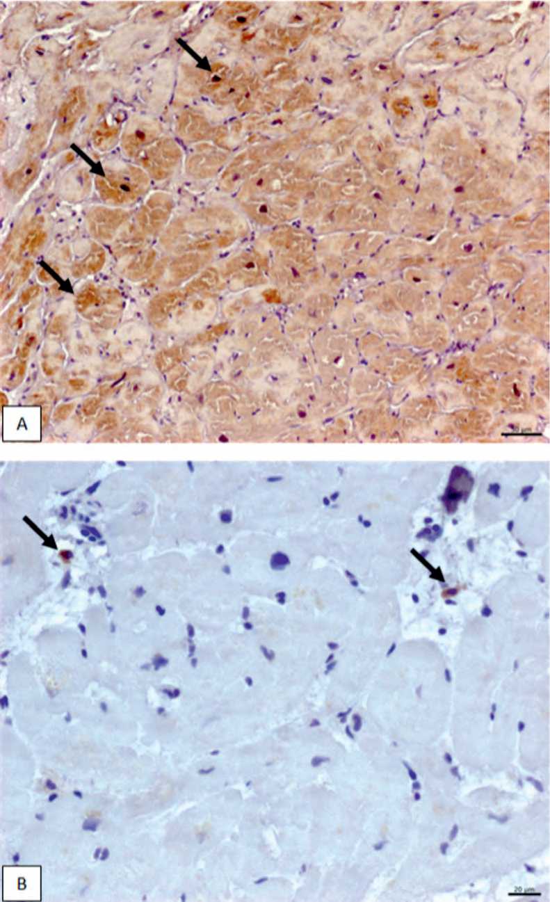
Fig. 1. Immunohistochemical detection of cardiotropic viral antigens in myocardial tissue. (A) Diffuse cytoplasmic immunopositivity for Enterovirus antigens in cardiomyocytes (arrows) (× 200; scale bar = 50 µm). (B) Cytoplasmic immunopositivity for Human Herpesvirus type 6 (HHV-6) in myocardial tissue macrophages (arrows) (× 400; scale bar = 20 µm)
Рис. 1. Иммуногистохимическое выявление антигенов кардиотропных вирусов в ткани миокарда. (A) Диффузное цитоплазматическое иммунное окрашивание антигенов энтеровируса в кардиомиоцитах (стрелки) (увеличение × 200; масштабная линейка – 50 мкм). (B) Цитоплазматическое иммунное окрашивание антигенов вируса герпеса человека 6-го типа (HHV-6) в макрофагах миокарда (стрелки) (увеличение × 400; масштабная линейка – 20 мкм)
Table 3 . Echocardiographic and laboratory parameters in patients with and without myocarditis
Таблица 3 . Лабораторные и ультразвуковые данные пациентов с миокардитом и без него
Parameter With myocarditis, n = 18 Without myocarditis, n = 8 p-value
The ultrasound examination of heart data, Me ( Q25 ; Q75 )
|
Left ventricular ejection fraction, % |
24.5 (19.0; 35.0) |
22.2 (18.0; 29.7) |
0.55 |
|
End-systolic volume index, ml/m2 |
83.9 (63.9; 99.1) |
77.7 (58.1; 85.7) |
0.52 |
|
End-diastolic volume index, ml/m2 |
114.3 (93.0; 129.0) |
97.8 (87.1; 105.8) |
0.23 |
|
Stroke volume index, ml/m2 |
28.4 (18.8; 31.2) |
18.8 (17.2; 28.1) |
0.76 |
|
Cardiac index, L/min/m2 |
1.9 (1.6; 2.4) |
1.7 (1.1; 2.4) |
0.53 |
|
Wall motion score index |
1.9 (1.8; 2.2) |
2.0 (1.8; 2.1) |
0.93 |
|
Left atrium index, ml/m2 |
51.0 (40.9; 73.6) |
53.6 (39.0; 61.6) |
0.44 |
|
Left ventricular myocardial mass index, g/m2 |
160.7 (136.0; 180.3) |
166.1 (126.4; 232.9) |
0.24 |
|
Right ventricle, mm |
33.0 (25.0; 39.0) |
36.0 (31.0; 37.5) |
0.62 |
|
Right ventricular systolic pressure, mmHg |
50.0 (37.0; 65.0) |
47.0 (46.0; 52.0) |
0.63 |
|
Mitral regurgitation grade 1-2 / grade 3-4 |
9 (50) / 7 (39) |
7 (88) / 1 (13) |
0.99 / – |
|
Tricuspid regurgitation grade 1-2 / grade 3-4 |
8 (44) /1 (6) |
1 (13) /2 (25) |
– |
Laboratory data, Me ( Q25 ; Q75 )
|
Troponin I, ng/mL |
0.3 (0.2; 0.7) |
0.1(0.0; 0.2) |
0.05 |
|
CRP, mg/L |
4.8 (1.0; 10.0) |
5.3 (1.7; 9.7) |
0.85 |
|
NT-proBNP level, pg/mL |
403.1 (307.2; 2424.7) |
392.3 (194.5; 665.2) |
0.12 |
Abbreviations: CRP – C-reactive protein, NT-proBNP – N-terminal pro b-type natriuretic peptide.
consistent with prior reports demonstrating frequent detection ofcardiotropicvirusesinpatientswithleftventriculardysfunction and HF [8–10]. Enterovirus and human herpesvirus type 6 were the most commonly identified viral antigens, in line with existing data on viral persistence within myocardial tissue [8, 9]. Importantly, viral antigen expression was also detected in a substantial proportion of patients without histological signs of myocarditis. Similar findings have been described previously and suggest that viral persistence may occur in the absence of overt inflammatory myocardial disease [6, 10]. Parvovirus B19 deserves particular consideration in this context. Although it has frequently been implicated in the development of HF symptoms and myocardial dysfunction, its pathogenic significance remains debated, especially when active myocardial inflammation is absent [15]. In the present cohort, parvovirus B19 was not detected, indicating that other cardiotropic viruses may have a more prominent role in inflammatory myocardial involvement among patients with advanced ischemic heart disease. The absence of parvovirus B19 in our cohort may reflect regional characteristics of the study population and methodological differences, as immunohistochemical detection may underestimate latent viral persistence compared with PCR-based approaches.
Several mechanisms may account for the presence of viral antigen without histological myocarditis, including latent infection, impaired viral clearance, or low-grade inflammatory activity below established diagnostic thresholds [6]. In the setting of advanced ischemic cardiomyopathy, chronic myocardialinjury, fibrosis, and alterationsinimmune regulation may further promote viral persistence. These considerations emphasize the need to interpret endomyocardial biopsy findings in an integrated manner, combining histological, immunohistochemical, and clinical data rather than relying solely on viral antigen detection.
Despite the high prevalence of myocarditis, echocardiographic parameters did not differ significantly between patients with and without histologically confirmed myocarditis. This likely reflects the advanced degree of ventricular dysfunction shared by all patients in the cohort, as severe systolic impairment was a prerequisite for hospitalization with ADHF. Within this context, myocarditis may act primarily as a trigger for acute decompensation rather than as the main determinant of baseline ventricular function. The observed trend toward higher troponin I levels in patients with myocarditis supports the presence of ongoing myocardial injury, although the difference did not reach conventional statistical significance.
Clinically, most patients presented with advanced HF and were classified as New York Heart Association functional class III or IV at admission. In addition, “cold” hemodynamic profiles predominated at the time of inclusion, reflecting marked impairment of cardiac output. Such hemodynamic vulnerability may increase susceptibility to additional inflammatory or infectious insults and contribute to clinical instability. From a clinical perspective, these findings suggest that myocarditis should be considered in patients with ADHF following myocardial infarction, particularly when coronary angiography does not reveal active ischemia. Failure to recognize concomitant myocarditis may delay appropriate diagnostic evaluation and limit opportunities for individualized management. At the same time, detection of viral antigens alone should be interpreted with caution, as it does not in itself justify antiviral or immunomodulatory therapy in the absence of histological inflammation [6].
The identification of histologically confirmed myocarditis in patients with post-infarction cardiosclerosis inevitably raises the question of pathogenetic therapy. However, the role of immunosuppressive treatment in this specific population remains poorly defined and is supported mainly by isolated clinical observations rather than prospective trials. Given the coexistence of ischemic myocardial injury and inflammatory processes, therapeutic decisions should be individualized and based on careful risk–benefit assessment.
Limitations
This study has several important limitations. The singlecenter design and relatively small sample size restrict the generalizability of the findings and preclude robust analysis of clinical outcomes. In addition, viral genome quantification by polymerase chain reaction was not performed, limiting assessment of viral load and replication activity. Nonetheless, the systematic use of endomyocardial biopsy combined with detailed immunohistochemical analysis represents a key strength of the study and provides valuable insight into the inflammatory substrate of acute HF decompensation in patients with ischemic heart disease.
Conclusion
Myocarditis was frequently identified as a concomitant pathological finding in patients hospitalized with acute decompensated heart failure, reduced left ventricular ejection fraction, prior myocardial infarction, and a history of coronary revascularization. In the absence of angiographic evidence of ongoing myocardial ischemia, inflammatory myocardial involvement may contribute to clinical deterioration in this patient population.
These findings indicate that ADHF in patients with advanced ischemic heart disease cannot always be explained by ischemic mechanisms alone. Integration of clinical assessment with histological and immunohistochemical evaluation may facilitate more accurate identification of concomitant myocarditis in selected patients.