Reliability of veterinarians routine cattle meat inspection tool on specific identification of cysticerci in bovine muscle tissue

Author: Rutaganira J., Glamazdin I.
Journal: Вестник Воронежского государственного университета инженерных технологий @vestnik-vsuet
Section: Пищевая биотехнология
Article in issue: 3 (97) т.85, 2023.
Free access
Cysticercus bovis and macroscopic Sarocysts detected from bovine meat look similar and specific identification of each of these cysts is important for addressing proper preventive, and control measures of tCysticercosis and Sarcocystosis diseases.Therefore, it is sometimes a challenge for Vets to distinguish these two species of parasites during their daily routine meat inspection using traditional methods. Cysticerci and Sarcocysts are responsible of Bovine Cysticercosis and Cattle Sarcocystosis respectively. These Veterinarians can sometimes make errors in identification of these two parasitic species from Cattle meat muscle tissues. The study was conducted from Abattoir in Russian Federation with intention of the problem addressed confirmation. During laboratory experimental examination, it was confirmed that out of 20 cattle muscles tissues sampled from 20 infected bovines reported to contain cysticerci parasites, 5 out of them were Sarcocysts. Therefore, it is obvious for veterinarians to make errors during their routine cattle meat inspection due to the morphological behaviour of these organisms.
Cestode parasites, cysticercus bovis, foodborne parasites, protozoa, sarcocystis spp, taenia saginata
Short address: https://sciup.org/140303243
IDR: 140303243 | UDC: 640 | DOI: 10.20914/2310-1202-2023-3-48-51
Надежность рутинного инструмента ветеринарных врачей для контроля мяса крупного рогатого скота по специфической идентификации цистицерков в мышечной ткани
Цистицерки и макроскопические саркоцисты, обнаруженные в тушах крупного рогатого скота, выглядят одинаково, и специфическая идентификация каждой из этих цист важна для принятия надлежащих мер профилактики и борьбы с цистицеркозом и саркоцистозом. Поэтому для ветеринарных врачей иногда представляет трудность различать эти два вида паразитов при ежедневном осмотре мяса традиционными методами. Цистицерки и саркоцисты ответственны за цистицеркоз крупного рогатого скота и саркоцистоз крупного рогатого скота соответственно. Ветеринары иногда допускают ошибки при идентификации этих двух видов паразитов в мышечных тканях мяса крупного рогатого скота. Исследование проводилось на скотобойне в Российской Федерации с целью подтверждения актуальности проблемы. В ходе лабораторно-экспериментального исследования было установлено, что из 20 исследованных мышечных тканей крупного рогатого скота, содержащих паразитов цистицерков, 5 из них оказались саркоцистами. Таким образом, очевидна возможность ошибок ветеринарных врачей при проведении рутинного осмотра мяса крупного рогатого скота, связанных с морфологическим поведением этих организмов
Text of the scientific article Reliability of veterinarians routine cattle meat inspection tool on specific identification of cysticerci in bovine muscle tissue
DOI: Research article
Bovine cysticercosis is a Cattle disease caused by the intermediate (larval) stage of Taenia Saginata called Cysticercus bovis . Taenia Saginata (Macrelli et al., 2020) is the human beef tapeworm (El-Sayad et al., 2021). Cysticercus bovis parasitizes the intermuscular connective tissue of striated muscles. Bovine cysticercosis has been reported in Russia, Benin, Cameroon, Central African Republic, Chad, Democratic Republic of Congo, Ghana, Guinea, Mali, Niger, Nigeria, Senegal, etc. (Uchendu et al., 2021).
Bovine sarcocystosis is either zoonotic or not and it is caused by single-celled protozoa, Sar-cocystis Species including: S.hominis, S. heydorni, S. cruzi, S. rommeli, S. hirsuta and S. bovifelis. Sarcocystosis has a worldwide distribution, especially where livestock are raised (Rosenthal, 2021). The pathogen forms a tissue cyst inside the muscle fiber of the striated muscle. The presence of macroscopic sarcocysts can cause cattle meat culling. Microscopically, the cysts are divided by septa into chambers that contain thousands of banana-shaped bradysoites. Sarcocystosis, cause significant economic losses, including death and forced slaughter of animals, loss of offspring, and a decrease in the quality and nutritional value of meat. In the Russian Federation, sarcocystosis is registered in 85 % of cattle and sheep, 5 % of pigs, 35 % of horses (Novak and Novak, 2020). This disease of many animal species is widespread in many countries (Fayer et al., 2015).
Bovine cysticercosis(Assefa and Bihon, 2019), is caused by cysticercus bovis , larval form, which is hosted in Cattle muscle tissues but humans have risk of hosting the adult form of this cestode parasite “Taenia Saginata” in the small intestine(Lopes et al., 2011), when they eat poorly heat-treated beef (Leal Filho et al., 2022). Despite the fact that Cysticercosis and Sarcocystosis are caused by completely different types of parasites, they may have negative effect on Cattle as well on Humans (Dubey, 2015). From Cysticercus and Sarcocystis spp, Figure 1 illustrates all possible steps and sources of negative effects faced by humans and Cattle.
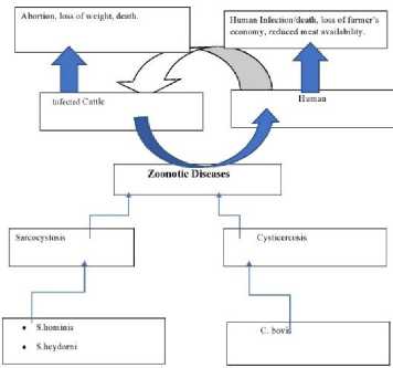
Figure 1. Flow Chart showing the effect of cysticercosis and Sarcocystosis on Infected Cattle as well as on Humans. Sarcocysts and Cysticerci identified in the bovine muscle tissues are all responsible of zoonotic diseases that can be transmitted between cattle
This is an open access article distributed under the terms of the Creative Commons Attribution 4.0 International License
Objective
post@vestnik-vsuet.ruResults and discussion
The objective is to conduct morphological study of the 20 bovine meat samples reported by Vets to host only cysticerci and identify if Sarco-cysts may be misnamed Cysticerci. The extracted information may also reveal tentative prevalence of these two species in Russian Federation
Material and methods
According to the informations given from food markets and Auchan stores chain in Russia, the meat tested by vets during their routine examination were collected from 875 infected slaughtered cattle carcasses(Ahmed et al., 2016) in Russian Federation and 20 were tested cysterci positive. These 20 cases that were reported to be infected by Cys-tercus bovis were objects of our Laboratory study. The 20 samples were in total bovine meat muscle tissues putatively infected by cysticerci parasites. During Laboratory examination, tissue cysts were allocated layer-by-layer cutting of the muscles, followed by examination of the internal content. The cysts were cut with a razor blade. necessary smears-prints were made, fixed and stained according to Romanovsky-Giemsa, microscoped.
15 cases were found to be muscle inclusions of gray color, oval shape and length from 1.5 to 2.0 cm. Covered by the connective tissue capsule of the host. After dissection of each of the 15 host capsules, a translucent oval-shaped bubble of about 1.0 cm in length, containing a transparent liquid was found inside. This was real cysticercus bovis. On the inner shell of the cysticercus there was a scolex, in which no hooks were found under microscopy.
-
5 cases were observed to be light gray in color, oval in shape, and were about 1.5 cm long. When the host capsule was cut with a razor blade, a clear or slightly cloudy liquid was found inside the cyst. In these cases, a smear was made on the slide and painted according to Romanovsky-Giemsa. Under microscopy with magnification of X400 or X800, banana-shaped structures were observed. This fact indicated that the studied cysts were formed by Sarcocysts .
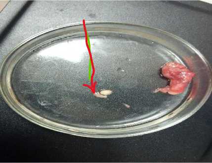
Figure 2. Cyst isolated from bovine meat before observation under microscopy
On the basis of Study examination results illustrated by Figure 2, the morphological study of 20 samples that were reported by Veterinarians to host only cysterci , w e found several inconsistencies. 15 cases were found to be real cysticerci larvae. According to our observations, the banana-like structures (endozoites) that were found preserved inside of each of the 50 cysts with different forms of liquid content (transparent, cloudy, different consistency), indicated that these 5 cases, were Sarcocysts .
Thus, a thorough study of 20 carcasses that were reported positive for C. bovis during routine veterinary examination revealed errors in 25 % of cases.
Table 1.
Results from Microscopy examination with a sample size of 20 cases reported putatively to be cysticerci only.s
|
S/N |
Cyst name |
Number Of Cases found |
|
1 |
Cysticercus bovis |
15 |
|
2 |
Cattle Sarcocyst |
5 |
|
Sample size |
20 |
|
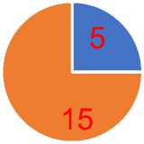
■ 1■2
Figure 3. Illustration of the results found after thorough micropy examination determine 15 cysticerci and 5 Sarcocysts
Conclusion
Cysticercosis and Sarcocystosis diseases are threat to cattle as well to human beings. These infections can be responsible of loss of weight, abortion and death of bovines and consequently cause loss of farmer’s economy, and reduced meat which are served to human as important food. Furthermore, due to the ability of these diseases to be transmitted to human they are known to threaten human health owing to infection in Cattle. Therefore, specific identication of cysticerci from sarcocysts will help in proper threatment of the above-mentioned diseases.
The identification of Cysticercus bovis from Sarcocystis spp. In routine apparent meat inspection by Veterinarians is not trustworthy. Out of 20 cases reported by Veterinarians to host putatively only cysticerci it was found that 5 cases of them were Sarcocysts. Thus, the error was found to be 25 % . These data are not enough to confirm whether it can be taken as a serious issue in Russian
Federation but it is indication of not relying on meat inspection data. Meat inspection should be used as preliminary screening tool for cysticerci. For more thorough researches with molecular methods for specific Cystercicus bovis and Sarco-cystis spp. identification are recommended.
post@vestnik-vsuet.ruAknowledgement
The authors thank the authority of Russian University of Biotechnology formerly Moscow State University of Food Production, for providing suitable facilities for this research work
References Reliability of veterinarians routine cattle meat inspection tool on specific identification of cysticerci in bovine muscle tissue
- Ahmed A.M., Elshraway N.T., Youssef A.I. Survey on Sarcocystis in bovine carcasses slaughtered at the municipal abattoir of El-Kharga, Egypt. Veterinary World. 2016. vol. 9. no. 12. pp. 1461. https://doi.org/10.14202/vetworld.2016.1461-1465
- Assefa A., Bihon A. Bovine cysticercosis in Ethiopia: a systematic review and meta-analysis of prevalence from abattoir-based surveys. Preventive veterinary medicine. 2019. vol. 169. pp. 104707. https://doi.org/10.1016/j.prevetmed.2019.104707
- Dubey J.P. Foodborne and waterborne zoonotic sarcocystosis. Food and Waterborne Parasitology. 2015. vol. 1. no.1. pp. 2-11. https://doi.org/10.1016/j.fawpar.2015.09.001
- El-Sayad M.H., Farag H., El-Taweel H., Fadly R. et al. Cysticercus bovis in cattle slaughtered in North Egypt: Overestimation by the visual inspection method. Veterinary World. 2021. vol. 14. no. 1. pp. 155. https://doi.org/10.14202/vetworld.2021.155-160
- Fayer R., Esposito D.H., Dubey J.P. Human Infections with Sarcocystis Species. Clin Microbiol Rev. 2015. vol. 28. pp. 295-311. https://doi.org/10.1128/CMR.00113-14
- Leal Filho W., Ternova L., Parasnis S.A., Kovaleva M. et al. Climate Change and Zoonoses: A Review of Concepts, Definitions, and Bibliometrics. IJERPH. 2022. vol. 19. pp. 893. https://doi.org/10.3390/ijerph19020893
- Lopes W.D.Z., Santos T.R., Soares V.E., Nunes J.L.N. et al. Preferential infection sites of Cysticercus bovis in cattle experimentally infected with Taenia saginata eggs. Research in Veterinary Science. 2011. vol. 90. pp. 84-88. https://doi.org/10.1016/j.rvsc.2010.04.014
- Macrelli M., Brena C., Reichel R., Boufana B. et al. Bovine cysticercosis outbreak in an indoor beef finisher farm in the North of England. Veterinary Record Case Reports. 2020. vol. 8. no. 3. pp. e001178. https://doi.org/10.1136/vetreccr-2020-001178
- Novak A., Novak M. Determining the quality of bovine meat in sarcocystosis. theory and practice of parasitic disease control. 2020. pp. 295-300. https://doi.org/10.31016/978-5-9902341-5-4.2020.21.295-300
- Rosenthal B.M. Zoonotic Sarcocystis. Research in Veterinary Science. 2021. vol. 136. pp. 151-157. https://doi.org/10.1016/j.rvsc.2021.02.008
- Uchendu G.O., Aganga A.O., Ama N.O., Madibela O.R. Investigation of Cysticercosis bovis prevalence using passive abattoir post-mortem inspection and active administration of structured non-participatory questionnaire to farmers in Botswana. Trop Anim Health Prod. 2021. vol. 53. pp. 49. https://doi.org/10.1007/s11250-020-02447-8
- Picard B., Gagaoua M. Muscle fiber properties in cattle and their relationships with meat qualities: An overview. Journal of Agricultural and Food Chemistry. 2020. vol. 68. no. 22. pp. 6021-6039. https://doi.org/10.1021/acs.jafc.0c02086
- Joo S.T., Kim G.D., Hwang Y.H., Ryu Y.C. Control of fresh meat quality through manipulation of muscle fiber characteristics. Meat science. 2013. vol. 95. no. 4. pp. 828-836. https://doi.org/10.1016/j.meatsci.2013.04.044
- Weber R., Herold C., Hollert H., Kamphues J. et al. Reviewing the relevance of dioxin and PCB sources for food from animal origin and the need for their inventory, control and management. Environmental Sciences Europe. 2018. vol. 30. pp. 1-42.
- Mughini-Gras L., Enserink R., Friesema I., Heck M. et al. Risk factors for human salmonellosis originating from pigs, cattle, broiler chickens and egg laying hens: a combined case-control and source attribution analysis. PloS one. 2014. vol. 9. no. 2. pp. e87933. https://doi.org/10.1371/journal.pone.0087933
- Blagojevic B., Antic D. Assessment of potential contribution of official meat inspection and abattoir process hygiene to biological safety assurance of final beef and pork carcasses. Food Control. 2014. vol. 36. no. 1. pp. 174-182. https://doi.org/10.1016/j.foodcont.2013.08.018
- Tizioto P.C., Decker J.E., Taylor J.F., Schnabel R.D. et al. Genome scan for meat quality traits in Nelore beef cattle. Physiological genomics. 2013. vol. 45. no. 21. pp. 1012-1020.
- Autio T., Tuunainen E., Nauholz H., Pirkkalainen H. et al. Overview of control programs for non-EU-regulated cattle diseases in Finland. Frontiers in Veterinary Science. 2021. vol. 8. pp. 688936. https://doi.org/10.3389/fvets.2021.688936
- Agus A., Widi T.S.M. Current situation and future prospects for beef cattle production in Indonesia-A review. Asian-Australasian journal of animal sciences. 2018. vol. 31. no. 7. pp. 976.
- Loiko M.R., de Paula C.M., Langone A.C., Rodrigues R.Q. et al. Genotypic and antimicrobial characterization of pathogenic bacteria at different stages of cattle slaughtering in southern Brazil. Meat Science. 2016. vol. 116. pp. 193-200. https://doi.org/10.1016/j.meatsci.2016.01.010
