
Obtaining and properties of bi-layer polyelectrolyte films based on chitosan and sodium hyaluronate
Journal: Bulletin of Voronezh State University of Engineering Technologies @vestnik-vsuet
Section: Химическая технология
Article in issue: 1 (107) т.88, 2026.
Free access
In this study, a method for preparing bilayer films based on a polyelectrolyte complex of sodium hyaluronate and chitosan is presented, with a view toward biomedical applications. The effects of the component mass ratio, layer deposition sequence, and film thickness on the structure, morphology, and mechanical properties of the resulting films were investigated. Formation of the polyelectrolyte complex was confirmed by FTIR spectroscopy, which revealed characteristic changes in the absorption bands of the polymers’ functional groups. The morphological features of the polymer films were evaluated by optical microscopy, and tensile testing was used to determine their mechanical performance. The optimal film properties were achieved at a hyaluronic acid (HA-Na) to chitosan (CH) mass ratio of 1:7 and a layer deposition sequence of HA-Na → CH. Increasing film thickness by increasing the volume of the deposited layers resulted in a pronounced improvement in tensile strength and elongation at break. Overall, the findings demonstrate that the properties of sodium hyaluronate/chitosan bilayer films can be purposefully tuned by varying processing parameters, making these materials promising biomedical film matrices for the treatment of injuries and tissue damage.
Similar articles in the section Pharmacology. Therapeutics. Toxicology
Short address: https://sciup.org/140314834
IDS: 140314834 | DOI: 10.20914/2310-1202-2026-1-306-315
Получение и свойства двухслойных полиэлектролитных плёнок на основе хитозана и гиалуроната натрия
В работе представлена методика получения двухслойных плёнок на основе полиэлектролитного комплекса гиалуроната натрия и хитозана, ориентированных на применение в биомедицинских целях. Изучено влияние массового соотношения компонентов, последовательности нанесения слоёв и толщины плёночного материала на структуру, морфологию и механические свойства получаемых плёнок. Формирование полиэлектролитного комплекса подтверждено методом ИК-Фурье спектроскопии, выявившим характерные изменения полос поглощения функциональных групп полимеров. В ходе работы определены морфологические характеристики полученных полимерных плёнок методом оптической микроскопии и физико-механические характеристики при растяжении. Показано, что оптимальные свойства плёночного материала достигаются при массовом соотношении гиалуроновой кислоты и хитозана 1:7 и последовательности нанесения слоёв HA-Na → CH. Установлено, что увеличение толщины плёнки за счёт повышения объёма наносимых слоев приводит к существенному росту прочностных характеристик и относительного удлинения. Полученные результаты свидетельствуют о возможности целенаправленного регулирования свойств двухслойных плёнок на основе гиалуроната натрия и хитозана путём варьирования технологических параметров, что делает разработанные материалы перспективными для использования в качестве биомедицинских плёночных матриксов при травмах и повреждениях.
Text of the scientific article Obtaining and properties of bi-layer polyelectrolyte films based on chitosan and sodium hyaluronate
The development of biocompatible and biodegradable polymer materials with controlled physicochemical and mechanical properties represents a critical frontier in modern biomedicine [1]. This is particularly evident in the fields of tissue engineering [2] and drug delivery systems [3], where the demand for advanced functional interfaces between synthetic materials and biological tissues is observed. The engineering of hydrophilic films and coatings capable of modulating the healing environment, for example by maintaining a moist wound bed, facilitating gas exchange, and serving as a barrier against infection, plays a crucial role in regenerative medicine [4].
Chitosan (CH), a natural linear polysaccharide, is derived from the deacetylation of chitin, a polymer found in crustacean shells, insect exoskeletons, and fungal cell walls [5]. It has been widely applied in medicine, pharmacology, and biotechnology [6,7]. As a cationic polyelectrolyte, its positive charge stems from the protonation of free amino groups in acidic conditions [8].
This unique structure confers a broad spectrum of biological activities, including inherent filmforming ability and broad-spectrum antimicrobial properties, which are attributed to its capacity to disrupt microbial membranes and bind toxins [9]. Its biodegradability by human enzymes like lysozyme and its ability to stimulate macrophage activity further enhance its profile for medical use [10]. Chitosan forms viscous solutions in weak organic acids, which can be cast into robust, transparent films upon drying [7]. Application films of pure chitosan are often limited by the brittle nature, necessitating the formation of composites for durable applications [11].
Hyaluronic acid (HA) is a naturally occurring anionic glycosaminoglycan, playing a crucial structural and regulatory role in the extracellular matrix of connective tissues [12]. It is integral to processes such as cell proliferation, migration, and signaling, which are fundamental to wound repair [13]. Its primary biological function is associated with exceptional hydrophilicity and waterbinding capacity, making it a key moisturizing agent in numerous biomedical and cosmetic
This is an open access article distributed under the terms of the Creative Commons Attribution 4.0 International License applications [14]. A notable limitation of high-molecular-weight HA is its large hydrodynamic size, which restricts deep tissue penetration, often causing it to remain on the surface [15]. To overcome this, its sodium salt, sodium hyaluronate (HA-Na), is frequently used. HA-Na offers improved stability and, due to its typically lower molecular weight or altered conformation, demonstrates enhanced ability to penetrate tissues. Furthermore, the molecular weight of hyaluronic acid (HA) and its sodium salt (HA-Na) is a critical parameter, since low-molecular-weight fragments are known to elicit pro-angiogenic and pro-inflammatory signaling responses, whereas high-molecular-weight species predominantly exhibit anti-angiogenic and structural space-filling functions [16].
The electrostatic interaction between the cationic amino groups of chitosan (–NH₃⁺) and the anionic carboxylate groups of hyaluronate (–COO⁻) causes the spontaneous formation of a polyelectrolyte complex (PEC) [17]. This selfassembly mechanism, based on charge neutralization, has advantages over chemical crosslinking, since it does not require the use of potentially cytotoxic reagents and therefore maintains the biocompatibility of the components. [18]. The resulting PEC exhibits enhanced mechanical stability in aqueous environments and reduced solubility compared to the individual polymers, making it highly suitable for obtaining biomedical films. The structure and properties of such complexes are highly sensitive to the polymer ratio, рН, ionic strength and the order of component assembly, which dictates the final stoichiometry and spatial organization of the network [19].
The layer-by-layer (LbL) deposition technique is particularly applicable for fabricating such complex-based materials. This method, based on the alternate adsorption of oppositely charged polyelectrolytes, enables the construction of thin films with nanometer-scale control over thickness and composition. A key advantage is the ability to regulate the final material's structure and properties by altering the sequence of layer deposition, which directly influences interdiffusion and complexation kinetics. The study of such two-layer systems is highly relevant, as they represent a fundamental and tunable model for more sophisticated multicomponent coatings. While the LbL technique is well-established for obtaining multilayered nano-assemblies, its application to fabricate macroscopic, free-standing bilayer films with a defined polyelectrolyte complex interface offers a simplified model for understanding structure-property relationships [20].
Systematic investigation of influence of the layer deposition subsequence on the morphology, intermolecular interaction and resulting functional properties of two-layer РЕСs is not well-study. Most studies focus on blended composites or multilayered systems, leaving a gap in understanding the asymmetric properties of a bilayer. Secondly, there is a notable scarcity of comprehensive data on the properties of bilayer PEC films from this pair, as opposed to blended composites or multilayered assemblies. Finally, there is a clear need for a detailed optimization of critical process parameters – such as polymer solution concentrations, volumes, and post-processing conditions – to achieve reproducible films with controlled characteristics for specific biomedical applications, such as wound dressings capable of stimulating regeneration and providing hemostatic and antibacterial functions [21, 22].
The purpose of this study is to develop, optimize, and comprehensively characterize two-layer films based on a PEC of sodium hyaluronate and chitosan fabricated by sequential LbL application, with a focused investigation into the effects of layer sequence, polymer concentrations on their mechanical properties.
Materials and Methods
The following reagents were used in this research: sodium hyaluronate (HA-Na, molecular weight 1.29 МDа) and chitosan (CH, molecular weight 0.2 МDа) were supplied by Bioprogress LLC (Russia), acetic acid (2% v/v aqueous solution) was purchased from Formula (Russia), sodium hydroxide (NаОН) and hydrochloric acid (НСl) solutions were obtained from LenReactiv JSC (Russia).
Sodium hyaluronate solutions were prepared by dissolving the polymer in distilled water at room temperature to achieve final concentrations of 0.25% and 0.5% (w/w). The mixtures were stirred on a magnetic stirrer at 400–600 rpm for 2–3 hours until homogenous gels were formed.
Chitosan solutions were obtained by dispersing the polymer in a 2% (v/v) aqueous acetic acid solution to reach final concentrations of 1.75% and 3.5% (w/w). The dispersions were stirred on a magnetic stirrer at room temperature (400–600 rpm) for 2–3 hours until complete dissolution.
The films were fabricated using a sequential layer-by-layer casting technique onto a plastic Petri dish. Initially, a layer of either Na-HA or CH solution at a specific concentration was applied and dried in an oven at 37 °C for 24 hours. Subsequently, a solution of the other polymer was applied onto the first dried layer and dried under identical conditions (37 °C, 24 hours).
After the drying process, all film samples underwent a short-term heat treatment in an oven at 100 °C for 5 minutes to stabilize the structure and enhance its strength via recrystallization of the polymer matrix.
The dried and heat-treated films were stored in dessicators over saturated aqueous potassium chloride (КСl) solutions to maintain a constant relative humidity of 70%, which prevented overdrying and ensured the stability of their characteristics.
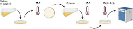
Figure 1. The process of forming a film of a polyelectrolyte complex based on sodium hyaluronate and chitosan
Mechanical properties of the samples were evaluated using a universal testing machine Instron 5966 (Instron, USA). The maximum static load of the testing system was 10 kN. The machine provided a crosshead travel of up to 1140 mm and a test speed range from 0.001 to 1500 mm • min⁻¹. The width of the test space was 418 mm, and the vertical test space was 1256 mm. Load measurements were performed with an accuracy of ± 0.5%.
Morphological characterization was carried out by optical microscopy using an Olympus SТМ6 microscope (Olympus, Tokyo, Japan). The microscope was equipped with a three-axis (XYZ) positioning system, providing a measurement range of up to 127 mm and a positioning accuracy of 0.1 μm.
Sample thickness was measured using a standard digital micrometer МКТs-25 (Tekhrim, Russia) with a measurement range of 0.001–25 mm.
Fourier transform infrared (FTIR) spectroscopy was employed to analyze the chemical structure of the samples. FTIR spectra were recorded using a Tensor 37 spectrometer (Bruker, Germany) at a spectral resolution of 2 cm⁻¹ with 32 scans per measurement. The spectral range covered 7500–375 cm⁻¹, corresponding to wavelengths of 1.33–26.7 μm. Measurements in the near-infrared (NIR) region were performed using the instrument extension in the range of 15,000–4000 cm⁻¹. The maximum achievable spectral resolution of the spectrometer was 0.5 cm⁻¹.
Results
The results demonstrate that the sequence of polymer layer deposition has a significant influence on film morphology and mechanical properties, as evidenced by optical microscopy (Fig. 2) and tensile testing (Fig. 3). At the optimal hyaluronic acid to chitosan mass ratio of 1:7, films were prepared by sequential deposition of a 0.5 wt.% sodium hyaluronate (HA-Na) solution as the first layer followed by a 3.5 wt.% chitosan (CH) solution (sequence: HA-Na → CH). This deposition sequence resulted in the formation of relatively homogeneous films with a smooth surface and a low aggregation degree. This material exhibited high tensile properties, characterized by a tensile strength of 28.59 МРа, a strength at break of 28.60 МРа, and a maximum strain of 27.37%. In contrast, the inverse deposition sequence, where a 3.5% (w/w) CH solution was the first layer and a 0.5% (w/w) HA-Na solution was the second (sequence: CH → HA-Na), led to films with significant heterogeneity, the presence of aggregates, and microdefects on the surface (Fig. 3). This morphology corresponded to substantially lower mechanical characteristics: tensile strength of 10.62 МРа, strength at break of 10.62 МРа, and maximum strain of 18.33%. Thus, the sequence HA-Na → CH provides superior mechanical properties and film integrity.
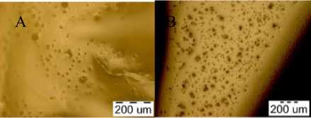
Figure 2. Surface morphology of bilayer films formed by different deposition sequences: A – sequential deposition of sodium hyaluronate followed by chitosan (HA-Na → CH); B – inverse deposition sequence, chitosan followed by sodium hyaluronate (CH → HA-Na)
Mechanical testing showed that the properties of the bilayer films strongly depend on the component ratio, layer deposition sequence, and film thickness. The use of a sodium hyaluronate solution with a concentration of 0.25% (w/w) resulted in the formation of a thin base layer with a thickness of 0.06 mm. Such films were characterized by a tensile strength of 15.25 МРа and a tensile strength a break of 15.35 МРа with a low maximum relative elongation of 1.81%. Increasing the volume of applied solutions led to an increase in the film thickness to 0.07 mm and a slight decrease in mechanical properties (to 13.98 and 14.01 МРа, respectively), accompanied by a moderate increase in the maximum relative elongation to 2.38%. At the same time, a sodium hyaluronate concentration of 0.5% (w/w) was found to be optimal for the formation of a uniform base layer. However, at a HA-Na to CH concentration ratio of 1:7 and a film thickness of 0.1 mm, the resulting films exhibited low mechanical properties, with a tensile strength of 0.81 МРа and a strength at break of 0.82 МРа. Film thickness was found to be directly dependent on the volume of the applied solutions. Thus, at a film thickness of 1.3 mm, the best mechanical performance was achieved, characterized by a high tensile strength of 16.20 МРа, a strength at break of 16.20 МРа, and a significantly increased maximum strain of 17.74%.
— НА-Na 0,5% (w/w) 20 ml, CH 3,5% (w/w) 20 ml CH 3,5% (w/w) 20 ml, HA-Na 0,5% (w/w) 20 ml
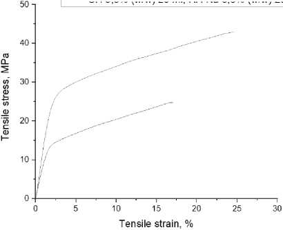
Figure 3. Mechanical testing of films with the sequence of layers CH → HA-Na and HA-Na → CH (1:7 и 7:1)
HA-Na 0,25% (w/w) 10 ml, CH 1,75% (w/w) 10 ml
HA-Na 0,25% (w/w) 20 ml, CH 1,75% (w/w) 20 ml
HA-Na 0,5% (w/w) 10 ml, CH 3,5% (w/w) 10 ml
HA-Na 0,5% (w/w) 20 ml, CH 3,5% (w/w) 20jml
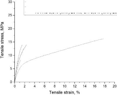
Figure 4. Tensile testing of films with various concentrations
The IR spectrum of pure sodium hyaluronate (HA-Na) is characterized by an intense absorbtion band at approximately 1410 cm⁻¹, corresponding to the symmetric stretching vibrations of carboxylate groups (–COO⁻), as well as bands in the 1000–1100 cm⁻¹ region associated with vibrations of the C–O–C and C–O bonds of the polysaccharide chain. The spectrum of pure chitosan (CH) exhibits a prominent distribution band at 1650 cm⁻¹, associated with the stretching vibrations of amino groups (–NH₂), as well as a band at approximately 1590 cm⁻¹, corresponding to the deformation vibrations of N–H bonds.
When examining bilayer films based on sodium hyaluronate and chitosan, the characteristic bands of both polymers undergo noticeable changes. The spectra of the samples show a shift in the chitosan amino group band around 1650 cm⁻¹, as well as a decrease in intensity of the sodium hyaluronate carboxylate band at approximately 1410 cm⁻¹, compared to the spectra of the individual polymers. These changes indicate the involvement of functional groups in intermolecular interactions.
In the FTIR spectra of bilayer films with a hyaluronic acid to chitosan mass ratio of 1:1, changes in the characteristic absorption bands of both polymers are also observed compared to the spectra of the pure components. A moderate shift of the chitosan amino group band in the region of 1650 cm⁻¹ is detected, along with a partial decrease in the intensity of the carboxylate band of sodium hyaluronate at approximately 1410 cm⁻¹. These spectral features indicate the onset of electrostatic interactions between – NH₂ groups of chitosan and – COO⁻ groups of sodium hyaluronate.
However, in contrast to the bilayer film with a hyaluronic acid to chitosan ratio of 1:7, the spectral changes observed for the 1:1 composition are less pronounced. The shifts of the characteristic bands are smaller, and the reduction in carboxylate band intensity is limited, suggesting incomplete charge compensation and a lower degree of ionic interaction between the oppositely charged functional groups. This indicates that, at a 1:1 mass ratio, the formation of the polyelectrolyte complex occurs to a lesser extent and is not optimal in terms of maximizing electrostatic interactions between the polymer components.
The most pronounced changes were recorded for the bilayer film with a 1:7 hyaluronic acid to chitosan mass ratio. For this example, the maximum band shift corresponding to the stretching vibrations of the amino groups are observed, as well as the widest distribution boundary for the carboxylate groups. Compared to the spectra of pure HA-Na and CH, this indicates more complete charge shielding and a greater degree of electrostatic bonding between oppositely charged functional units of the resulting polymers, indicating the most effective polyelectrolyte complex structures for these components.
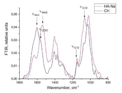
Figure 5. FTIR spectra of pure sodium hyaluronate (HANa) and chitosan (CH)
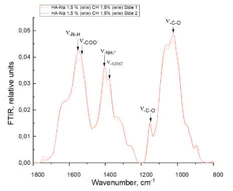
Figure 6. FTIR spectroscopy: sodium hyaluronate to chitosan ratio of 1:1
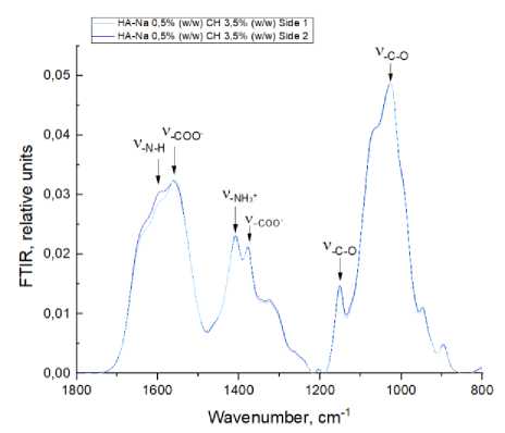
Figure 7. FTIR spectroscopy: sodium hyaluronate to chitosan ratio of 1:7
Discussion
The obtained results indicate that the formation of a sodium hyaluronate–chitosan polyelectrolyte complex, as well as the morphology and mechanical properties of the bilayer films, are largely governed by the component ratio, the sequence of layer deposition, and the film thickness.
The foundation for obtaining stable film structures is the complexation process between oppositely charged macromolecules. From a thermodynamic perspective, PEC formation is a spontaneous process driven by an increase in the system’s entropy due to the electrostatic interaction of ionic polymer groups [7]. However, the kinetics of this process and the morphology of the resulting complex critically depend on parameters such as the polymer ratio, the рН of the medium, and the order of layers [23]. The spectroscopic confirmation of PEC formation, characterized by the most pronounced shifts in the FTIR bands for the 1:7 HA-Na: CH ratio, underscores the importance of achieving an optimal stoichiometric balance.
This observation can be discussed in the context of charge density and the cooperative nature of polyelectrolyte interactions, where an excess of one component can drive the reaction to completion and stabilize the complex network [24].
The selection of the HA-Na: CH mass ratio of 1:7 is based on the principle of charge compensation in the forming complex [25]. This ratio ensures a stoichiometric balance between the carboxylate anions of HA-Na and the protonated amino groups of chitosan [6]. The excess of chitosan serves several important functions: firstly, it ensures the complete binding of carboxyl groups of HA-Na, preventing hydration and premature dissolution of the complex under physiological conditions; secondly, it obtains a dense structural framework due to the ability of chitosan molecules to form extensive hydrogen bonds and crystalline domains [26]; thirdly, it provides antimicrobial activity of the materials due to the high concentration of free amino groups not involved in complexation [27]. This structural advantage is directly reflected in the superior mechanical performance of films with a CH-rich composition.
The рН value of 5.5, selected for the preparation of solutions, is a parameter determining the degree of ionization of the functional groups of both polymers. For chitosan, with рКа = 6.3 – 6.5, at рН = 5.5, almost complete protonation of amino groups is ensured, which is necessary both for its dissolution and for subsequent electrostatic interaction with the anionic groups of HA-Na [7]. For hyaluronic acid (рКа = 2.5 – 3), this рН achieves an optimal balance between the degree of ionization of the carboxyl groups (ensuring reactivity) and the conformational stability of the polymer chain [28]. It should be noted that deviation from the optimal value in either direction disrupts the complexation process. At рН < 4, excessive protonation of the carboxyl groups of HA-Na is observed, reducing their capacity for electrostatic interaction. At рН > 6, deprotonation of chitosan amino groups begins, leading to its precipitation and disruption of the homogeneity of the formed film [27]. Thus, the рН value of 5.5 represents a compromise that ensures maximum PEC formation efficiency while maintaining the stability of individual polymer solutions, directly impacting the reproducibility and quality of the final films.
The method of sequential layer-by-layer polymer deposition with intermediate drying has proven crucial for determining final film properties. This approach allows for the formation of anisotropic structures with a controlled property gradient across the film thickness. From a physicochemical viewpoint, this approach separates the processes of primary polymer matrix formation and subsequent interphase complexation. Upon application and drying of the first HA-Na layer, a porous hydrophilic matrix is formed, characterized by a relatively low packing density of polymer chains due to steric hindrances associated with the hydrated hyaluronate macromolecules [29]. The subsequent application of the chitosan solution leads to its capillary diffusion into the pores of the first layer and volumetric complexation, which is confirmed by microscopy data showing the homogeneous structure and superior mechanical strength (tensile strength 28.6 МРа, strain 27.4%) of such films (HA-Na → CH sequence). The formation of PEC throughout the volume of the first layer obtains a unique biphasic structure combining mechanical strength (provided by dense complex domains) and elasticity (retained by regions with a predominance of HA-Na).
The inverse application sequence (chitosan as the first layer) leads to a fundamentally different morphology. The rapid gelation of chitosan during drying forms a dense, low-porosity matrix with a limited capacity for subsequent diffusion of HA-Na macromolecules [30]. Consequently, complexation occurs predominantly at the phase interface, obtaining a sharp property gradient and leading to structural heterogeneity, as observed in microscopy. This explains the observed defects and the significantly lower mechanical characteristics (tensile strength 10.6 МРа, strain 18.3%) of such samples. This finding aligns with principles of diffusionlimited reactions in polymer matrices [31].
The investigation into solution concentration and volume revealed another critical layer of control. While a 0.5% HA-Na base layer was optimal for uniformity, the applied volume was determinative for mechanical performance. The very low strength observed with 10 ml volumes of 0.5% HA-Na and 3.5% CH suggests the formation of an incomplete or discontinuous layer, perhaps too thin to form a coherent matrix. Crucially, increasing the volume to 20 ml of each solution yielded films with an optimal balance of high strength (16.2 МРа) and flexibility (17.7% strain), outperforming films made from lower-concentration solutions. This indicates that sufficient polymer mass per unit area is required to build an interpenetrated network during the sequential casting process. The data suggests that mechanical properties are not solely a function of concentration but of the absolute amount of polymer deposited, influencing layer thickness and the depth of inter-diffusion and complexation. For the PEC, thermal exposure stabilizes the complex by enhancing not only electrostatic but also hydrophobic interactions between polymer chains [32]. This could contribute to increased complex stability in aqueous environments and modulate biodegradation kinetics, which is particularly important for biomedical applications such as wound dressings that function in moist environments [33].
The obtained mechanical characteristics of the optimal films (e.g., HA-Na 0.5% (w/w) → CH 3.5% (w/w), 20 ml, tensile strength > 16 МРа, strain > 17%) demonstrate their promising suitability for use in regenerative medicine, particularly as wound dressings or scaffold materials. Comparable mechanical performance ranges have been reported for hyaluronic acid-based biomedical films intended for transdermal and wound-healing applications, where a balance between strength and elasticity is critical [34]. These values indicate a material that is strong enough to provide structural support and handle mechanical stress during application, yet sufficiently flexible. The retained elasticity is crucial for ensuring patient comfort and for withstanding dynamic movements in the application area. Furthermore, the inherent properties of the components – chitosan's antimicrobial and hemostatic activity combined with HA-Na's biocompatibility and hydrating capacity – obtain a multifunctional material whose physical properties are tailored through controlled fabrication.
Thus, a comprehensive analysis confirms the profound interdependence of stoichiometry, deposition sequence, solution parameters (concentration and volume), and post-processing conditions on the structure and function of HA-Na/CH PEC films. The systematic optimization described here provides a framework for designing materials with precisely controlled functional characteristics, from highly elastic to more rigid films, by adjusting these key variables. This opens significant prospects for tailoring such bilayer systems for specific clinical applications, ranging from elastic skin substitutes to more rigid guided tissue regeneration membranes. Future work should focus on correlating these in vitro properties with in vivo performance, including degradation profiles, bioactivity, and tissue integration.
Conclusion
This study developed and characterized two-layer polyelectrolyte complex (PEC) films based on sodium hyaluronate (HA-Na) and chitosan (CH) fabricated by a sequential layer-by-layer casting technique. The comprehensive investigation of key technological parameters – layer deposition sequence, polymer solution concentrations, and applied volumes – has elucidated fundamental structureproperty relationships and established a reproducible framework for engineering films with tailored characteristics for biomedical applications.
Fourier-transform infrared (FTIR) spectroscopy provided evidence of electrostatic interaction between the cationic amino groups of chitosan and the anionic carboxylate groups of hyaluronate.
The most pronounced spectral shifts were observed for the HA-Na: CH mass ratio of 1:7, indicating this to be the optimal stoichiometry for effective and complete complex formation. The excess chitosan secures complete binding of HA-Na and also contributes a robust structural framework and reserves free amino groups beneficial for antimicrobial functionality.
The sequence of polymer application is a decisive factor governing the internal structure and macroscopic properties of the bilayer films. Depositing HA-Na as the foundational layer followed by CH (HA-Na → CH sequence) resulted in homogeneous films with superior mechanical integrity (tensile strength 28.6 МРа, strain 27.4%). This is attributed to the formation of a porous, hydrophilic HA-Na matrix that facilitates the subsequent capillary diffusion and volumetric interpenetration of chitosan, leading to a deeply interdiffused PEC throughout the film thickness. Conversely, the inverse sequence (CH → HA-Na) produced heterogeneous films with surface defects and inferior mechanical properties (tensile strength 10.6 МРа), as the initially formed dense chitosan layer limits HA-Na diffusion, confining complexation primarily to a sharp interface and obtaining structural weaknesses.
The study demonstrates that film properties are governed not by concentration alone but by the synergistic effect of concentration and the absolute volume (mass) of polymer deposited per unit area. While a 0.5% (w/w) HA-Na solution was optimal for forming a uniform base layer, sufficient applied volume (20 ml in this setup) was crucial to achieve adequate layer thickness and polymer mass. The combination of 20 ml of 0.5% HA-Na and 20 ml of 3.5% CH produced films with an optimal balance of strength (16.2 МРа) and flexibility (17.7% strain). Lower volumes or concentrations resulted in films that were either too brittle or mechanically weak, highlighting the necessity of depositing
The optimized protocol proved essential for achieving reproducible films with enhanced stability. The selected рН ensures optimal ionization of both polymers for effective complexation. The thermal treatment likely enhances structural stability via hydrophobic interactions and recrystallization. Storage at 70% RH maintains an optimal water content, acting as a plasticizer to prevent embrittlement without causing over-hydration and dissolution of the PEC.
The mechanical properties of the optimal films (tensile strength > 16 МРа, elongation at break > 17%) align with the requirements for durable yet flexible biomaterials. The inherent bioactivity of the components makes this bilayer PEC system highly promising. It offers a versatile platform for obtaining functional materials, such as wound dressings that can provide moist wound healing, antimicrobial protection (from CH), enhanced tissue regeneration (from HA-Na), and appropriate mechanical support.
Thus, a comprehensive analysis confirms the profound interdependence of stoichiometry, deposition sequence, solution parameters (concentration and volume), and post-processing conditions on the structure and function of HA-Na/CH PEC films. The systematic optimization described here provides a framework for designing materials with precisely controlled functional characteristics, from highly elastic to more rigid films, by adjusting these key variables. This opens significant prospects for tailoring such bilayer systems for specific clinical applications, ranging from elastic skin substitutes to more rigid guided tissue regeneration membranes. Future work should focus on correlating these in vitro properties with in vivo performance, including degradation profiles, bioactivity, and tissue integration.